Review
. 2024 Nov 27;13(23):7217.
doi: 10.3390/jcm13237217.

Temporoparietal Fascia Flap (TPFF) in Extended Endoscopic Transnasal Skull Base Surgery: Clinical Experience and Systematic Literature Review

Affiliations
Review

Temporoparietal Fascia Flap (TPFF) in Extended Endoscopic Transnasal Skull Base Surgery: Clinical Experience and Systematic Literature Review

Martina Offi et al. J Clin Med. .

Abstract

Background and Objectives: The temporoparietal fascia flap (TPFF) has recently emerged as an option for skull base reconstruction in endoscopic transnasal surgery when vascularized nasal flaps are not available. This study provides a systematic literature review of its use in skull base surgery and describes a novel cohort of patients. Methods: PRISMA guidelines were used for the review. Patients undergoing skull base reconstruction with TPFF in our center from May 2022 to April 2024 were retrospectively included. Data were collected on pre- and post-operative clinical and radiological features, histology, surgical procedures, and complications. Results: Sixteen articles were selected, comprising 42 patients who underwent TPFF reconstruction for treatment of complex skull base pathologies. In total, 5 of 358 patients (0.9%) who underwent tumor resection via endoscopic transanal surgery in the study period in our institution required TPFF. All had been previously treated with surgery and radiation therapy for different pathologies (three chordomas, one giant pituitary neuroendocrine tumor (PitNET), and one sarcoma). Post-operative complications included CSF leak, which resolved after flap revision, and an internal carotid artery pseudoaneurysm requiring endovascular embolization. Conclusions: TPFF is an effective option for skull base reconstruction in complex cases and should be part of the armamentarium of the skull base surgeon.

Keywords: cranial base reconstruction; endoscopic transnasal surgery; neurosurgery; skull base reconstruction; temporo-parietal flap; vascularized flap.


Conflict of interest statement

The authors declare no conflicts of interest.

Figures

Figure 1
Figure 1
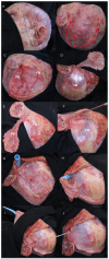
First and final nasal phases. (A) Opening of the maxillary sinus. (B) Opening of the anterior wall of the maxillary sinus. (C) Opening of the posterior wall of the maxillary sinus. (D) Identification of the maxillary artery. (E) Sectioning of the maxillary artery. (F) Identification of the temporal muscle, the infraorbital nerve and artery. (G) Passage of the Blue Rhino® guide (see text for details) for flap transposition. (H) Positioning of the folded TPFF to cover the bone defect. Abbreviations: IN, infraorbital nerve; MA, maxillary artery; MSm, medial wall of maxillary sinus; MSp, posterior wall of maxillary sinus; TM, temporal muscle.
Figure 2
Figure 2
Cranial phases. (A) The lines depict the different landmarks on the skin. Blue: Pitanguy’s line, Yellow: Temporal branch of the facial nerve, Black: Zygomatic arch, Red: STA. (B) Red dashed line: Incision. (C) After subcutaneous dissection, identification of STA, frontal (FB) and posterior branches (PB). (D) Incision of the flap utilizing the posterior branch of the STA with dissection from the temporal muscle fascia (TM). (E) TPFF flap with identification of FB and PB branches of the STA. (F) Subfascial dissection. (G) Placement of the Rhino percutaneous tracheostomy dilator in the infratemporal fossa. (H) Suturing of the TPFF to the guide. (I) Progressive transposition of the TPFF through the infratemporal fossa, pterygoid fossa, and into the nasal cavity by pulling the guide from the maxillary sinus into the nasal cavity. (J) Complete transposition of the TPFF without compression or bending of the flap base. Abbreviations: FB, frontal branch of superficial temporal artery; FC, facial nerve; PB, parietal branch of superficial temporal artery; STA, superficial temporal artery; ZA, zygomatic arch; TM, temporal muscle.
Figure 3
Figure 3
PRISMA flowchart of the systematic literature review.
Figure 4
Figure 4
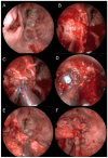
First and final nasal phases. (A) Defect of the previous surgery, with necrosis of the previous Hadad septal nose flap and recurrent CSF fistula. (B) Opening of the posterior wall of the maxillary sinus. (C) Identification and coagulation of the maxillary artery. (D) Passage of the Blue Rhino® guide for flap transposition. (E) Preparation of the folded TPFF. (F) Positioning of the folded TPFF to cover the bone defect. Abbreviations: MA, maxillary artery; MSp, posterior wall of maxillary sinus.
Figure 5
Figure 5
Cranial phases. (A) Preparation and incision of the flap utilizing the posterior branch of the STA with dissection from the temporal muscle fascia (TM) and placement of the Rhino percutaneous tracheostomy dilator in the infratemporal fossa. (B) Suturing of the TPFF to the guide. (C) Progressive transposition of the TPFF into the nasal cavity by pulling the guide until the guide completely comes out of the nasal cavity, then the suture is cut. (D) Complete transposition of the TPFF without compression or bending of the flap base (white arrow). Abbreviations: TM, temporal muscle.

References

    1. Prevedello D.M., Doglietto F., Jane J.A., Jagannathan J., Han J., Laws E.R. History of Endoscopic Skull Base Surgery: Its Evolution and Current Reality. J. Neurosurg. 2007;107:206–213. doi: 10.3171/JNS-07/07/0206. - DOI - PubMed
    1. Kassam A., Snyderman C.H., Mintz A., Gardner P., Carrau R.L. Expanded Endonasal Approach: The Rostrocaudal Axis. Part II. Posterior Clinoids to the Foramen Magnum. Neurosurg. Focus. 2005;19:E4. doi: 10.3171/foc.2005.19.1.5. - DOI - PubMed
    1. Kassam A., Snyderman C.H., Mintz A., Gardner P., Carrau R.L. Expanded Endonasal Approach: The Rostrocaudal Axis. Part I. Crista Galli to the Sella Turcica. Neurosurg. Focus. 2005;19:E3. doi: 10.3171/foc.2005.19.1.4. - DOI - PubMed
    1. Prevedello D.M., Ditzel Filho L.F.S., Solari D., Carrau R.L., Kassam A.B. Expanded Endonasal Approaches to Middle Cranial Fossa and Posterior Fossa Tumors. Neurosurg. Clin. N. Am. 2010;21:621–635. doi: 10.1016/j.nec.2010.07.003. - DOI - PubMed
    1. Dehdashti A.R., Ganna A., Witterick I., Gentili F. Expanded Endoscopic Endonasal Approach for Anterior Cranial Base and Suprasellar Lesions: Indications and Limitations. Neurosurgery. 2009;64:677–689. doi: 10.1227/01.NEU.0000339121.20101.85. - DOI - PubMed

LinkOut - more resources