

Assessment of the Mandibular Osseous Architecture in Cleft Lip and Palate Using Fractal Dimension Analysis: A Pilot Study
- PMID: 39685792
- PMCID: PMC11641932
- DOI: 10.3390/jcm13237334
Assessment of the Mandibular Osseous Architecture in Cleft Lip and Palate Using Fractal Dimension Analysis: A Pilot Study
Abstract
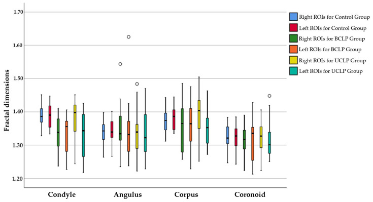
Background/Objectives: Although there has been extensive research on the orofacial morphologic effects of cleft lip and palate (CLP), the effects of CLP on mandibular structures remain largely unknown. The aim of this study was to investigate the trabeculation differences in the mandibular osseous architecture of patients with bilateral CLP (BCLP) and left-sided unilateral CLP (UCLP) using fractal dimension (FD) analysis and to compare these findings with healthy controls without CLP. Methods: A total of 63 patients (27 females, 36 males) with a mean age of 9.69 ± 1.5 years in the pre-peak growth stage were divided into three groups (n = 21 per group): the control group (CG), the BCLP group, and the UCLP group. The FD analysis was conducted on selected regions of interest (ROIs) from the mandibular condyle, angulus, corpus, and coronoid areas in TIFF-formatted panoramic radiographs. Statistical analyses were performed using the paired t-test and ANOVA for parametric data, and the Wilcoxon and Kruskal-Wallis tests for nonparametric data. Statistical significance was set at p < 0.05. Results: The FD values obtained from the ROIs of the right condyle were found to be significantly lower in the BCLP group compared to the CG and UCLP groups (p < 0.05). Conversely, the FD values for the left condyle were significantly higher in the CG group (p < 0.05), while no significant differences were observed between the BCLP and UCLP groups (p > 0.05). The FD value of the left condyle in the UCLP group was found to be significantly lower than that of the right condyle (p < 0.05). In the CG group, the FD values for both the right and left mandibular condyle and corpus were significantly higher than those for the angulus and coronoid regions; in the UCLP group, only the FD values of the right mandibular condyle and corpus were significantly higher than those for the same regions (p < 0.05). Conclusions: The reduced FD values in the mandibular condyle of CLP patients during the pre-peak growth stage suggest a loss of trabeculation and lower metabolic activity, while similarly, reduced FD values in the corpus region contribute to delayed tooth eruption timing, likely due to decreased masticatory forces during the intercuspal position and altered occlusal relationships. Clinical Relevance: In treating CLP patients, particularly with orthopedic face masks, the reduction in metabolic activities in these areas should be considered to achieve the optimal mandibular growth and development, and dental eruptions during the distribution of force from the chin to the corpus and condyle.
Keywords: bilateral cleft lip and palate; fractal dimension analysis; metabolic activity; trabeculation; unilateral cleft lip and palate.
Conflict of interest statement
The authors declare no conflicts of interest.
Figures


Similar articles
-
Mandibular Radiomorphometric Characteristics of Individuals with Bilateral or Unilateral Cleft Lip and Palate.Cleft Palate Craniofac J. 2024 Oct;61(10):1663-1669. doi: 10.1177/10556656231178504. Epub 2023 May 25. Cleft Palate Craniofac J. 2024. PMID: 37229644
-
Evaluation of the mandibular volume and correlating variables in patients affected by unilateral and bilateral cleft lip and palate: a cone-beam computed tomography study.Clin Oral Investig. 2016 Sep;20(7):1741-6. doi: 10.1007/s00784-015-1651-9. Epub 2015 Nov 10. Clin Oral Investig. 2016. PMID: 26556574
-
Bite force of children with repaired unilateral and bilateral cleft lip and palate.Arch Oral Biol. 2016 Aug;68:83-7. doi: 10.1016/j.archoralbio.2016.03.019. Epub 2016 Mar 31. Arch Oral Biol. 2016. PMID: 27107381
-
Evaluation of temporomandibular fossa and mandibular condyle in adolescent patients affected by bilateral cleft lip and palate using cone beam computed tomography.Scanning. 2016 Nov;38(6):720-726. doi: 10.1002/sca.21320. Epub 2016 Apr 22. Scanning. 2016. PMID: 27103610
-
Cranial Base Angle in Patients With Cleft Lip and Palate-A Systematic Review and Meta-Analysis.Cleft Palate Craniofac J. 2023 Jan;60(1):39-54. doi: 10.1177/10556656211053545. Epub 2021 Nov 17. Cleft Palate Craniofac J. 2023. PMID: 34787478
Cited by
-
Fractal Dimension Analysis of Mandibular Trabecular Architecture in Gingival Recession During Orthodontic Retention: A Cross-Sectional Study.Diagnostics (Basel). 2025 Apr 16;15(8):1013. doi: 10.3390/diagnostics15081013. Diagnostics (Basel). 2025. PMID: 40310431 Free PMC article.
-
Evaluation of the efficacy of clear aligners in mandibular advancement and their effect on mandibular trabecular structures using fractal dimension analysis.BMC Oral Health. 2025 Aug 21;25(1):1351. doi: 10.1186/s12903-025-05925-3. BMC Oral Health. 2025. PMID: 40841642 Free PMC article.
-
Assessment of bone mineral density by fractal dimension in OI patients treated with bisphosphonates.BMC Oral Health. 2025 Aug 23;25(1):1367. doi: 10.1186/s12903-025-06687-8. BMC Oral Health. 2025. PMID: 40849484 Free PMC article.
References
-
- Cash A.C. Orthodontic treatment in the management of cleft lip and palate. Front. Oral Biol. 2012;16:111–123. - PubMed
-
- Semer N.B. Practical Plastic Surgery for Nonsurgeons. Hanley & Belfus, Inc.; Philadelphia, PA, USA: 2001. Cleft Lip/Palate; pp. 235–243.
LinkOut - more resources
Full Text Sources
Miscellaneous
