Review
. 2024 Dec 7;13(23):7458.
doi: 10.3390/jcm13237458.

Biochemical Sensors for Personalized Therapy in Parkinson's Disease: Where We Stand

Affiliations
Review

Biochemical Sensors for Personalized Therapy in Parkinson's Disease: Where We Stand

Davide Ciarrocchi et al. J Clin Med. .

Abstract

Since its first introduction, levodopa has remained the cornerstone treatment for Parkinson's disease. However, as the disease advances, the therapeutic window for levodopa narrows, leading to motor complications like fluctuations and dyskinesias. Clinicians face challenges in optimizing daily therapeutic regimens, particularly in advanced stages, due to the lack of quantitative biomarkers for continuous motor monitoring. Biochemical sensing of levodopa offers a promising approach for real-time therapeutic feedback, potentially sustaining an optimal motor state throughout the day. These sensors vary in invasiveness, encompassing techniques like microdialysis, electrochemical non-enzymatic sensing, and enzymatic approaches. Electrochemical sensing, including wearable solutions that utilize reverse iontophoresis and microneedles, is notable for its potential in non-invasive or minimally invasive monitoring. Point-of-care devices and standard electrochemical cells demonstrate superior performance compared to wearable solutions; however, this comes at the cost of wearability. As a result, they are better suited for clinical use. The integration of nanomaterials such as carbon nanotubes, metal-organic frameworks, and graphene has significantly enhanced sensor sensitivity, selectivity, and detection performance. This framework paves the way for accurate, continuous monitoring of levodopa and its metabolites in biofluids such as sweat and interstitial fluid, aiding real-time motor performance assessment in Parkinson's disease. This review highlights recent advancements in biochemical sensing for levodopa and catecholamine monitoring, exploring emerging technologies and their potential role in developing closed-loop therapy for Parkinson's disease.

Keywords: Parkinson’s disease (PD); biochemical sensors; closed-loop therapy; levodopa.


Conflict of interest statement

Davide Ciarrocchi has no disclosures. Alessandro Zompanti has no disclosures. Giorgio Pennazza has no disclosures. Marco Santonico has no disclosures. Pasquale Maria Pecoraro has no disclosures. Lazzaro di Biase is the scientific director and one of the shareholders of Brain Innovations Srl, a University spinoff of Campus Bio-Medico University of Rome. The authors have no other relevant affiliations or financial involvement with any organization or entity with a financial interest in or conflict with the subject matter or materials discussed in this manuscript apart from those disclosed.

Figures
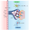
Figure 1
Figure 1
Synaptic metabolic pathways of levodopa. Abbreviations: AADC = aromatic L-amino acid decarboxylase; L-DOPA = levodopa; COMT = catechol-O-methyltransferase; 3-O-M-DOPA = 3-O-Methyldopa; DBH = Dopamine-β-hydroxylase; DAT = dopamine transporter; DR = dopamine receptor; D2-AR = dopamine autoreceptor; DOPAC = 3,4-Dihydroxyphenylacetic acid; MAO-B = monoamine oxidase-B; NE = norepinephrine; HVA = homovanilic acid.
Figure 2
Figure 2
Pharmacokinetic profiles of single doses of levodopa for early-stage PD (A) and advanced-stage PD (B). Top row (A): In early-stage PD, the pharmacokinetic profile of each levodopa dose (Dose 1 to 4) shows a gradual rise and fall in plasma levels. The concentration remains consistently between the OFF–ON transition threshold (lower dashed line) and the peak-dose dyskinesia threshold (upper dashed line), ensuring effective symptom control without inducing peak-dose or biphasic dyskinesias. The lower-right graph in this row represents the combined net effect of the four doses, showing a controlled and steady plasma levodopa concentration within the therapeutic range. Bottom row (B): In advanced-stage PD, while the pharmacokinetic behavior of each single dose (Dose 1 to 4) appears preserved, significant pharmacodynamic changes lead to a narrowing of the therapeutic window. The plasma concentrations more frequently exceed the peak-dose dyskinesia threshold, causing dyskinesias, and drop below the OFF–ON threshold, resulting in motor fluctuations such as “wearing-off”, “No-ON”, or “delayed ON” periods. The lower-right graph in this row illustrates the combined net effect of the four doses, characterized by increased variability and greater difficulty maintaining plasma levels within the therapeutic range. Legend: Lower dashed line: Plasma levodopa level [L-Dopa] threshold for OFF–ON transition (symptom improvement). Upper dashed line: Plasma levodopa level [L-Dopa] threshold for peak-dose dyskinesia (motor complications).
Figure 3
Figure 3
Configuration of IMU sensor data collection and implementation of a seven-layer convolutional neural network model. Figure drawn by Dr. Pfister, and reproduced, under the terms of the Creative Commons Attribution 4.0 License, from [36].
Figure 4
Figure 4
Schematic diagram illustrating biosensors for catecholamine detection, differentiated by surface modification. The sensors are categorized based on their applicability: minimally invasive, tested in vitro, and portable. Additionally, they are classified according to the type of sample analyzed.
Figure 5
Figure 5
Working process of two portable and wearable biosensor examples for L-Dopa sensing. (A) An SPCE with enzyme functionalization and hydrogel as the exchange interface for the biological fluid to be analyzed. The enzymatic reaction occurs on aSPCE modified with tyrosinase, where L-Dopa is oxidized to a dopaquinone layer, releasing two electrons (2e). The hydrogel layer plays a crucial role in facilitating the diffusion of L-Dopa to the electrode surface while maintaining a hydrated environment for the enzymatic reaction. (B) Enzyme-based microneedles with Nafion coating as antifouling layer. The microneedle tips are coated with a carbon paste electrode and modified with an enzyme layer of tyrosinase for the detection of L-Dopa. A Nafion coating is applied to enhance selectivity and stability. The enzyme catalyzes the oxidation of L-Dopa to dopaquinone, releasing electrons (2e), which are detected by the underlying electrode, generating an electrochemical signal for real-time monitoring. Abbreviations: SPCE = screen-printed carbon electrode; L-Dopa = Levodopa.
Figure 6
Figure 6
Overview of the DBS sampling process. 1. Blood collection is performed via a fingerstick. 2. The blood is applied onto specialized filter paper. 3. The process advances with the shipment of the dried blood spots to the laboratory. 4. The last step is the subsequent analysis of the samples through MS-ESI. Abbreviations: DBS = dried blood spot; MS-ESI = mass spectrometry–electrospray ionization.
Figure 7
Figure 7
The construction of a polymer-based electrochemical sensor utilizing PANI and WO3 NPs. 1. The process begins with the incorporation of WO3 NPs into the aniline polymerization solution, facilitating the synthesis of a composite material. 2. Subsequently, the PANI-WO3 composite is deposited onto the surface of a GCE, forming the active sensing layer for electrochemical detection. Abbreviations: PANI = polyaniline; NPs = nanoparticles; GCE = glassy carbon electrode; WO3 = tungsten trioxide.
Figure 8
Figure 8
Development of a carbon nanomaterial-based electrochemical sensor utilizing a biocompatible polymer, chitosan, and fCNTs. 1. The process begins with the solution-based synthesis of fCNTs, which are functionalized with carboxylic groups at 70 °C for 24 h. 2. Following synthesis, the fCNTs are dispersed into a chitosan solution to create a composite material. 3. Finally, the chit-fCNTs composite is deposited onto the surface of a GCE, forming the active layer for electrochemical sensing applications. This setup improves sensitivity for detecting specific analytes in biochemical sensing applications. Abbreviations: MWCNTs = multi-walled carbon nanotubes; fCNTs = functionalized multi-walled carbon nanotubes; GCE = glassy carbon electrode.
Figure 9
Figure 9
Fabrication of a metal nanoparticle-based electrochemical sensor utilizing Pd and Ni-MOF. 1. The process begins with the solvothermal synthesis of amine-terminated Ni-MOF using 2-aminoterephthalic acid. Ni(NO3)2 is added to the solution, which is then heated at 160 °C for 24 h. 2. PdCl2 and NaBH4 are dispersed into the Ni-MOF nanocomposite, which acts as a structural and conductive scaffold for the sensor to facilitate the reduction and incorporation of Pd nanoparticles. 3. Finally, the Pd@Ni-MOF composite is deposited onto the surface of a GCE, creating the active sensing layer for electrochemical detection. Abbreviations: Pd = palladium; Ni-MOF = nickel-based metal–organic framework; PdCl2 = palladium chloride; NaBH4 = sodium borohydride; GCE = glassy carbon electrode; Ni(NO3)2 = nickel nitrate.
Figure 10
Figure 10
Stepwise fabrication of an electrochemical enzyme biosensor. 1. First, the polishing of the GCE ensures a smooth and clean surface. 2. Then, AuNPs are electrodeposited onto the GCE using the amperometry technique, enhancing the electrode’s surface properties. 3. Finally, the amperometric polymerization of T3BA with PPO occurs on the surface of the AuNPs/GCE, forming a functional layer that facilitates enzyme immobilization and enhances biosensor performance. Abbreviations: AuNPs = gold nanoparticles; PT3BA = poly(3-thiophene boronic acid); PPO = polyphenol oxidase.
Figure 11
Figure 11
Steps involved in the fabrication of an electrochemical aptasensor. (1) The process begins by polishing the Au electrode to achieve a smooth and clean surface. (2) Then, spindle-shaped gold nanostructures are electrodeposited onto the Au electrode using the chronoamperometry technique, enhancing the electrode’s surface area and properties. (3) A thiolated RNA aptamer with dopamine binding sites is then deposited onto the surface, with MCH employed to minimize nonspecific attachment of aptamer molecules. (4) Finally, MB is deposited to further enhance electron transfer, optimizing the sensor’s electrochemical response. Abbreviations: Au = gold; MB = methylene blue; MCH = mercaptohexanol.
Figure 12
Figure 12
Multimodal adaptive closed-loop therapy system in advanced PD. Advanced therapeutic strategies—such as deep brain stimulation, oral medications, intestinal infusion therapy, and subcutaneous infusion therapy, used individually or in combination—can be delivered via an adaptive closed-loop framework. This system incorporates a modular, multiparametric sensing platform capable of monitoring local field potentials, biochemical markers, and kinematic data for personalized therapy optimization. Reproduced, under the terms of the Creative Commons Attribution 4.0 License, from [43].

References

    1. Cotzias G.C., Van Woert M.H., Schiffer L.M. Aromatic amino acids and modification of parkinsonism. N. Engl. J. Med. 1967;276:374–379. doi: 10.1056/NEJM196702162760703. - DOI - PubMed
    1. Abrams W.B., Coutinho C.B., Leon A.S., Spiegel H.E. Absorption and metabolism of levodopa. JAMA. 1971;218:1912–1914. doi: 10.1001/jama.1971.03190260028007. - DOI - PubMed
    1. Bryant B., Knights K. Pharmacology for Health Professionals Ebook. Elsevier Health Sciences; Philadelphia, PA, USA: 2014.
    1. Del Amo E.M., Urtti A., Yliperttula M. Pharmacokinetic role of L-type amino acid transporters LAT1 and LAT2. Eur. J. Pharm. Sci. 2008;35:161–174. doi: 10.1016/j.ejps.2008.06.015. - DOI - PubMed
    1. Masato A., Plotegher N., Boassa D., Bubacco L. Impaired dopamine metabolism in Parkinson’s disease pathogenesis. Mol. Neurodegener. 2019;14:35. doi: 10.1186/s13024-019-0332-6. - DOI - PMC - PubMed

LinkOut - more resources