

Exploring novel markers for coronary heart disease associated with systemic lupus erythematosus: A review
- PMID: 39686502
- PMCID: PMC11651451
- DOI: 10.1097/MD.0000000000040773
Exploring novel markers for coronary heart disease associated with systemic lupus erythematosus: A review
Abstract
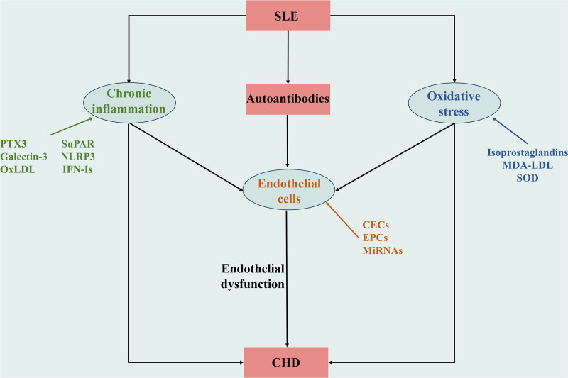
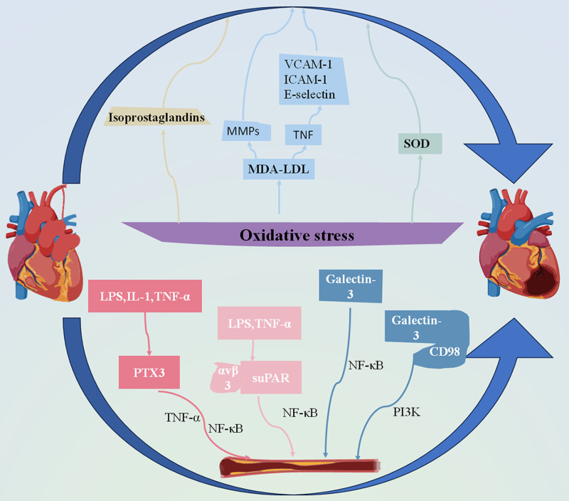
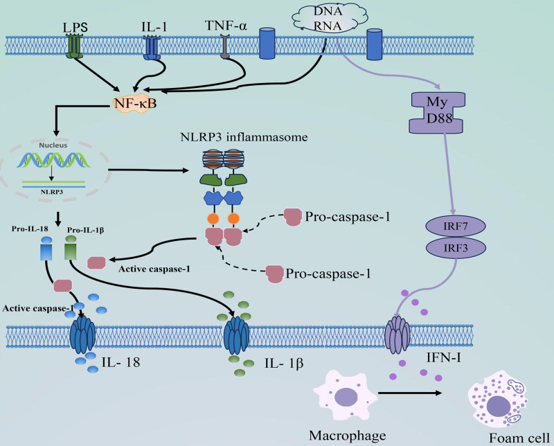
Systemic lupus erythematosus (SLE) is an autoimmune condition that is characterized by the production of autoantibodies and sustained inflammatory damage. Coronary heart disease (CHD) is a common complication of SLE, significantly increases CHD-related mortality in SLE patients. Despite conventional risk factors, the mechanisms contributing to a higher CHD risk require further investigation, with the immune and inflammatory aspects of SLE playing a significant role. Endothelial cell damage and dysfunction are key factors in the progression of coronary atherosclerosis in SLE patients. This review specifically focuses on endothelial dysfunction and the role of specific microRNAs in the context of SLE and CHD. In addition, we discuss the effects and functions of oxidative stress markers, endothelial progenitor cells, and circulating endothelial cells in individuals with both SLE and CHD. We also explored the typical inflammatory markers associated with SLE and CHD, addressing their clinical significance and limitations.
Copyright © 2024 the Author(s). Published by Wolters Kluwer Health, Inc.
Conflict of interest statement
The authors have no conflicts of interest to disclose.
Figures
Similar articles
-
Systemic lupus erythematosus: an independent risk factor for endothelial dysfunction in women.Circulation. 2004 Jul 27;110(4):399-404. doi: 10.1161/01.CIR.0000136807.78534.50. Epub 2004 Jul 19. Circulation. 2004. PMID: 15262847
-
'Not only...but also': factors that contribute to accelerated atherosclerosis and premature coronary heart disease in systemic lupus erythematosus.Rheumatology (Oxford). 2005 Dec;44(12):1492-502. doi: 10.1093/rheumatology/kei142. Epub 2005 Oct 18. Rheumatology (Oxford). 2005. PMID: 16234277 Review.
-
Risk factors for clinical coronary heart disease in systemic lupus erythematosus: the lupus and atherosclerosis evaluation of risk (LASER) study.J Rheumatol. 2010 Feb;37(2):322-9. doi: 10.3899/jrheum.090306. Epub 2009 Dec 1. J Rheumatol. 2010. PMID: 19955047
-
Risk factors for coronary heart disease in women with systemic lupus erythematosus: the Toronto Risk Factor Study.Arthritis Rheum. 2003 Nov;48(11):3159-67. doi: 10.1002/art.11296. Arthritis Rheum. 2003. PMID: 14613278
-
IFN-I Mediates Dysfunction of Endothelial Progenitor Cells in Atherosclerosis of Systemic Lupus Erythematosus.Front Immunol. 2020 Nov 11;11:581385. doi: 10.3389/fimmu.2020.581385. eCollection 2020. Front Immunol. 2020. PMID: 33262760 Free PMC article. Review.
References
-
- Durcan L, O’Dwyer T, Petri M. Management strategies and future directions for systemic lupus erythematosus in adults. Lancet. 2019;393:2332–43. - PubMed
-
- Fasano S, Milone A, Nicoletti GF, Isenberg DA, Ciccia F. Precision medicine in systemic lupus erythematosus. Nat Rev Rheumatol. 2023;19:331–42. - PubMed
-
- Wang L, Luqmani R, Udalova IA. The role of neutrophils in rheumatic disease-associated vascular inflammation. Nat Rev Rheumatol. 2022;18:158–70. - PubMed
Publication types
MeSH terms
Substances
Grants and funding
LinkOut - more resources
Full Text Sources
Medical
