. 2025 Jan 6;42(1):msae260.
doi: 10.1093/molbev/msae260.

Less, but More: New Insights From Appendicularians on Chordate Fgf Evolution and the Divergence of Tunicate Lifestyles

Affiliations

Less, but More: New Insights From Appendicularians on Chordate Fgf Evolution and the Divergence of Tunicate Lifestyles

Gaspar Sánchez-Serna et al. Mol Biol Evol. .

Abstract

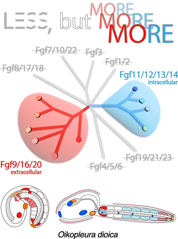
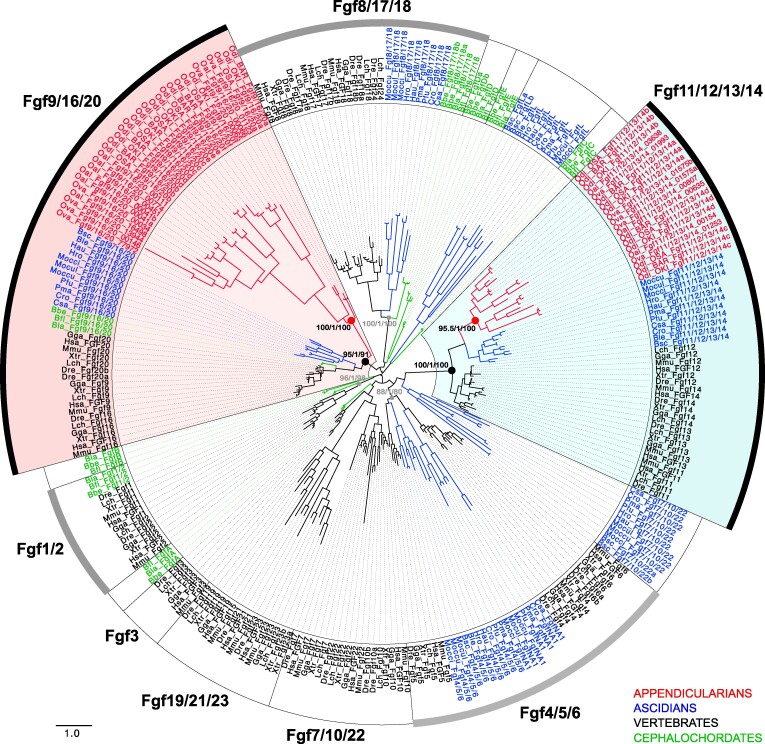
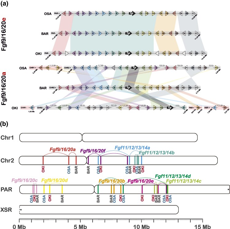
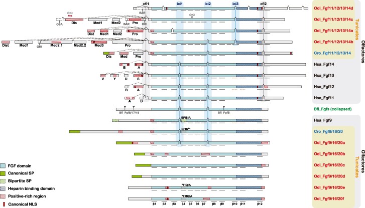
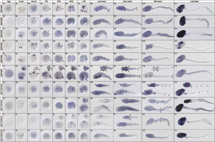
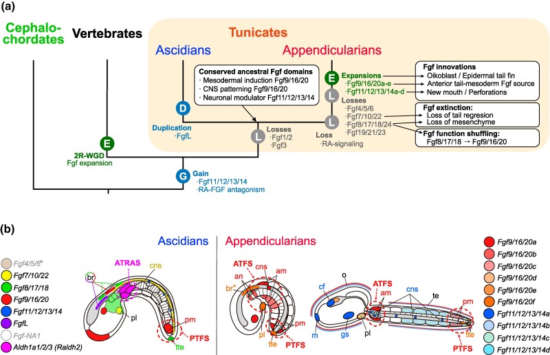
The impact of gene loss on the diversification of taxa and the emergence of evolutionary innovations remains poorly understood. Here, our investigation on the evolution of the Fibroblast Growth Factors (FGFs) in appendicularian tunicates as a case study reveals a scenario of "less, but more" characterized by massive losses of all Fgf gene subfamilies, except for the Fgf9/16/20 and Fgf11/12/13/14, which in turn underwent two bursts of duplications. Through phylogenetic analysis, synteny conservation, and gene and protein structure, we reconstruct the history of appendicularian Fgf genes, highlighting their paracrine and intracellular functions. An exhaustive analysis of developmental Fgf expression in Oikopleura dioica allows us to identify four associated evolutionary patterns characterizing the "less, but more" conceptual framework: conservation of ancestral functions; function shuffling between paralogs linked to gene losses; innovation of new functions after the duplication bursts; and function extinctions linked to gene losses. Our findings allow us to formulate novel hypotheses about the impact of Fgf losses and duplications on the transition from an ancestral ascidian-like biphasic lifestyle to the fully free-living appendicularians. These hypotheses include massive co-options of Fgfs for the development of the oikoblast and the tail fin; recruitment of Fgf11/12/13/14s into the evolution of a new mouth, and their role modulating neuronal excitability; the evolutionary innovation of an anterior tail FGF signaling source upon the loss of retinoic acid signaling; and the potential link between the loss of Fgf7/10/22 and Fgf8/17/18 and the loss of drastic metamorphosis and tail absorption in appendicularians, in contrast to ascidians.

Keywords: Oikopleura dioica; FGF signaling; alternative splicing; appendicularian; embryo development; gene duplication; gene loss; tunicate evolution; “less, but more”.


Figures

Graphical Abstract
Graphical Abstract
Fig. 1.
Fig. 1.
Evolutionary tree of the Fgf subfamilies in chordates. ML phylogenetic tree of the Fgf family in chordates reveals that the 10 Fgf genes found Oikopleura dioica, together with all other genes found in other appendicularian species (in red) group in two clusters with high support values (nodes with red solid circles). The tree topology indicates that the two clusters belong with high support (nodes with black solid circles) to two subfamilies: Fgf9/16/20 (red background) and Fgf11/12/13/14 (blue background). The presence of Fgfs from ascidians (in blue), vertebrates (in black), and cephalochordates (in green) allowed to infer that appendicularians have lost subfamilies Fgf8/17/18, Fgf19/21/23, Fgf7/10/22, and Fgf4/5/6. The absence of Fgf11/12/13/14 in cephalochordates suggests that this subfamily might be a synapomorphy of the olfactores. Well-supported nodes of other Fgf subfamilies (aBayes = 1) with members of more than one subphylum are indicated with gray solid circles. Node support values correspond to likelihood-based method aLRT-SH-like/aBayes/uf-boostrap. The scale bar indicates amino-acid substitutions. Ascidian Fgf names have been maintained according to previous works (Satou et al. 2002; Oulion et al. 2012), despite some of them show ambiguities with the tree topology (i.e. Fgf7/10/22, Fgf-NA1-19/21/23, and FgfL) and the lack of high support for those nodes. Species abbreviations: Vertebrates (in black): Danio rerio (Dre), Gallus gallus (Gga), Homo sapiens (Hsa), Latimeria chalumnae (Lch), Mus musculus (Mmu), Xenopus tropicalis (Xtr); Ascidian tunicates (in blue): Botrylloides leachii (Ble), Botrylloides schlosseri (Bsc), Ciona robusta (Cro), Ciona savignyi (Csa), Halocynthia aurantium (Hau), Halocynthia roretzi (Hro), Molgula occidentalis (Mocci), Molgula occulta (Moccu), Molgula oculata (Mocul), Phallusia fumigata (Pfu), Phallusia mammillata (Pma); Appendicularian tunicates (in red): Oikopleura albicans (Oal), Oikopleura dioica (Odi), Oikopleura vanhoeffeni (Ova); Cephalochordates (in green): Branchiostoma belcheri (Bbe), Branchiostoma floridae (Bfl), Branchiostoma lanceolatum (Bla).
Fig. 2.
Fig. 2.
Comparative synteny analysis of Fgf genes among the three Oikopleura dioica cryptic species from Barcelona (BAR), Osaka (OSA), and Okinawa (OKI). a) Comparison of microsynteny conservation between the genomic neighborhoods of Fgf genes (black arrow, and ten adjacent genes on each side). The BAR genome was used as the reference. Here, we show two illustrative examples, in which the Fgf9/16/20e neighborhood represents a case of high level of microsynteny conservation, and the Fgf9/16/20a neighborhood represents a case of low level of microsinteny conservation, especially when compared with OKI. The microsynteny comparison of the full O. dioica Fgf catalogue is provided in supplementary fig. S3, Supplementary Material online. b) Macrosynteny analysis comparing the position of Fgf genes at chromosome arm level. Each Fgf gene is labeled with a distinctive color.
Fig. 3.
Fig. 3.
Comparative gene structures of O. dioica and other chordate Fgf genes. Exon–intron organization supports phylogenetic classification of Fgf9/16/20 and Fgf11/12/13/14 paralogs of O. dioica. “cfi” denotes conserved core-flanking introns, and “ici” denotes conserved internal core introns. Bfl_Fgfs represent the common structure of cephalochordate Fgf genes, featuring two internal core introns (ici) within the FGF domain coding sequence. Gene-specific introns are depicted as arrowheads and dashed lines at their respective locations. Predicted functional motifs are indicated as described in the legend. Black underlines highlight the presence and location of β-sheets as predicted by AlphaFold2. Orange dashed underlines highlight the presence and location of β-sheets that have been empirically determined, even though the AlphaFold2 software does not predict them (Goetz et al. 2009; Olsen et al. 2003; Plotnikov et al. 2001). For comparative purposes, genes and motifs are not drawn to scale. Dashed lines indicate alternative splicing variants of Fgf11/12/13/14 (Dis, distal; Med, medial; Pro, proximal), and black dashed lines boxes indicate exon length differences between O. dioica cryptic species.
Fig. 4.
Fig. 4.
Developmental expression atlas of O. dioica Fgf genes. Whole-mount in situ hybridization images of O. dioica at various developmental stages: eggs (a-j1), 8-cell embryos (a-j2), 64-cell embryos (a-j3), incipient tailbud (ITB) embryos (a-j4), early tailbud (ETB) embryos (a-j5), mid tailbud (MTB) embryos (a-j6), late tailbud (LTB) embryos (a-j7), just hatchlings (a-j8), early hatchling larvae (a-j9), mid hatchling larvae (a-j10), and late-hatchling larvae (a-j11). Central images in each panel are left lateral views, oriented anterior to the left and dorsal to the top. Upper-right image insets (‘) are dorsal views of optical cross-sections at the levels indicated by black dashed lines. Black arrowheads label an stained cortical spot in unfertilized eggs; black double arrowheads label the A pair blastomeres in 8-cell embryos; orange arrowheads mark ingressing vegetal blastomeres in the 64-cell embryos; blue arrowheads label neural derivatives (cyan-blue labels the neural plate in 64-cell and ITB embryos, and the nerve cord in later stages; dark-blue labels the caudal ganglion, and pale-blue labels the anterior brain); light green arrowheads label epidermal domains in the trunk and light green double arrowheads label the area of the Langerhans receptors primordia; dark green arrowheads label epidermal domains in the tailbud tip; green dashed lines mark the lateral epithelium of the tail and the fins; purple arrowheads label undetermined endomesodermal domains in the trunk; magenta arrowheads mark the mouth primordium; magenta double arrowheads mark the pharyngeal slits; yellow arrowheads label notochord cells; red arrowheads label muscle precursor cells and muscle cells in the tail.
Fig. 5.
Fig. 5.
Evolutionary scenario of Fgf subfamilies in chordates. a) Evolutionary tree of chordate subphyla indicating main events of losses (L), gains (G), duplications (D), or expansions by burst of duplications (E), as well as main associated patterns of conservation, innovation, function shuffling and extinction of Fgf expression domains. 2R-WGD: two rounds of whole-genome duplication. b) Comparative schematic representation of the main expression domains of Fgf subfamily members between ascidians and appendicularians (tailbud stage in the left, and hatchling in the right). Distinct colors are assigned to each Fgf as indicated in the figure legend. FgfNA1 expression has not been described in ascidians, and Fgf4/5/6* has been detected maternally and widely throughout development with no obvious tissue-specific domains (Imai et al. 2004). In appendicularians, the expression of Fgf9/16/20 paralogs is the most abundant in tailbud stages, and the expression of Fgf11/12/13/14 paralogs is more obvious at hatchling stages. Asterisk (in br*) denotes that expression was found in a slightly earlier stage than the one represented in the figure. Ascidians show a characteristic PTFS (Posterior-Tail FGF Source) and ATRAS (Anterior-Tail RA Source), while in appendicularians the loss of RA signaling (Cañestro and Postlethwait 2007; Martí-Solans et al. 2016) might be related to the innovation of an ATFS (Anterior-Tail FGF Source), considering that the conserved RA-FGF antagonistic action emerged at the base of olfactores (Pasini et al. 2012; Bertrand et al. 2015). Abbreviations: an, anterior notochord; am, anterior muscle; br, brain; cf, ciliary funnel; cns, central nervous system; gs, gill slit; m, mouth; o, oikoblastic epithilium; pl, placode; pm, posterior muscle; te, tail epidermis; tte, terminal tail epidermis.

Similar articles

Cited by

References

    1. Albalat R, Cañestro C. Evolution by gene loss. Nat Rev Genet. 2016:17(7):379–391. 10.1038/nrg.2016.39. - DOI - PubMed
    1. Antoine M, Reimers K, Wirz W, Gressner AM, Müller R, Kiefer P. Fibroblast growth factor 3, a protein with a dual subcellular fate, is interacting with human ribosomal protein S2. Biochem Biophys Res Commun. 2005:338(2):1248–1255. 10.1016/j.bbrc.2005.10.079. - DOI - PubMed
    1. Bassham S, Cañestro C, Postlethwait JH. Evolution of developmental roles of Pax2/5/8 paralogs after independent duplication in urochordate and vertebrate lineages. BMC Biol. 2008:6(1):35. 10.1186/1741-7007-6-35. - DOI - PMC - PubMed
    1. Bassham S, Postlethwait J. Brachyury (T) expression in embryos of a larvacean urochordate, Oikopleura dioica, and the ancestral role of T. Dev Biol. 2000:220(2):322–332. 10.1006/dbio.2000.9647. - DOI - PubMed
    1. Bassham S, Postlethwait JH. The evolutionary history of placodes: a molecular genetic investigation of the larvacean urochordate Oikopleura dioica. Development. 2005:132(19):4259–4272. 10.1242/dev.01973. - DOI - PubMed

Substances

LinkOut - more resources