. 2024 Dec 13;38(24):e70235.
doi: 10.1096/fj.202400508RR.

Time-of-day control of mitochondria regulates NLRP3 inflammasome activation in macrophages

Affiliations

Time-of-day control of mitochondria regulates NLRP3 inflammasome activation in macrophages

James R O'Siorain et al. FASEB J. .

Abstract

Macrophages are innate immune cells that orchestrate the process of inflammation, which varies across time of day. This ensures appropriate biological timing of the immune response with the external environment. The NLRP3 inflammasome mediates IL-1-family cytokine release via pyroptosis. Mitochondria play a multifaceted role regulating NLRP3 inflammasome activity. Mitochondria exhibit distinct metabolic changes across time of day, which are influenced by clock genes. However, whether the macrophage clock regulates the NLRP3 inflammasome via mitochondrial control remains unclear. We find heightened mitochondrial membrane potential (Δψm) and enhanced NLRP3 inflammasome activation from peritoneal exudate cells (PECs) isolated at circadian time (CT) 12 compared to CT 0. In vitro time-of-day synchronization of bone-marrow derived macrophages (BMDMs) induced time-dependent differences in NLRP3 inflammasome activation. Myeloid-specific Bmal1-deletion enhanced NLRP3 inflammasome activity in PECs at CT0 and in unsynchronized BMDMs compared to controls. Pharmacologically disrupting Δψm in synchronized cells reduced NLRP3 inflammasome activation to comparable levels, and the same occurred with Bmal1-deletion. These results further demonstrate circadian clock timing of the NLRP3 inflammasome, which is dependent on mitochondrial function and driven through the circadian gene Bmal1.

Keywords: NLRP3 inflammasome; circadian rhythms; macrophages; mitochondria; pyroptosis.


Figures

FIGURE 1
FIGURE 1
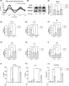
Clock timing impacts IL‐1 family cytokine release and cell death. (A) PER2::luciferase bioluminescent counts of synchronized BMDMs assessed for 48 h (n = 3). (B, C) BMAL1 protein expression from synchronized BMDMs (n = 3). (D–I) Bmal1, Nr1d1, Cry1, Cry2, Per1, and Per2 mRNA expression of BMDMs 16 h and 28 h post‐synchronization (PS) (n = 3). (J) IL‐1β released from NLRP3 inflammasome activated BMDMs at 16 h PS versus 28 h PS (n = 3). (K) IL‐18 released from NLRP3 inflammasome activated BMDMs at 16 h PS versus 28 h PS (n = 4). (L) Cell death assayed via release of LDH from NLRP3 inflammasome activated BMDMs at 16 h PS versus 28 h PS (n = 4). Data are expressed as mean ± SEM. N numbers represent biological samples with technical duplicates. (A) Circadian parameters measured with MetaCycle JTK cycle analysis (period = 23.9 h, p < .001). (C–I) Statistical analyses were conducted using Kruskal–Wallis with Dunn's multiple comparisons test. (J–L) Statistical analyses were conducted using two‐factor analysis of variance (ANOVA) with Tukey's multiple comparisons test. *p < 0.05, **p < 0.01, ***p < 0.001 and ****p < 0.0001 and ns, non significant.
FIGURE 2
FIGURE 2
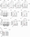
Time of day transcriptionally regulates levels of pro‐IL‐1β, independent of mitochondrial membrane potential. (A–D) Nlrp3, Il1b, Casp1, and Gsdmd mRNA expression of synchronized BMDMs treated with LPS (100 ng/mL, 3 h) 16 h PS and 28 h PS (n = 3). (E–G) NLRP3 and pro‐IL‐1β protein expression from LPS treated (100 ng/mL, 3 h) synchronized BMDMs at 16 h PS versus 28 h PS, with corresponding densitometry of relative quantity (RQ) normalized to β‐Actin (n = 3). (H, I) NLRP3 and pro‐IL‐1β protein expression from LPS (100 ng/mL, 3 h) and FCCP (100 nM, 1 h) treated synchronized BMDMs at 16 h PS versus 28 h PS, with corresponding densitometry of relative quantity (RQ) normalized to β‐Actin (n = 4). (K) Cellular reactive oxygen species (ROS) abundance measured via CellROX from synchronized BMDMs 16 h and 28 h PS treated with LPS (100 ng/mL, 3 h) followed by FCCP (100 nM, 1 h) (n = 4). Data are expressed as mean ± SEM. N numbers represent biological samples with minimum technical duplicates. (A–J) Statistical analyses were conducted using Kruskal–Wallis with Dunn's multiple comparisons test. (K) Statistical analyses were conducted using two‐factor analysis of variance (ANOVA) with Tukey's multiple comparisons test. *p < 0.05, **p < 0.01, ***p < 0.001 and ns, non significant.
FIGURE 3
FIGURE 3
NLRP3 inflammasome activation is time‐of‐day regulated and dependent on mitochondrial membrane potential. (A, B) Caspase‐1 (CASP1) p45 and p20 supernatant expression following NLRP3 inflammasome activation at 16 h and 28 h PS (n = 3). (C, D) Gasdermin‐D (GSDMD) p53 and p30 supernatant expression following NLRP3 inflammasome activation at 16 h and 28 h PS (n = 3). (E, F) Caspase‐1 (CASP1) p45 and p20 supernatant expression following NLRP3 inflammasome activation with FCCP inhibition at 16 h and 28 h PS (n = 3). (G, H) Gasdermin‐D (GSDMD) p53 and p30 supernatant expression following NLRP3 inflammasome activation with FCCP inhibition at 16 h and 28 h PS (n = 3). (I) IL‐1β released from NLRP3 inflammasome activated synchronized BMDMs with FCCP inhibition at 16 h PS versus 28 h PS (n = 3). (J) Cell death assayed via release of LDH from NLRP3 inflammasome activated synchronized BMDMs with FCCP inhibition at 16 h PS versus 28 h PS (n = 4). (K) TNFα release from synchronized BMDMs 16 h and 28 h PS treated with LPS (100 ng/mL, 3 h) followed by FCCP (100 nM, 1 h) (n = 4). Data are expressed as mean ± SEM. N numbers represent biological samples with minimum technical duplicates. (A–H) Statistical analyses were conducted using Kruskal–Wallis with Dunn's multiple comparisons test. (I–K) Statistical analyses were conducted using two‐factor analysis of variance (ANOVA) with Tukey's multiple comparisons test. *p < 0.05, **p < 0.01, ***p < 0.001 and ****p < 0.0001 and ns, non significant.
FIGURE 4
FIGURE 4
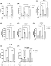
NLRP3 inflammasome activation is enhanced with Bmal1 deletion and dependent on mitochondrial membrane potential. (A) IL‐1β protein released from NLRP3 inflammasome activated unsynchronized Bmal1 +/+ and Bmal1 −/− BMDMs (n = 3/4). (B) Cell death assayed via release of lactate dehydrogenase from NLRP3 inflammasome activated unsynchronized Bmal1 +/+ and Bmal1 −/− BMDMs (n = 3). (C–F) Nlrp3, Il1b, Casp1 and Gsdmd mRNA expression of control and LPS (100 ng/mL, 3 h) treated Bmal1 +/+ and Bmal1 −/− BMDMs (n = 3/4). (G) IL‐1β release from NLRP3 inflammasome activated Bmal1 +/+ and Bmal1 −/− BMDMs with FCCP inhibition (n = 3). (H) Cell death assayed via release of LDH from NLRP3 inflammasome activated Bmal1 +/+ and Bmal1 −/− BMDMs with FCCP inhibition (n = 3). Data are expressed as mean ± SEM. N numbers represent biological samples. (A, B, G, H) Statistical analysis carried out by two‐factor ANOVA with Tukey's multiple comparisons test. (C–F) Statistical analyses were conducted using Kruskal–Wallis with Dunn's multiple comparisons test. *p < 0.05, **p < 0.01, ***p < 0.001 and ****p < 0.0001 and ns = non significant.

References

    1. Ko CH, Takahashi JS. Molecular components of the mammalian circadian clock. Hum Mol Genet. 2006;15(2):271‐277. doi:10.1093/hmg/ddl207 - DOI - PubMed
    1. O'Siorain JR, Curtis AM. Circadian control of redox reactions in the macrophage inflammatory response. Antioxid Redox Signal. 2022;37(10–12):664‐678. doi:10.1089/ars.2022.0014 - DOI - PubMed
    1. Ruan W, Yuan X, Eltzschig HK. Circadian rhythm as a therapeutic target. Nat Rev Drug Discov. 2021;20(4):287‐307. doi:10.1038/s41573-020-00109-w - DOI - PMC - PubMed
    1. Scheer FA, Hilton MF, Evoniuk HL, et al. The endogenous circadian system worsens asthma at night independent of sleep and other daily behavioral or environmental cycles. Proc Natl Acad Sci U S A. 2021;118(37):e2018486118. doi:10.1073/pnas.2018486118 - DOI - PMC - PubMed
    1. Hand LE, Hopwood TW, Dickson SH, et al. The circadian clock regulates inflammatory arthritis. FASEB J. 2016;30(11):3759‐3770. doi:10.1096/fj.201600353R - DOI - PMC - PubMed

MeSH terms

Substances

LinkOut - more resources