. 2024 Nov 15;27(12):111400.
doi: 10.1016/j.isci.2024.111400. eCollection 2024 Dec 20.

RNPS1 in PSAP complex controls periodic pre-mRNA splicing over the cell cycle

Affiliations

RNPS1 in PSAP complex controls periodic pre-mRNA splicing over the cell cycle

Kazuhiro Fukumura et al. iScience. .

Abstract

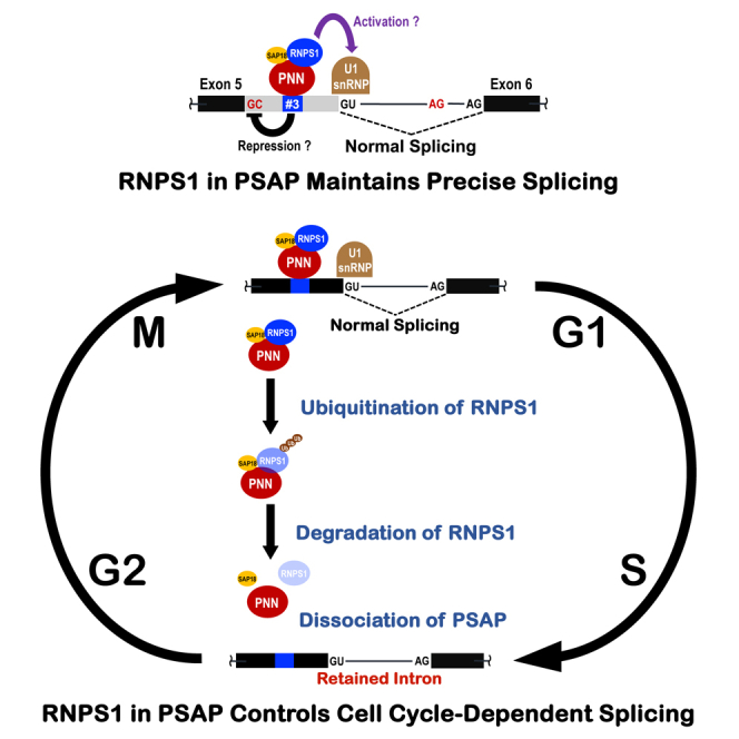
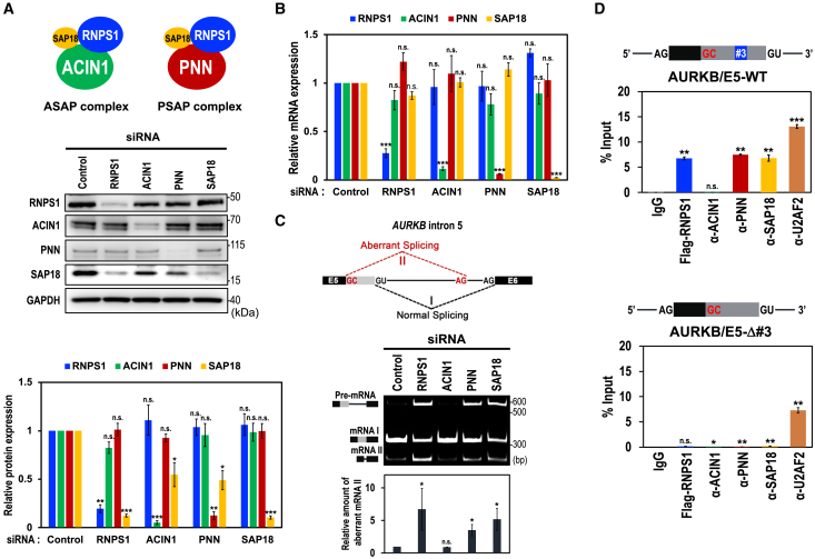
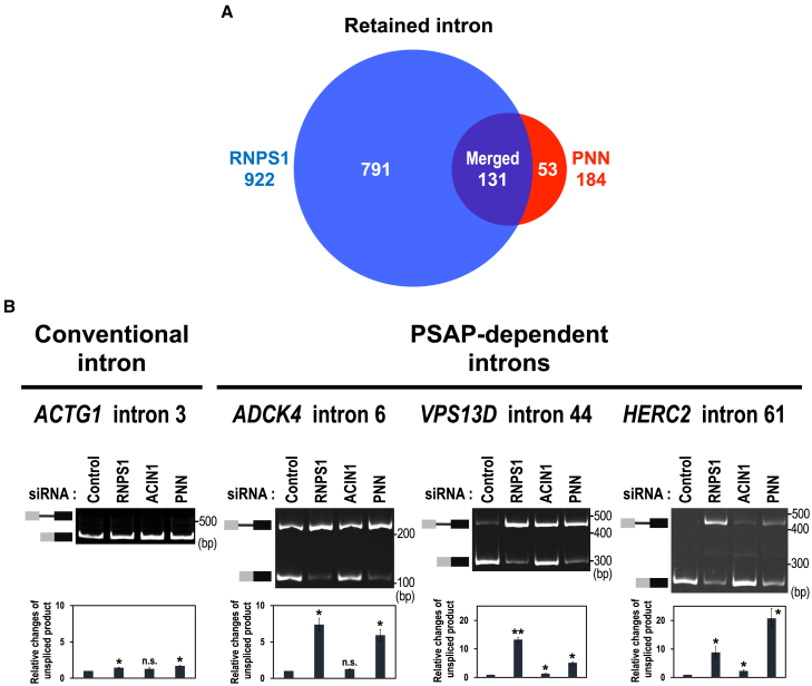
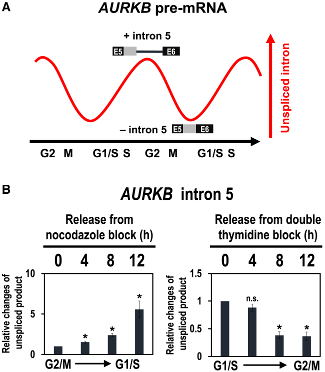
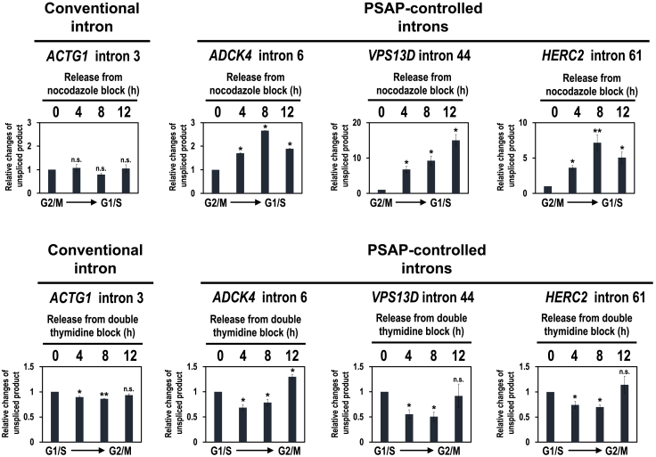
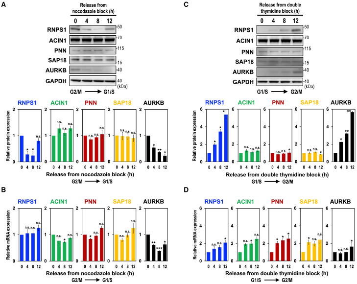
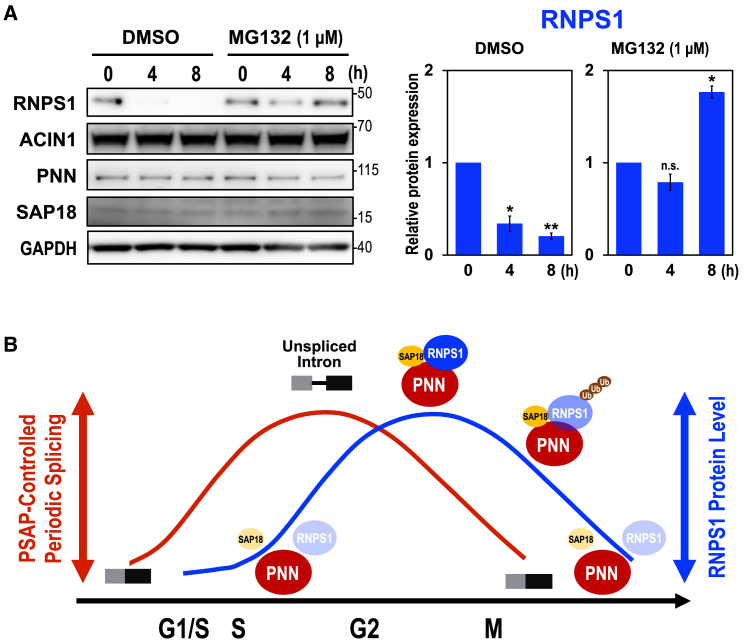
Cell cycle progression requires periodic gene expression through splicing control. However, the splicing factor that directly controls this cell cycle-dependent splicing remains unknown. Cell cycle-dependent expression of the AURKB (aurora kinase B) gene is essential for chromosome segregation and cytokinesis. We previously reported that RNPS1 is essential to maintain precise splicing in AURKB intron 5. Here we show that RNPS1 plays this role in PSAP complex with PNN and SAP18, but not ASAP complex with ACIN1 and SAP18. Whole-transcriptome sequencing of RNPS1- and PNN-deficient cells indicated that RNPS1, either alone or as PSAP complex, is an essential splicing factor for a subset of introns. Remarkably, protein expression of RNPS1, but not PNN, is coordinated with cyclical splicing in PSAP-controlled introns including AURKB intron 5. The ubiquitin-proteasome pathway is involved in the periodic decrease of RNPS1 protein level. RNPS1 is a key factor that controls periodic splicing during the cell cycle.

Keywords: Cell biology; Cellular physiology; Molecular biology; Properties of biomolecules.


Conflict of interest statement

The authors declare no competing interests.

Figures

None
Graphical abstract
Figure 1
Figure 1
PSAP component binds to a specific site that promotes normal splicing of the AURKB pre-mRNA (A) Schematic structures of ASAP and PSAP complexes (upper scheme). siRNA-mediated depletion of the indicated ASAP/PSAP components in HeLa cells were analyzed by Western blotting with indicated antibodies (middle panel). Individual bands on the Western blots were quantified and the relative values were standardized to that in the control siRNA (lower graph). Means ± standard errors (SE) are given for three independent experiments and Welch’s t-test values were calculated (∗p < 0.05, ∗∗p < 0.005, ∗∗∗p < 0.0005, n.s. p > 0.05). See also Figure S2A. (B) The mRNA levels of ASAP/PSAP components in (A) were analyzed by RT–qPCR using specific primer sets. See (A) for the statistical analysis. See also Figure S2B. (C) Splicing defect and aberrant splicing in the endogenous AURKB gene, induced by siRNA-mediated depletion of PSAP proteins, were detected by RT–PCR, visualized by PAGE, and individual bands on the PAGE gel were quantified. The ratios of the aberrantly spliced mRNA value (II) to the sum of the spliced mRNAs value (I + II) were standardized to that of the control siRNA and plotted. See (A) for the statistical analysis. See also Figures S1 and S3. (D) RNA immunoprecipitation assay was performed using HeLa cells co-transfected with Flag-RNPS1 expression plasmids and the indicated reporter AURKB-Exon 5 mini-genes. After immunoprecipitation using the indicated antibodies, immunoprecipitated RNAs were quantified by RT–qPCR using specific primers. See (A) for the statistical analysis.
Figure 2
Figure 2
RNPS1 and PNN are general splicing factors for individual subsets of introns (A) Venn diagram of retained introns that are generated by RNPS1- and PNN-knockdown HEK293 cells. See also Figure S4 and Table S2. The list of splicing changes (alternative 3′ splice site, alternative 5′ splice site, mutually exclusive exons, intron retention and cassette exon) in RNPS1-deficient HEK293 cells (related to Figures 2A, S4), Table S3. The list of splicing changes (alternative 3′ splice site, alternative 5′ splice site, mutually exclusive exons, intron retention and cassette exon) in PNN-deficient HEK293 cells (related to Figures 2A, S4), Table S4. The list of overlapping splicing changes (alternative 3′ splice site, alternative 5′ splice site, mutually exclusive exons, intron retention and cassette exon) in RNPS1- and PNN-deficient HEK293 cells (related to Figures 2A, S4). (B) In cellulo splicing assays of the pre-mRNAs including a conventional intron and three representative PSAP-dependent introns. After the indicated siRNA-mediated knockdown in HEK293 cells, indicated endogenous splicing was analyzed by RT–PCR followed by PAGE (upper panel). The unspliced RNA products were quantitated by RT–qPCR and the relative values were standardized to that in the control siRNA (lower graph). Means ± SE are given for three independent experiments and Welch’s t-test values were calculated (∗p < 0.05, ∗∗p < 0.005, n.s. p > 0.05).
Figure 3
Figure 3
Reported periodic AURKB pre-mRNA splicing is recapitulated in synchronized cells during the cell cycle (A) Schematic representation of cyclical splicing in AURKB intron 5 (modified from Dominguez et al.19). (B) HEK293 cells were synchronized in G2/M phase by treating with nocodazole and synchronized in G1/S phase by treating with double thymidine. Cells were washed, released, and harvested at the indicating time points. Endogenous splicing in the AURKB intron 5 was analyzed by RT–qPCR and the unspliced products were standardized to that at the start time, 0 h. Means ± SE are given for three independent experiments and Welch’s t-test values were calculated (∗p < 0.05, n.s. p > 0.05). See also Figures S5 and S6.
Figure 4
Figure 4
Periodic modulation of unspliced introns across the cell cycle was observed in PSAP-controlled introns but not in conventional intron HEK293 cells were synchronized in G2/M phase by nocodazole block (upper panels) and synchronized in G1/S phase by double thymidine block (lower panels). Endogenous splicing in the indicated conventional intron and PSAP-controlled introns was analyzed by RT–qPCR. Quantification and the statistical analysis were performed as described in Figure 3B (∗p < 0.05, ∗∗p < 0.005, n.s. p > 0.05). See also Figures S5 and S6.
Figure 5
Figure 5
The level of RNPS1 protein, but not other ASAP/PSAP proteins, is coordinated with AURKB protein level during the cell cycle (A) HEK293 cells were synchronized in G2/M phase by nocodazole block and released cells were harvested at indicated time points. ASAP/PSAP proteins were analyzed by Western blotting with indicated antibodies (upper panel). Individual bands on the Western blots were quantified and the relative values were standardized to that at the start time, 0 h (lower graph). Means ± SE are given for three independent experiments and Welch’s t-test values were calculated (∗p < 0.05, ∗∗p < 0.005, ∗∗∗p < 0.0005, n.s. p > 0.05). See also Figure S6. (B) mRNA levels in (A) were analyzed by RT–qPCR and the relative values were standardized to that at the start time, 0 h. See (A) for the statistical analysis. (C) HEK293 cells were synchronized at G1/S phase by double thymidine block and ASAP/PSAP proteins were analyzed as described in (A). See (A) for the statistical analysis. See also Figure S6. (D) mRNA levels in (C) were analyzed by RT–qPCR and the relative values were standardized to that at the start time, 0 h. See (A) for the statistical analysis.
Figure 6
Figure 6
PSAP-mediated cyclical splicing is controlled by RNPS1 protein level through the ubiquitin-proteasome pathway (A) HEK293 cells were cultured for 4 h and 8 h with proteasome inhibitor MG132 and the protein levels were analyzed by Western blotting using the antibody against each ASAP/PSAP protein. A common solvent, dimethyl sulfoxide (DMSO), was added to the medium (final concentration at 0.1%). The RNPS1 band on the Western blots was quantified and the relative values were standardized to that at the start time, 0 h (right graph). Means ± SE are given for three independent experiments and Welch’s t-test values were calculated (∗p < 0.05, ∗∗p < 0.005, n.s. p > 0.05). (B) A schematic model of RNPS1-mediated periodic splicing during cell cycle. RNPS1 protein, but neither PNN nor SAP18 protein, in PSAP plays an essential role to control periodic splicing over the cell cycle progression. The ubiquitin-mediated proteolysis of RNPS1 also destabilizes SAP18 that induces dissociation of PSAP complex, leading to the splicing defect.

References

    1. Will C.L., Lührmann R. Spliceosome structure and function. Cold Spring Harbor Perspect. Biol. 2011;3 doi: 10.1101/cshperspect.a003707. - DOI - PMC - PubMed
    1. Kastner B., Will C.L., Stark H., Lührmann R. Structural insights into nuclear pre-mRNA splicing in higher eukaryotes. Cold Spring Harbor Perspect. Biol. 2019;11 doi: 10.1101/cshperspect.a032417. - DOI - PMC - PubMed
    1. Mayeda A., Badolato J., Kobayashi R., Zhang M.Q., Gardiner E.M., Krainer A.R. Purification and characterization of human RNPS1: a general activator of pre-mRNA splicing. EMBO J. 1999;18:4560–4570. doi: 10.1093/emboj/18.16.4560. - DOI - PMC - PubMed
    1. Sakashita E., Tatsumi S., Werner D., Endo H., Mayeda A. Human RNPS1 and its associated factors: a versatile alternative pre-mRNA splicing regulator in vivo. Mol. Cell Biol. 2004;24:1174–1187. doi: 10.1128/MCB.24.3.1174-1187.2004. - DOI - PMC - PubMed
    1. Trembley J.H., Tatsumi S., Sakashita E., Loyer P., Slaughter C.A., Suzuki H., Endo H., Kidd V.J., Mayeda A. Activation of pre-mRNA splicing by human RNPS1 is regulated by CK2 phosphorylation. Mol. Cell Biol. 2005;25:1446–1457. doi: 10.1128/MCB.25.4.1446-1457.2005. - DOI - PMC - PubMed

LinkOut - more resources