. 2024 Oct 7:26:867-878.
doi: 10.1016/j.reth.2024.09.017. eCollection 2024 Jun.

Vitamin A-loaded decellularized kidney capsule promoted wound healing in rat

Affiliations

Vitamin A-loaded decellularized kidney capsule promoted wound healing in rat

Mozafar Khazaei et al. Regen Ther. .

Abstract

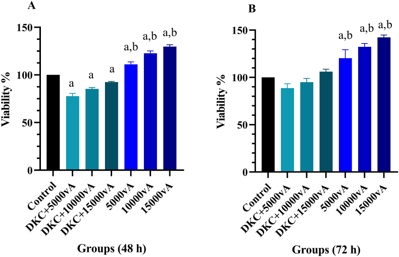
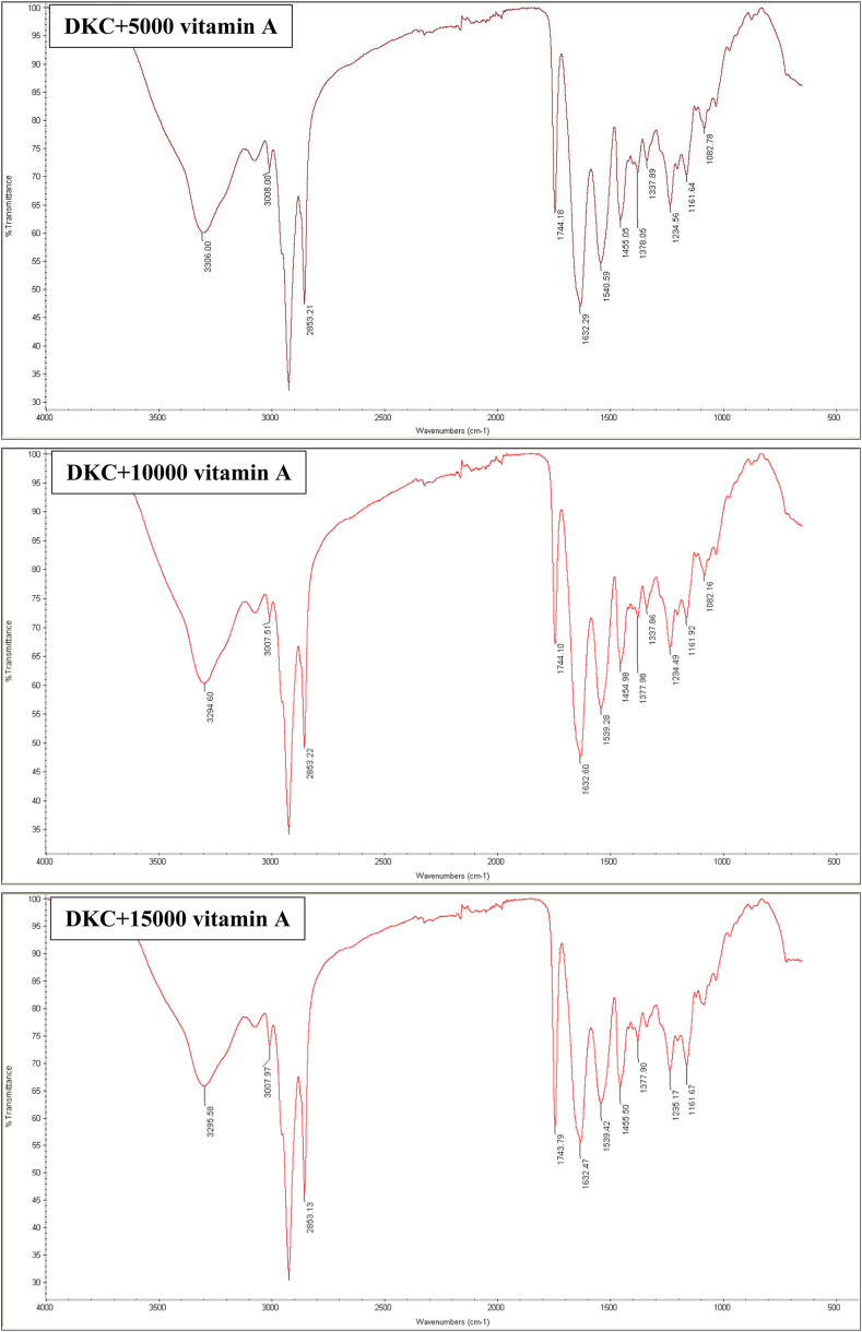
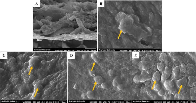
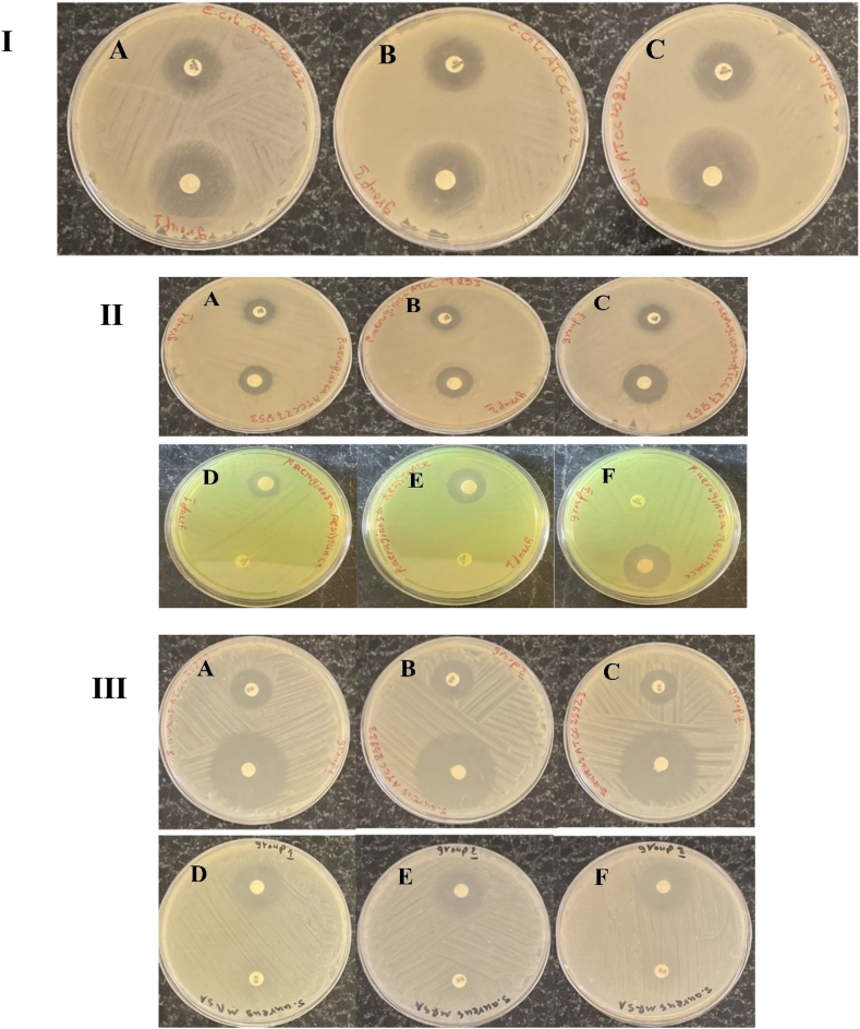
Tissue regeneration in many skin defects is progressing with new treatments in recent years. Tissue engineering with the use of scaffolds offers more versatile and faster solutions in treatment. Extracellular matrix (ECM) and its three-dimensional (3D) network structure as a biological bond by imitating the tissue microstructure has been used for tissue repair, which can answer many existing challenges. Vitamin A, which comes in several forms such as retinols, retinals, and retinoic acids, is a necessary vitamin that is crucial for wound healing. In this research, sheep kidney capsule tissue decellularized with sodium dodecyl sulfate (SDS) containing different doses of vitamin A has been used as an ECM in skin tissue engineering. The above scaffold was evaluated in terms of properties such as biocompatibility, analysis of mechanical properties, attenuated total reflection Fourier transform infrared spectroscopy (ATR-FTIR), hydrophilicity, antibacterial, and cell adhesion. The findings reported suitable properties for wound dressing, especially at a dose of 15,000 U/ml vitamin A for this scaffold. Then, the above scaffold was evaluated on the full-thickness wound model in rat, which showed good wound contraction, and increased VEGF factor. It showed a decrease in IL-1β level. Therefore, the use of the above-mentioned decellularized scaffold in combination with medicinal agents effective in wound healing can be introduced for further pre-clinical studies.

Keywords: Decellularization; Kidney capsule; Vitamin A; Wound healing.


Conflict of interest statement

The authors declare no conflicts of interest with respect to the research, authorship, and/or publication of this article.

Figures

Fig. 1
Fig. 1
A) MTT assay in 48 h and B) MTT assay in 72 h, Cytotoxicity after 48 and 72 h reported good cell survival in all experimental groups, survival at 72 h was reported higher than 48 h, %, a: Significant compared to the control group, b: Significant compared to DKCs containing vitamin A, the data presented are mean ± SD, n = 3, DKC: decellularized kidney capsules, 5000vA: 5000 U/ml vitamin A, 10000vA: 10,000 U/ml vitamin A, 15000vA: 15,000 U/ml vitamin A.
Fig. 2
Fig. 2
ATR-FTIR spectrum of DKCs loaded with different amounts of vitamin A, DKC: decellularized kidney capsules, 5000vA: 5000 U/ml vitamin A, 10000vA: 10,000 U/ml vitamin A, 15000vA: 15,000 U/ml vitamin A.
Fig. 3
Fig. 3
Mechanical test of DKCs loaded with different amounts of vitamin A, DKC: decellularized kidney capsules, 5000vA: 5000 U/ml vitamin A, 10000vA: 10,000 U/ml vitamin A, 15000vA: 15,000 U/ml vitamin A.
Fig. 4
Fig. 4
SEM images, A) X-4.0K cross-section of DKC, B) cell adhesion on DKC surface without vitamin A, C) cell adhesion on DKC loaded with 5000 U/ml vitamin A, D) cell adhesion on DKC loaded with 10,000 U/ml vitamin A, E) cell adhesion on DKC loaded with 15,000 U/ml vitamin A, The magnification in the images is related to adhesion X-2.5K, cells are indicated by orange arrows.
Fig. 5
Fig. 5
To measure the hydrophilicity of the surface of the DKCs containing vitamin A, the contact angle test was performed in Fig. 6 A, and WRC in Fig. 6 B, DKC: decellularized kidney capsules, 5000vA: 5000 U/ml vitamin A, 10000vA: 10,000 U/ml vitamin A, 15000vA: 15,000 U/ml vitamin A.
Fig. 6
Fig. 6
Diameter of inhibition zone of around scaffolds containing vitamin A in three group, and antibiotic disks, I) antibacterial assay of scaffolds against E. coli ATCC25922. The scaffolds containing Vitamin A shown significant antibacterial activity against E. coli ATCC25922. Bactericidal efficiency of scaffold in all three groups was better than meropenem antibiotic. Antibacterial activity of the scaffold in group 1 (Plate. A), group 2 (Plate. B), and group 3 (Plate. C) was 35 mm, 40 mm, and 42 mm, respectively, II) significant antibacterial activity of scaffolds against P. aeruginosa ATCC27853. Diameter of the growth inhibition zone of the scaffold in all three groups was higher than that of the imipenem antibiotic. Antibacterial activity of the scaffold in group 1 (Plate. A), group 2 (Plate. B), and group 3 (Plate. C) was 21 mm, 24 mm, and 23 mm, respectively. Antibacterial activity against P. aeruginosa resistant to meropenem. The highest antibacterial effect was related to the in group 3 (Plate. F), which created an inhibition zone with a diameter of 25 mm, but group 1(Plate. D), and group 2 (Plate. E) created an inhibition zone with a diameter of 19 mm, and 20 mm, III) the scaffolds show significant antibacterial activity against S. aureus ATCC27853. Diameter of the growth inhibition zone of the scaffold in all groups was higher than that of the methicillin antibiotic. Antibacterial activity in group 1 (Plate. A), group 2 (Plate. B), and group 3 (Plate. C) was 35 mm, 38 mm, and 40 mm, respectively. The highest antibacterial effect against S. aureus resistant to methicillin in group 3 (Plate. F) was significantly higher than that of the group 1 (Plate. D), and group 2 (Plate. E).
Fig. 7
Fig. 7
Studies on in vivo wound healing, A) 7 and 14 days following the damage, the macroscopic state of the wounds was evaluated, B) A comparison of the wound closure on days 7 and 14 following wounding is shown in the diagram. The values represent the mean ± standard deviation (n = 5). a, a’: significant compared to the control group (days 7 and 14), b, b’: significant compared to the DKC group (day 7 and 14), c’: significant compared to day 7 of the same group.
Fig. 8
Fig. 8
Evaluations of wound healing in histopathology, A) day 7, B) day 14, The yellow lines show the boundary between the healthy tissue and the tissue being repaired, white arrows: scab skin, red arrows: immature collagens, pink arrows: collagen in Masson trichrome staining and orange arrow: hair follicle.
Fig. 9
Fig. 9
A) The effect of DKC and DKC + vitamin A treatments on VEGF levels and B) IL-1β level in skin wounds after 7 and 14 days, the values represent the mean ± standard deviation (n = 5). a, a’: significant compared to the control group (days 7 and 14), b’: significant compared to the DKC group (day 7), c’: significant compared to day 7 of the same group.

References

    1. Khavkin J., Ellis D.A. Aging skin: histology, physiology, and pathology. Facial Plast Surg Clin. 2011;19(2):229–234. - PubMed
    1. Nguyen A.V., Soulika A.M. The dynamics of the skin's immune system. Int J Mol Sci. 2019;20(8):1811. - PMC - PubMed
    1. Arda O., Göksügür N., Tüzün Y. Basic histological structure and functions of facial skin. Clin Dermatol. 2014;32(1):3–13. - PubMed
    1. Nourian Dehkordi A., Mirahmadi Babaheydari F., Chehelgerdi M., Raeisi Dehkordi S. Skin tissue engineering: wound healing based on stem-cell-based therapeutic strategies. Stem Cell Res Ther. 2019;10:1–20. - PMC - PubMed
    1. Asghari F., Faradonbeh D.R., Malekshahi Z.V., Nekounam H., Ghaemi B., Yousefpoor Y., et al. Hybrid PCL/chitosan-PEO nanofibrous scaffolds incorporated with A. euchroma extract for skin tissue engineering application. Carbohydr Polym. 2022;278:118926. - PubMed

LinkOut - more resources