

A Rare Malignancy of the Eyelid: Report A Case of Primary Periocular Histiocytoid Carcinoma
- PMID: 39687454
- PMCID: PMC11646197
- DOI: 10.30699/IJP.2024.2016655.3219
A Rare Malignancy of the Eyelid: Report A Case of Primary Periocular Histiocytoid Carcinoma
Abstract
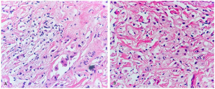
Primary periocular histiocytoid carcinoma is a very rare malignant tumor. Until now, less than 50 cases have been reported in the English literature. It is characterized by resistant epiphora, limitation in extraocular motility, and ptosis. The definitive diagnosis of this lesion is made based on detecting histological histiocytoid features along with tracing positivity of specific biomarkers using immunohistochemistry. However, pathologists may be faced with two major obstacles in the diagnosis of this tumor including distinguishing it from metastatic histiocytoid lesions and also from benign mimics such as reactive inflammatory lesions. Here, we describe a case of primary periocular histiocytoid carcinoma located on the eyelid as well as review the literature to clarify the histopathological and diagnostic features of this tumor.
Keywords: Eyelid malignancy; Histiocytoid carcinoma; Orbital malignancy; PPHC; Periorbital malignancy.
© 2024.
Conflict of interest statement
The authors declared no conflict of interest.
Figures

References
-
- Bernárdez C, Macías Del Toro E, Ramírez Bellver JL, et al. Primary Signet-Ring Cell/Histiocytoid Carcinoma of the Eyelid: A "Binocle" Presentation of the "Monocle Tumor". Am J Dermatopathol. 2016;38(8):623–7. - PubMed
-
- Ishida M, Okabe H. Primary signet-ring cell/histiocytoid carcinoma of the axilla. Pathol Int. 2013;63(7):374–376. - PubMed
-
- Berdugo J, Dumont-Mackay V, Brissy-Lachery S, et al. Cutaneous Apocrine Carcinoma With an In Situ Component and Histiocytoid and Signet-Ring Cells. Am J Dermatopathol. 2017;39(6):e76–8. - PubMed
-
- Warrick JI, Lewis JS Jr, Diaz JA. Pathology quiz case 2 Primary signet ring carcinoma of the eyelid. Arch Otolaryngol Head Neck Surg. 2010;136(11):1146–9. - PubMed
-
- Jakobiec FA, Stagner AM, Homer N, Yoon MK. Periocular Breast Carcinoma Metastases: Predominant Origin From the Lobular Variant. Ophthalmic Plast Reconstr Surg. 2017;33(5):361–6. - PubMed
Publication types
LinkOut - more resources
Full Text Sources