. 2024 Dec 2:15:1493946.
doi: 10.3389/fimmu.2024.1493946. eCollection 2024.

Entamoeba histolytica-induced NETs are highly cytotoxic on hepatic and colonic cells due to serine proteases and myeloperoxidase activities

Affiliations

Entamoeba histolytica-induced NETs are highly cytotoxic on hepatic and colonic cells due to serine proteases and myeloperoxidase activities

Fabian Jorge-Rosas et al. Front Immunol. .

Abstract

During intestinal and liver invasion by the protozoan parasite Entamoeba histolytica, extensive tissue destruction linked to large neutrophil infiltrates is observed. It has been proposed that microbicidal components of neutrophils are responsible for the damage, however, the mechanism by which they are released and act in the extracellular space remains unknown. In previous studies, we have shown that E. histolytica trophozoites induce NET formation, leading to the release of neutrophil granule content into extruded DNA. In this work, we evaluate the possible participation of NETs in the development of amoeba-associated pathology and analyze the contribution of anti-microbial components of the associated granules. E. histolytica-induced NETs were isolated and their effect on the viability and integrity of HCT 116 colonic and Hep G2 liver cultures were evaluated. The results showed that simple incubation of cell monolayers with purified NETs for 24 h resulted in cell detachment and death in a dose-dependent manner. The effect was thermolabile and correlated with the amount of DNA and protein present in NETs. Pretreatment of NETs with specific inhibitors of some microbicidal components suggested that serine proteases, are mostly responsible for the damage caused by NETs on HCT 116 cells, while the MPO activity was the most related to Hep G2 cells damage. Our study also points to a very important role of DNA as a scaffold for the activity of these proteins. We show evidence of the development of NETs in amoebic liver abscesses in hamsters as a preamble to evaluate their participation in tissue damage. In conclusion, these studies demonstrate that amoebic-induced NETs have potent cytotoxic effects on target cells and, therefore, may be responsible for the intense damage associated with tissue invasion by this parasite.

Keywords: Entamoeba histolytica; HCT 116; Hep G2; NETs; cell damage; neutrophils.


Conflict of interest statement

The authors declare that the research was conducted in the absence of any commercial or financial relationships that could be construed as a potential conflict of interest.

Figures
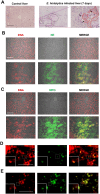
Figure 1
Figure 1
Amoebic liver abscesses contain NET-like structures. (A) Histological sections of a healthy liver and a liver infected with E. histolytica trophozoites after 7 days of infection stained with PAS. Black dotted lines in demarcate microabscesses filled with amoebae and necrotic tissue foci (asterisks), accompanied with a massive infiltration of immune cells surrounding these areas. A 40x magnification (purple dotted lines) also shows trophozoites (black arrows), inflammatory infiltrate (red dotted lines) and adjacent necrotic areas (asterisk). (B, C) Immunofluorescence images of amoebic liver microabscesses revealing the presence of neutrophil elastase (NE) and myeloperoxidase (MPO) (both in green), respectively, and their co-localization with DNA (in red), as seen in the merge boxes (yellow and orange). Areas of co-localization are considered as NETs. Chromatin decondensation was evidenced using the NUCLEAR-ID® Green Chromatin Condensation Detection Kit, showing neutrophils in early NETosis (D condensed nucleus colocalizing with NE) or in late NETosis (E decondensed nucleus colocalizing with NE). All scale bars: 100 μm. SnPMN: cell-free supernatant obtained from neutrophils culture only.
Figure 2
Figure 2
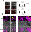
NETs induction by E. histolytica trophozoites and their effect on colon and liver cell monolayers. (A) Neutrophils (1 x 10⁴) and trophozoites (5 x 10³) were co-cultured on confluent (>90%) monolayers of HCT 116 or Hep G2 cells (72 h of grown) for 0.5, 1, and 5 h. Red dotted lines highlight areas of cell detachment. Samples were fixed, and images were captured using an inverted brightfield microscope (Nikon) at 5x magnification. (B) Quantification of detached area. (C) Neutrophils (1 x 10⁴) and trophozoites (5 x 10³) were cultured on a confluent (>90%) monolayer of HCT 116 or Hep G2 cells for 5 h. Samples were fixed, and DNA was stained with DAPI. Light blue boxes highlight areas of interaction between NETs, amoebae, and cell monolayer. Yellow arrows indicate neutrophils, arrowheads indicate NET-like structures, white arrows indicate trophozoites and, green arrow indicate nuclei of HCT 116 or Hep G2 cells. Images were obtained using an inverted fluorescence microscope (Nikon) at 20x magnification. For (A, C): scale bars represent 100 μm. E. h, E. histolytica trophozoites.
Figure 3
Figure 3
NETs induced by E. histolytica trophozoites affect the viability of colon and liver cell monolayers in vitro. (A) Human neutrophils (1 x 10⁵) were stimulated with viable E. histolytica trophozoites (5 x 10³, 2 x 10³, and 1 x 10³) at neutrophil:amoeba ratios of 20:1, 50:1, and 100:1, respectively, and extracellular DNA was quantified in relative fluorescence units (RFU) using SYTOX Green® (500 nM). PMA (50 nM) and A23187 (10 μM) were used as positive controls. NETs were purified from the previous samples, and the cell-free supernatant (SnNET) was added to confluent monolayers of HCT 116 cells (B) or Hep G2 cells (C) for 24 h. After this time, cell viability was assessed using the MTT assay. The amount of DNA and protein in the SnNET was quantified using a UV spectrometer (NanoDrop 2000) at 260 nm and 280 nm, respectively, before being added to cells. DMSO (20%) was used as a positive control for cytotoxicity and cell lines death. Data are shown as means from three independent experiments, each performed in triplicate. (#) indicates a statistical difference compared to the control (p < 0.05). *(p < 0.05), **(p < 0.01), and ***(p < 0.001). SnPMN, cell-free supernatant obtained from neutrophils culture only; SnAmb, cell-free supernatant obtained from amoebae culture only.
Figure 4
Figure 4
Effect of inhibitors and degraders of NETs components on the cytotoxic effect of NETs in HCT 116 cells. (A) Confluent HCT 116 monolayers were treated for 24 h with purified NETs (SnNET 20:1) or with NETs that were pretreated with sivelestat (ST; 10 µM), luminol (Lu; 50 µM), fluoromethylphenylsulfonyl fluoride (PMSF; 0.1 mM), DNase I (3 U), a combination of all treatments (Mix), or by heating to 60°C. Cell viability was determined using the MTT assay. Images of the treated monolayers after 24 h at 10x magnification are shown (middle). (B) NE activity in NETs (SnNET 20:1) without treatment or heating to 60°C, in the absence or presence of ST (10 µM). Data are presented from three independent experiments, each performed in triplicate. (#) indicates a statistical difference compared to the control (p < 0.05). *(p < 0.05), **(p < 0.01), and ***(p < 0.001). SnPMN: cell-free supernatant obtained from neutrophils culture only.
Figure 5
Figure 5
Effect of inhibitors and degraders of NETs components on the cytotoxic effect of NETs in Hep G2 cells. (A) NETs from neutrophils (3 x 10⁶) incubated with viable trophozoites (1.5 x 10⁵; SnNET 20:1) in the presence or absence of sivelestat (ST; 10 µM), luminol (Lu; 50 µM), fluoromethylphenylsulfonyl fluoride (PMSF; 0.1 mM), DNase I (3 U), and a combination of all NET inhibitors (Mix) was added to a confluent monolayer of Hep G2 cells for 24 h. Cell viability was assessed using the MTT assay. Images of the treated monolayers after 24 h at 10x magnification are shown (middle). (B) A monolayer of Hep G2 cells was pre-treated with the ROS indicator H2DCFDA for 10 min to measure ROS production over a 90-min interval. (C) A monolayer of Hep G2 cells was pretreated with a hydrogen peroxide detector for 30 min to measure hydrogen peroxide production using excitation and emission filters of 485/40 nm and 528/20 nm, respectively (D) or analyzed by fluorescence microscopy using an inverted fluorescence microscope (Nikon) at 10x magnification scale bar represents 100 μm. (E) A monolayer of Hep G2 cells was pretreated with a superoxide anion indicator to measure superoxide anion production. Absorbance was measured at 620 nm. (F) NETs from neutrophils (3 x 10⁶) incubated with viable trophozoites (1.5 x 10⁵; SnNET 20:1) in the presence or absence of iMPO (p-aminobenzohydrazide, 40 µM) was added to a monolayer of Hep G2 cells for 24 h. Cell viability was determined using the MTT assay. (G) MPO activity was measured in NETs from neutrophils (3 x 10⁶) incubated with viable trophozoites (1.5 x 10 5 ; SnNET 20:1) in the absence or presence of iMPO (40 µM) and after heating to 60°C in the absence or presence of iMPO. For (A, F) DMSO (20%) was used as a positive control for cytotoxicity and Hep G2 line cell death. (#) indicates a statistical difference compared to the control (p < 0.05). *(p < 0.05), **(p < 0.01), and ***(p < 0.001). SnAmb, cell-free supernatant obtained from amoebae culture only. SnPMN: cell-free supernatant obtained from neutrophils culture only.

References

    1. Carrero JC, Reyes-López M, Serrano-Luna J, Shibayama M, Unzueta J, León-Sicairos N, et al. . Intestinal amoebiasis: 160 years of its first detection and still remains as a health problem in developing countries. Int J Med Microbiol. (2020) 310:151358. doi: 10.1016/j.ijmm.2019.151358 - DOI - PubMed
    1. Nasrallah J, Akhoundi M, Haouchine D, Marteau A, Mantelet S, Wind P, et al. . Updates on the worldwide burden of amoebiasis: A case series and literature review. J Infect Public Health. (2022) 15:1134–41. doi: 10.1016/j.jiph.2022.08.013 - DOI - PubMed
    1. Bottazzi ME, Dumonteil E, Valenzuela JG, Betancourt-Cravioto M, Tapia-Conyer R, Hotez PJ. Bridging the innovation gap for neglected tropical diseases in Mexico: capacity building for the development of a new generation of antipoverty vaccines. Bol Med Hosp Infant Mex. (2011) 2:138–46.
    1. Roy M, Chakraborty S, Kumar S, Srivastava, Kaushik S, Jyoti A, Kumar V, Srivastava. Entamoeba histolytica induced NETosis and the dual role of NETs in amoebiasis. Int Immunopharmacol. (2023) 118:110100. doi: 10.1016/j.intimp.2023.110100 - DOI - PubMed
    1. Watanabe K, Gilchrist CA, Uddin MJ, Burgess SL, Abhyankar MM, Moonah SN, et al. . Microbiome-mediated neutrophil recruitment via CXCR2 and protection from amebic colitis. PloS Pathog. (2017) 13:e1006513. doi: 10.1371/journal.ppat.1006513 - DOI - PMC - PubMed

LinkOut - more resources