

Peroneal Tendon Tears: Four Simple-to-Complex Cases
- PMID: 39687829
- PMCID: PMC11647192
- DOI: 10.7759/cureus.73787
Peroneal Tendon Tears: Four Simple-to-Complex Cases
Abstract
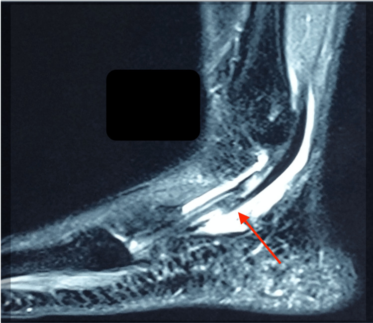
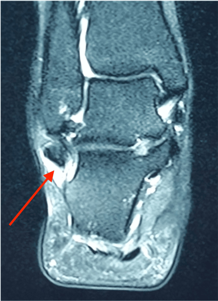
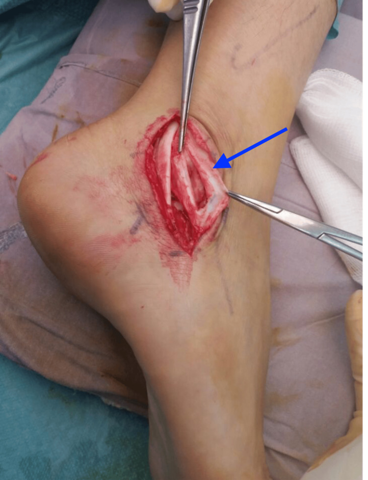
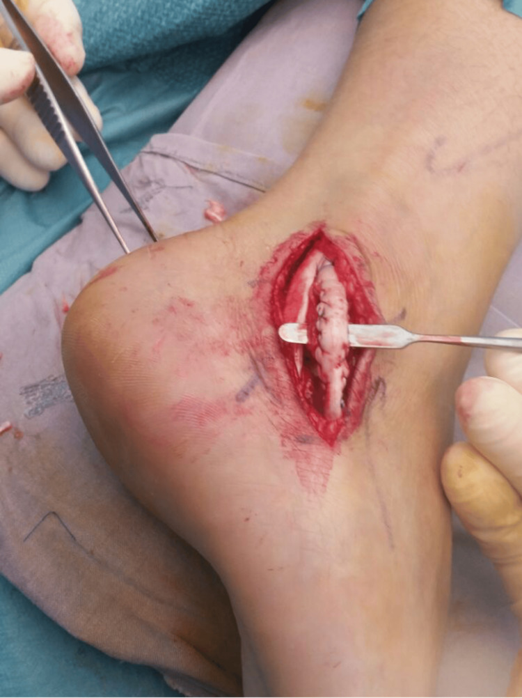
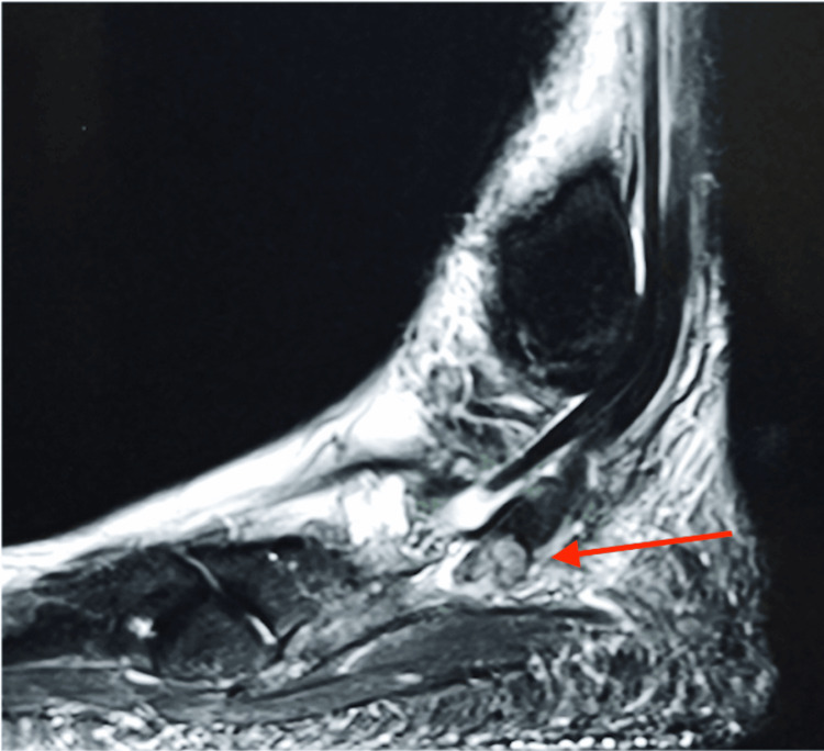
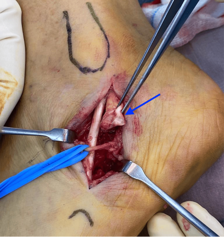
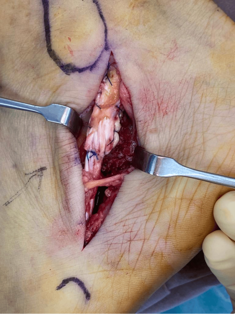
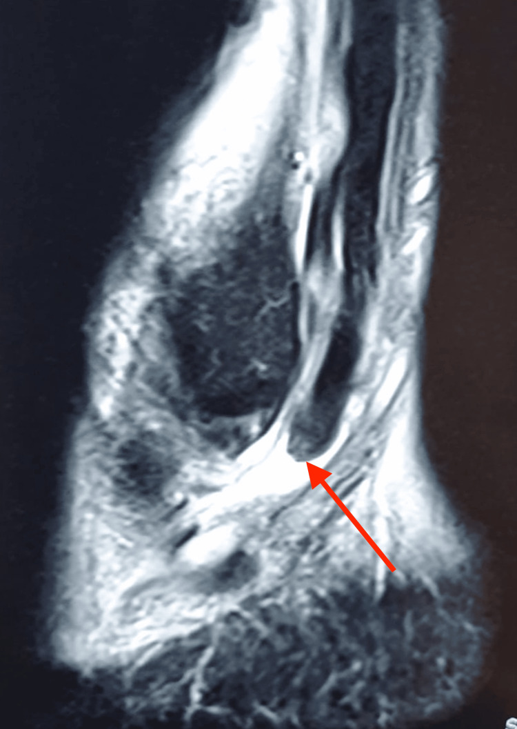
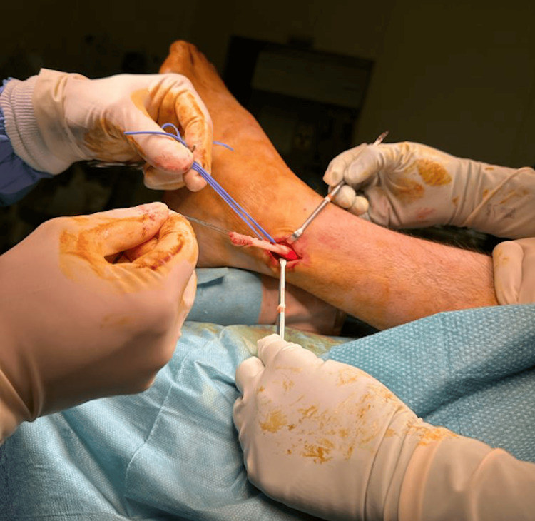
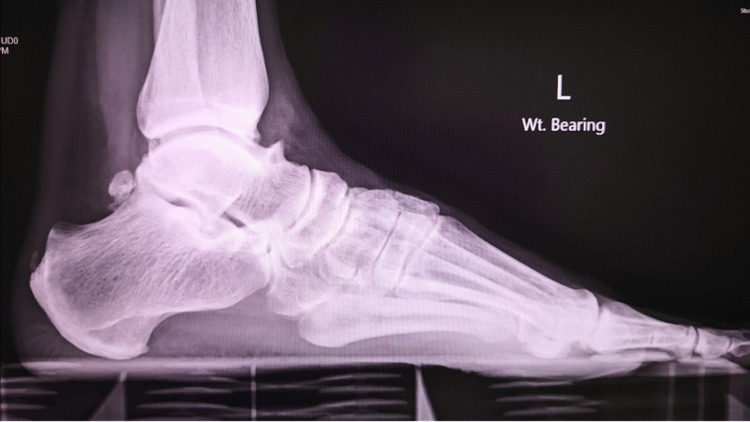
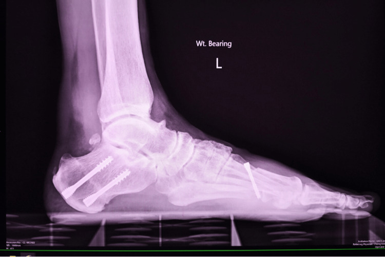
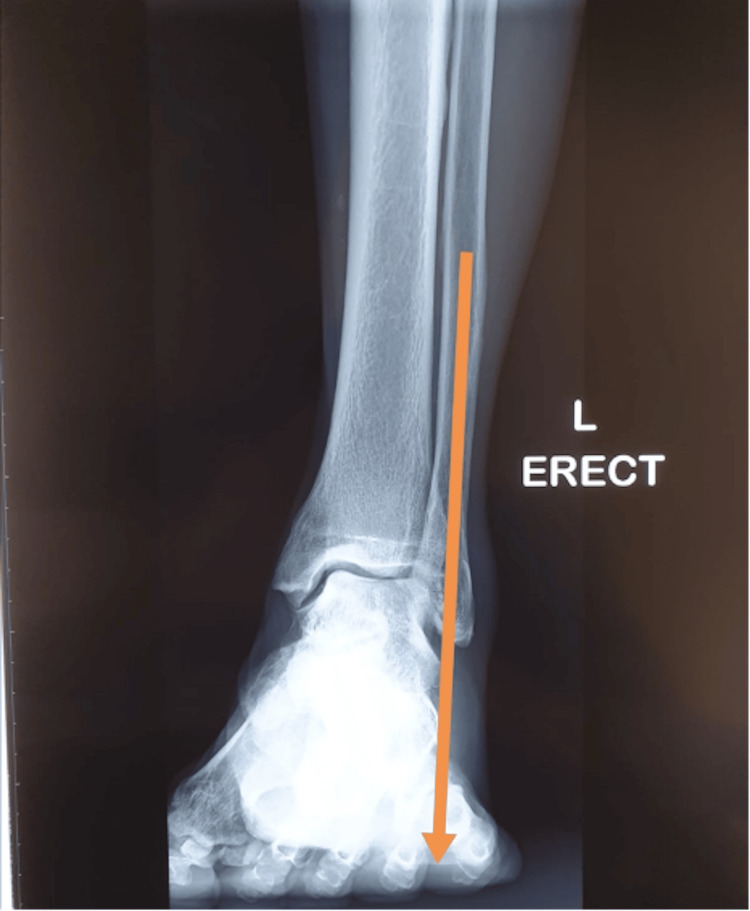
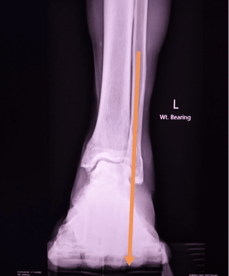
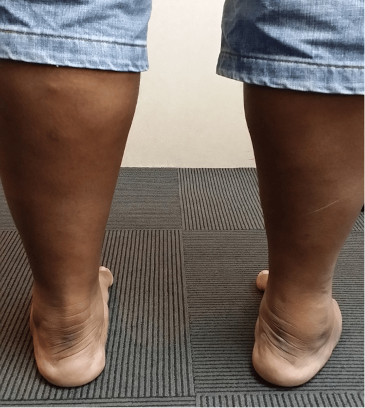
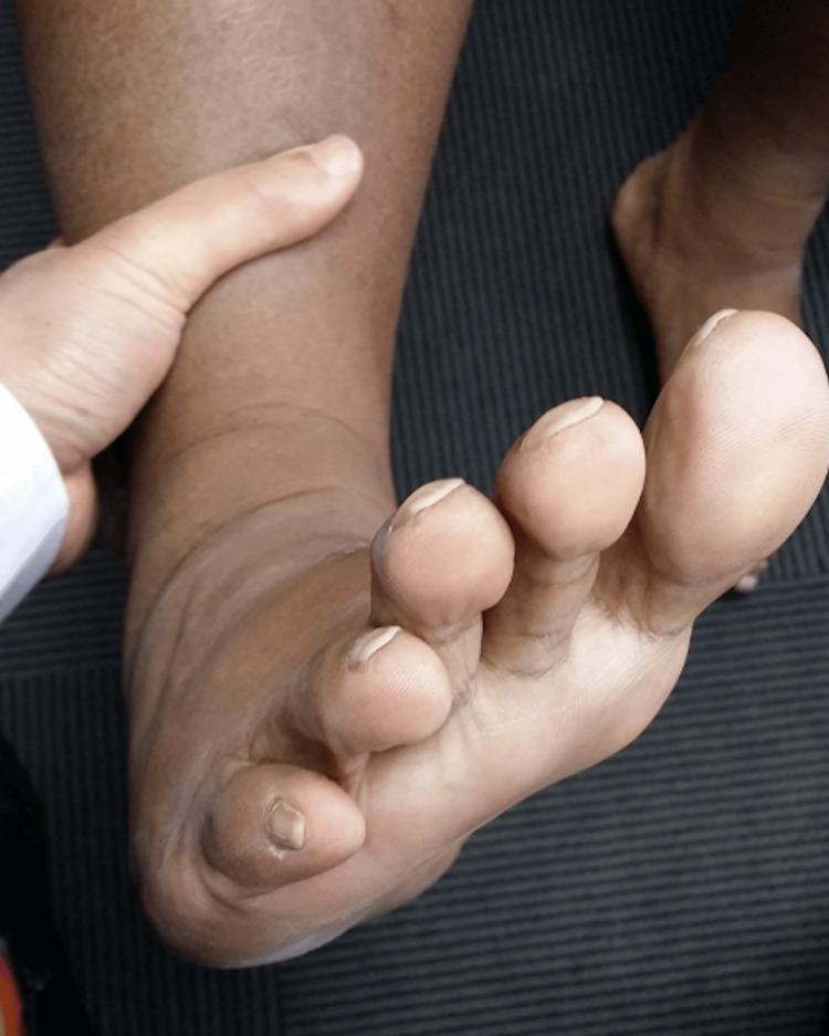
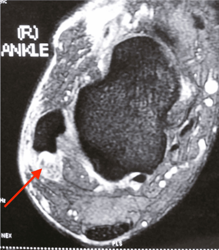
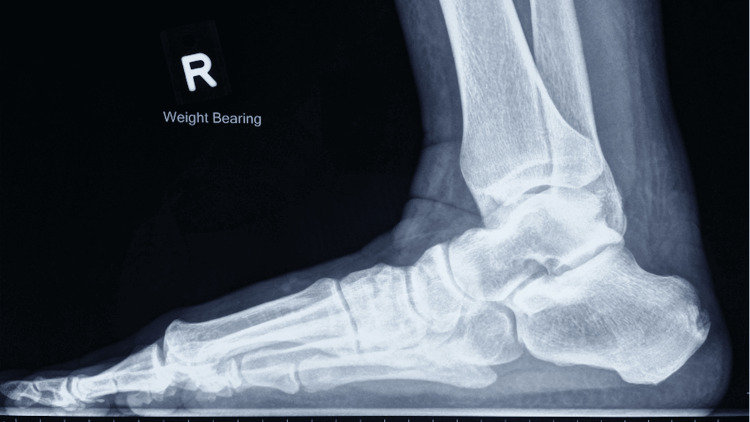
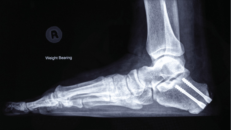
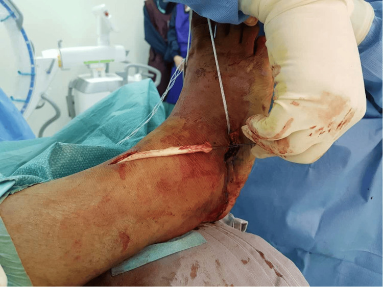
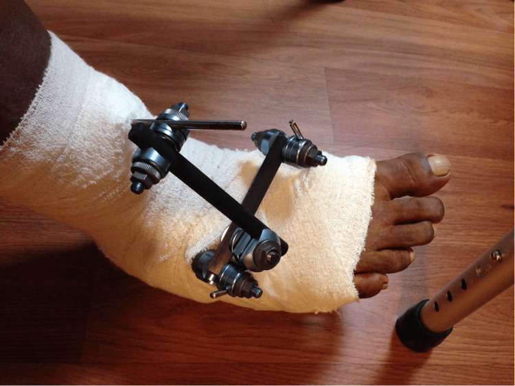
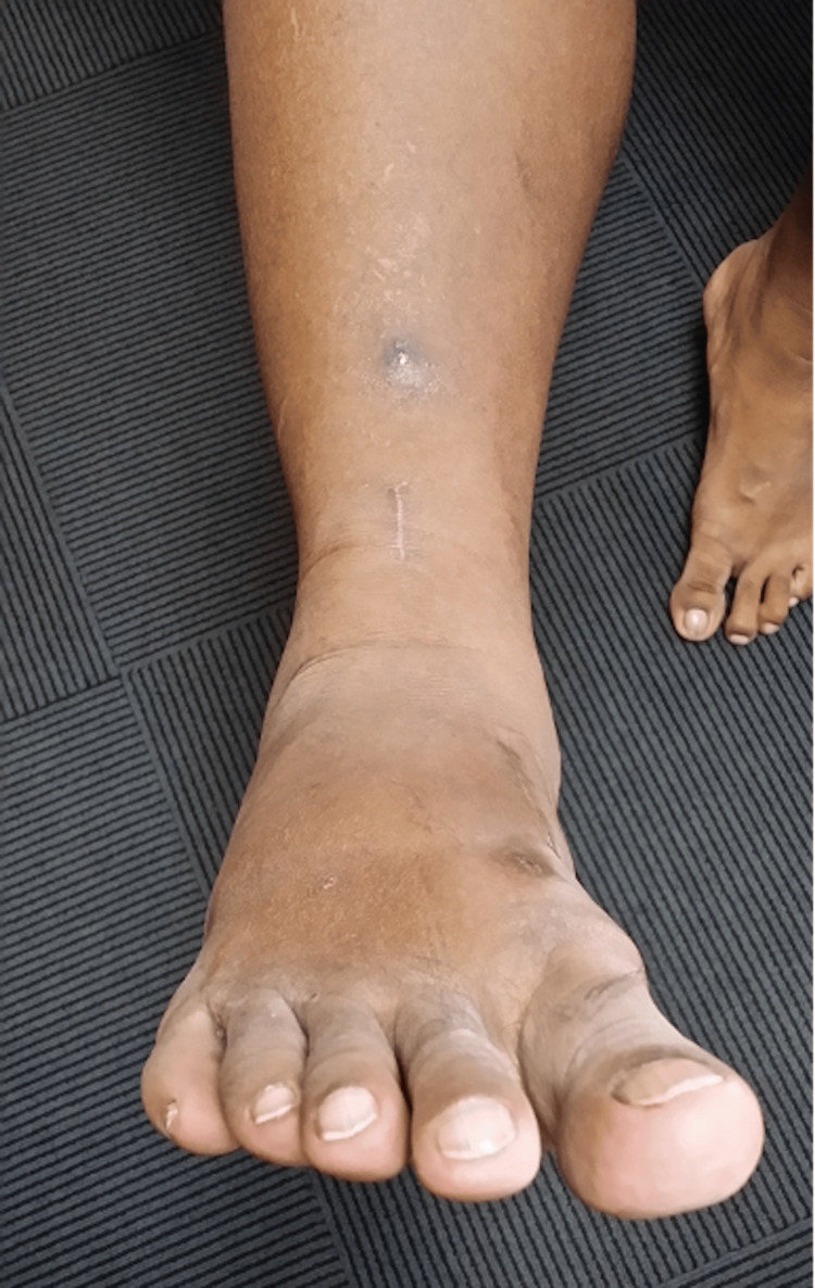
Peroneal tears are an important cause of lateral ankle pain and are often missed. Peroneal tears can present in different combinations requiring different surgical strategies. If the tears are symptomatic in patients in whom conservative treatment has failed, surgery is an option. We present the various types of surgical management of four patients, each with a different tear combination of the peroneal tendons. The first patient presented with a longitudinal split of the peroneal brevis tendon, which was repaired. The second patient had a tear of the peroneal longus tendon with a significant gap, while his peroneal brevis tendon was intact. His peroneal longus was tenodesed to the intact peroneal brevis. The third patient had ruptures of both his peroneal brevis and longus tendons with significant gaps. There was only a small peroneal brevis remnant left. The patient also had a cavovarus deformity of the same foot. His flexor hallucis longus tendon was harvested, routed, and sutured to the remnant peroneal brevis tendon. A lateralising calcaneal osteotomy and a dorsiflexion closing wedge osteotomy of his first metatarsal bone were also performed. The last patient had ruptures of both peroneal tendons with no remnant tendon remaining for repair. His anterior tibialis tendon was transferred from its insertion to his cuboid. A lateralising calcaneal osteotomy was performed, and an ankle-spanning external fixator was applied. A high index of suspicion for peroneal tears in lateral-sided ankle pain must be maintained. Peroneal tears can present in various combinations, with each combination requiring a different surgical treatment.
Keywords: cavovarus deformity; peroneal ruptures; peroneal tears; tendon repair; tendon transfer; tenodesis.
Copyright © 2024, Tang et al.
Conflict of interest statement
Human subjects: Consent for treatment and open access publication was obtained or waived by all participants in this study. Conflicts of interest: In compliance with the ICMJE uniform disclosure form, all authors declare the following: Payment/services info: All authors have declared that no financial support was received from any organization for the submitted work. Financial relationships: All authors have declared that they have no financial relationships at present or within the previous three years with any organizations that might have an interest in the submitted work. Other relationships: All authors have declared that there are no other relationships or activities that could appear to have influenced the submitted work.
Figures
References
-
- Recurrent subluxation of the peroneal tendons. Ferran NA, Oliva F, Maffulli N. Sports Med. 2006;36:839–846. - PubMed
-
- Operative reconstruction after transverse rupture of the tendons of both peroneus longus and brevis. Surgical reconstruction by transfer of the flexor digitorum longus tendon. Borton DC, Lucas P, Jomha NM, Cross MJ, Slater K. J Bone Joint Surg Br. 1998;80:781–784. - PubMed
-
- Traumatic subluxation/dislocation of the peroneal tendons. Brage ME, Hansen ST Jr. Foot Ankle. 1992;13:423–431. - PubMed
-
- Long-term results of debridement and primary repair of peroneal tendon tears. Demetracopoulos CA, Vineyard JC, Kiesau CD, Nunley JA 2nd. Foot Ankle Int. 2014;35:252–257. - PubMed
-
- Use of HyProCure in correcting flatfoot in adolescent and adult Asians: a radiographic study using a new method of measuring hindfoot alignment. Tang ZH, Chong KW. J Foot Ankle Surg (Asia-Pacific) 2023;10:56–60.
Publication types
LinkOut - more resources
Full Text Sources
Miscellaneous