. 2025 Jan 28;10(1):e0078024.
doi: 10.1128/msphere.00780-24. Epub 2024 Dec 17.

The transcriptional regulator Lrp activates the expression of genes involved in the biosynthesis of tilimycin and tilivalline enterotoxins in Klebsiella oxytoca

Affiliations

The transcriptional regulator Lrp activates the expression of genes involved in the biosynthesis of tilimycin and tilivalline enterotoxins in Klebsiella oxytoca

Miguel A De la Cruz et al. mSphere. .

Abstract

The toxigenic Klebsiella oxytoca strains secrete tilymicin and tilivalline enterotoxins, which cause antibiotic-associated hemorrhagic colitis. Both enterotoxins are non-ribosomal peptides synthesized by enzymes encoded in two divergent operons clustered in a pathogenicity island. The transcriptional regulator Lrp (leucine-responsive regulatory protein) controls the expression of several bacterial genes involved in virulence. In this work, we have uncovered novel findings that have significant implications. We determined the transcriptional expression of aroX and npsA, the first genes of each tilimycin (TM)/tilivalline (TV) biosynthetic operon in K. oxytoca MIT 09-7231 wild-type and its derivatives Δlrp mutant and complemented strains. Our results suggest that Lrp directly activates the transcription of both aroX and npsA genes by binding to the intergenic regulatory region in a leucine-dependent manner. Furthermore, the lack of Lrp significantly diminished the cytotoxicity of K. oxytoca on HeLa cells due to reduced production of TM and TV. Altogether, our data present a new perspective on the role of Lrp as a regulator in cytotoxin-producing K. oxytoca strains and how it controls the expression of genes involved in the biosynthesis of their main virulence factors.IMPORTANCETilimycin (TM) and tilivalline (TV) are enterotoxins that are a hallmark for the cytotoxin-producing Klebsiella oxytoca strains, which cause antibiotic-associated hemorrhagic colitis. The biosynthesis of TM and TV is driven by enzymes encoded by the aroX- and NRPS-operons. In this study, we discovered that the transcriptional regulator Lrp plays a crucial role in activating the expression of the aroX- and NRPS-operons, thereby initiating TM and TV biosynthesis. Our results underscore a molecular mechanism by which TM and TV production by toxigenic K. oxytoca strains is regulated and shed further light on developing strategies to prevent the intestinal illness caused by this enteric pathogen.

Keywords: Klebsiella oxytoca; aroX; citotoxicity; lrp; npsA.


Conflict of interest statement

The authors declare no conflict of interest.

Figures

Fig 1
Fig 1
Amino acid sequence alignment of Lrp from K. oxytoca and other Enterobacterales. The amino acid sequence of Lrp (KMV84644.1) from K. oxytoca MIT 09-7231, and Lrp homologs proteins: (BAH62617.1) from K. pneumoniae NTUH-K2044, (NP_415409.1) from E. coli K-12 MG1655, (ADF62302.1) from Enterobacter cloacae ATCC 13047, (AA069588.1) from Salmonella enterica serovar Typhi Ty2, (ABB62447.1) from Shigella dysenteriae Sd197, (CAL11603.1) from Yersinia enterocolitica 8081, (CAR41630.1) from Proteus mirabilis HI4320, and (CDG11553.1) from Serratia marcescens Db11 were aligned by using Clustal Omega (https://www.ebi.ac.uk/Tools/msa/clustalo/). The predicted regions to encode the HTH motif (DNA-binding), and RAM domain (Regulation of amino acid metabolism) are indicated. Secondary structural elements are shown by barrels (α-helix) and arrows (β-sheet). Hydrophobic, polar, positively charged, and negatively charged amino acids are represented in red, green, magenta, and blue, respectively. The asterisk (*), colon (:), and dot (.) indicate identical, conserved, and semi-conserved amino acids among all aligned sequences.
Fig 2
Fig 2
Growth curves of K. oxytoca wild-type (WT), Δlrp mutant, and Δlrp pT3-Lrp complemented strains. Cultures were grown for 12 h in TSB (nutrient-rich) (A, B) and N-MM (nutrient-limiting) (C, D) medium, in the absence and presence of l-leucine (Leu). The OD600nm values were recorded every hour. Data represent the mean of three independent experiments with standard deviations.
Fig 3
Fig 3
Transcriptional regulation of Lrp on aroX and npsA genes. Determination of gene expression by RT-qPCR of aroX and npsA of K. oxytoca wild-type (WT), Δlrp mutant, and Δlrp pT3-Lrp complemented strains grown in TSB (nutrient-rich) and N-MM (nutrient-limiting) medium at stationary phase (OD600nm = 1.6) at 37°C, in the absence and presence of l-leucine. Data represent the mean of three independent experiments performed in triplicate with standard deviations. Statistically significant: **P < 0.01; ***P < 0.001; ns: not significant. All P values were determined using unpaired two-tailed Student’s t test.
Fig 4
Fig 4
In silico analysis of the intergenic region of aroX and npsA genes. (A) Genetic organization of the aroX and NRPS operons. Regulatory regions of aroX (B) and npsA (C) indicating the initiaton codon (ATG, bold), the predicted promoter region (−35 and −10, bold), the transcription start site (+1, bold), and the Lrp-binding motif (highlighted in gray; nucleotides matching the consensus sequence from E. coli are bold).
Fig 5
Fig 5
Lrp binds to the intergenic region of aroX and npsA genes in the presence of leucine. Electrophoretic mobility shift assays (EMSAs) were carried out to find out the binding of the His6-Lrp purified recombinant protein to the DNA probe from the intergenic regulatory region of aroX and npsA in the absence (A) or presence (B) of leucine. DNA probes from K. oxytoca ilvI regulatory region (C) and M. tuberculosis fbpA coding region (D) were employed as positive and negative controls, respectively. 0.2 µM of DNA fragments was individually mixed and incubated with increasing concentrations of purified His6-Lrp. l-leucine was added at a final concentration of 7 mM. Arrows show free DNA or Lrp-DNA complex stained with ethidium bromide.
Fig 6
Fig 6
Analysis of sequences to determine the bind of Lrp to the intergenic region of aroX and npsA genes. Electrophoretic mobility shift assays (EMSAs) were performed to determine the binding of the His6-Lrp purified recombinant protein to three different fragments from the intergenic regulatory region of aroX and npsA. Schematic representation of the aroX-npsA intergenic regulatory region indicating the position of the two Lrp putative motifs for npsA and aroX, respectively, and the three fragments analyzed to determine the Lrp binding to DNA (A). DNA probes from fragment 1 (B), fragment 2 (C) and fragment 3 (D). 0.2 µM of DNA fragments were individually mixed and incubated with increasing concentrations of purified His6-Lrp. l-leucine was added at a final concentration of 7 mM. Arrows show free DNA or Lrp-DNA complex stained with ethidium bromide.
Fig 7
Fig 7
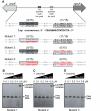
Lrp binds to two specific sequences on the intergenic region of aroX and npsA genes. Electrophoretic mobility shift assays (EMSAs) were realized to identify the binding of the His6-Lrp purified recombinant protein to three fragments from the intergenic regulatory region of aroX and npsA containing substitution mutations on the Lrp putative motifs. Schematic representation of the aroX-npsA intergenic regulatory region indicating the position of the two Lrp putative motifs for npsA and aroX, respectively, and the three fragments with the different substitution mutations analyzed (A). DNA probes from Mutant 1 (B), Mutant 2 (C) and Mutant 3 (D). 0.2 µM of DNA fragments were individually mixed and incubated with increasing concentrations of purified His6-Lrp. l-leucine was added at a final concentration of 7 mM. Arrows show free DNA or Lrp-DNA complex stained with ethidium bromide.
Fig 8
Fig 8
LDH cytotoxicity of K. oxytoca WT, Δlrp mutant, and Δlrp pT3-Lrp strains. HeLa cells were inoculated with TSB and N-MM culture media and with K. oxytoca supernatants (WT, Δlrp, Δlrp pT3-Lrp, and ΔnpsA) from cultures with an OD600nm = 1.6, in the absence and presence of leucine, for 48 h. After treatment, the measurement of extracellular LDH was quantified. Minimal and maximal measurable LDH release was determined by incubating HeLa cells with PBS (negative control) and lysis buffer (positive control). Statistically significant: **P < 0.01; ***P < 0.001; ns: not significant. All P values were determined using unpaired two-tailed Student’s t test.

References

    1. Alexander EM, Kreitler DF, Guidolin V, Hurben AK, Drake E, Villalta PW, Balbo S, Gulick AM, Aldrich CC. 2020. Biosynthesis, mechanism of action, and inhibition of the enterotoxin tilimycin produced by the opportunistic pathogen Klebsiella oxytoca ACS Infect Dis 6:1976–1997. doi:10.1021/acsinfecdis.0c00326 - DOI - PMC - PubMed
    1. Högenauer C, Langner C, Beubler E, Lippe IT, Schicho R, Gorkiewicz G, Krause R, Gerstgrasser N, Krejs GJ, Hinterleitner TA. 2006. Klebsiella oxytoca as a causative organism of antibiotic-associated hemorrhagic colitis. N Engl J Med 355:2418–2426. doi:10.1056/NEJMoa054765 - DOI - PubMed
    1. Herzog KAT, Schneditz G, Leitner E, Feierl G, Hoffmann KM, Zollner-Schwetz I, Krause R, Gorkiewicz G, Zechner EL, Högenauer C. 2014. Genotypes of Klebsiella oxytoca isolates from patients with nosocomial pneumonia are distinct from those of isolates from patients with antibiotic-associated hemorrhagic colitis. J Clin Microbiol 52:1607–1616. doi:10.1128/JCM.03373-13 - DOI - PMC - PubMed
    1. Maharshak N, Packey CD, Ellermann M, Manick S, Siddle JP, Huh EY, Plevy S, Sartor RB, Carroll IM. 2013. Altered enteric microbiota ecology in interleukin 10-deficient mice during development and progression of intestinal inflammation. Gut Microbes 4:316–324. doi:10.4161/gmic.25486 - DOI - PMC - PubMed
    1. Walker WA. 2017. Dysbiosis,p 227–232. In The microbiota in gastrointestinal pathophysiology. Elsevier.

MeSH terms

Substances

LinkOut - more resources