. 2025 Feb 5;16(2):e0337524.
doi: 10.1128/mbio.03375-24. Epub 2024 Dec 17.

Split-Cre-mediated GFP expression as a permanent marker for flagellar fusion of Trypanosoma brucei in its tsetse fly host

Affiliations

Split-Cre-mediated GFP expression as a permanent marker for flagellar fusion of Trypanosoma brucei in its tsetse fly host

Ruth Etzensperger et al. mBio. .

Abstract

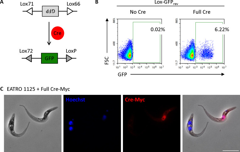
Trypanosomes have different ways of communicating with each other. While communication via quorum sensing, or by the release and uptake of extracellular vesicles, is widespread in nature, the phenomenon of flagellar fusion has only been observed in Trypanosoma brucei. We showed previously that a small proportion of procyclic culture forms (corresponding to insect midgut forms) can fuse their flagella and exchange cytosolic and membrane proteins. This happens reproducibly in cell culture. It was not known, however, if flagellar fusion also occurs in the tsetse fly host, and at what stage of the life cycle. We have developed a split-Cre-Lox system to permanently label trypanosomes that undergo flagellar fusion. Specifically, we engineered trypanosomes to contain a GFP gene flanked by Lox sites in the reverse orientation to the promoter. In addition, the cells expressed inactive halves of the Cre recombinase, either N-terminal Cre residues 1-244 (N-Cre) or C-terminal Cre residues 245-343 (C-Cre). Upon flagellar fusion, these Cre halves were exchanged between trypanosomes, forming functional full Cre and flipping reverse-GFP into its forward orientation. We showed that cells that acquired the second half Cre through flagellar fusion were permanently modified and that the cells and their progeny constitutively expressed GFP. When tsetse flies were co-infected with N-Cre and C-Cre cells, GFP-positive trypanosomes were observed in the midgut and proventriculus 28-34 days post-infection. These results show that flagellar fusion not only happens in culture but also during the natural life cycle of trypanosomes in their tsetse fly host.

Importance: We have established a procedure to permanently label pairs of trypanosomes that transiently fuse their flagella and exchange proteins. When this occurs, a reporter gene is permanently flipped from the "off" to the "on" position, resulting in the production of green fluorescent protein. Crucially, green trypanosomes can be detected in tsetse flies co-infected with the two cell lines, proving that flagellar fusion occurs in the host. To our knowledge, we are the first to describe a split-Cre-Lox system for lineage tracing and selection in trypanosomes. In addition to its use in trypanosomes, this system could be adapted for other parasites and in other contexts. For example, it could be used to determine whether flagellar fusion occurs in related parasites such as Leishmania and Trypanosoma cruzi or to monitor whether intracellular parasites and their hosts exchange proteins.

Keywords: Split-Cre; flagellar fusion; lineage tracing; trypanosoma; tsetse.


Conflict of interest statement

The authors declare no conflict of interest.

Figures

Fig 1
Fig 1
Lox-GFPrev cells become GFP-positive upon Cre recombinase expression. (A) Schematic representation of the Lox-Cre system. (B) Lox-GFPrev cells were analyzed by flow cytometry for GFP expression in the absence (left) or presence (right) of transient full-length Cre expression. Numbers indicate the percentage of GFP-positive cells. (C) Representative immunofluorescence image showing full-length Myc-tagged Cre expression in the nucleus following transient transfection. Scale bar: 10 µm.
Fig 2
Fig 2
The split-Cre system is functional in trypanosomes. (A) Schematic representation of split-Cre used to invert GFPrev. The reverse GFP sequence is flanked by Lox66 and Lox71 sites. The N-terminal and C-terminal fragments of Cre assemble to form functional Cre that recognizes the Lox sites and inverts reverse GFP to its forward orientation where GFP is expressed. The inversion leads to the conversion of the Lox66 and Lox71 sites to Lox72 and LoxP sites, respectively, preventing further inversions. (B) Flow cytometry analysis of EATRO 1125 procyclic forms stably transformed with Lox66-GFPrev-Lox71 and transiently transfected with either N-Cre1-244, C-Cre245-343 or N-Cre1-244 plus C-Cre245-343. Data were acquired 24 h after transient transfection. Percentages of GFP-positive cells are indicated. (C) Representative fluorescence microscopy images showing HA-tagged N-Cre1-244 and Myc-tagged C-Cre245-343 expression in stably transfected EATRO 1125 cells. The parental line serves as a control for background staining. Cells were stained with Hoechst to visualize nuclei and kinetoplasts. Scale bar: 10 µm.
Fig 3
Fig 3
In vitro fusion using the split-Cre system. (A) Schematic view depicting N- and C-Cre exchange between two trypanosomes and inversion of GFP. Electron microscopy image from Imhof et al. (12) (B) Flow cytometry analysis of GFP-positive cells after 0 h, 24 h, and 48 h of co-culture of GFPrev N-Cre1-244 and GFPrev C-Cre245-343 cells in fusion conditions (non-heat-inactivated serum). GFP is expressed in cells that have undergone flagellar fusion. Numbers indicate percentages of GFP-positive cells. (C) Microscopy images of GFPrev N-Cre1-244 and GFPrev C-Cre245-343 cells from a 48 h fusion culture. GFP is expressed in cells that have undergone flagellar fusion. Cells were stained with Hoechst dye for visualization of nuclei and kinetoplasts. Scale bars: 10 µm.
Fig 4
Fig 4
In vitro fusion and selection after inversion of HygroGFPrev by split-Cre. (A) Flow cytometry analysis after 0 h, 24 h, and 48 h of HygorGFPrev N-Cre1-244 and HygorGFPrev C-Cre245-343 cells in fusion culture. Numbers indicate percentages of GFP-positive cells. (B) After 48 h in fusion culture, hygromycin was added and the frequencies of GFP-positive cells were determined by flow cytometry at the time points indicated. Means ± SD of technical triplicates are shown. (C) PCR analysis of N-Cre (top) and C-Cre (bottom) in the genomic DNA of GFP-positive clones resulting from a fusion experiment. P: parental wild-type cells, N: N-Cre cells without fusion, C: C-Cre cells without fusion, M: marker, A1-3, B1-2, and C1-7: hygromycin-resistant cultures isolated by limiting dilution.
Fig 5
Fig 5
GFP-positive trypanosomes from the midgut and proventriculus of flies coinfected with Lox-GFPrev/N-Cre1-244 and Lox-GFPrev/C-Cre245-343 cells. (A) GFP-positive trypanosomes from a midgut sample. (B) GFP-positive trypanosome from a proventriculus sample. Cells were stained with Hoechst dye and analyzed by microscopy. The scale bar is 10 µm.
Fig 6
Fig 6
GFP-positive trypanosomes from the midgut and proventriculus of flies coinfected with Lox-HygroGFPrev/N-Cre1-244 and Lox-HygroGFPrev/C-Cre245-343 cells. (A) GFP-positive trypanosomes from a midgut sample. (B) GFP-positive trypanosomes from a proventriculus sample. Cells were stained with Hoechst dye and analyzed by microscopy. The scale bar is 10 µm.

References

    1. Roditi I, Schumann G, Naguleswaran A. 2016. Environmental sensing by African trypanosomes. Curr Opin Microbiol 32:26–30. doi:10.1016/j.mib.2016.04.011 - DOI - PubMed
    1. Roditi I. 2016. The languages of parasite communication. Mol Biochem Parasitol 208:16–22. doi:10.1016/j.molbiopara.2016.05.008 - DOI - PubMed
    1. McWilliam KR. 2023. Cell-cell communication in African trypanosomes. Microbiol (Reading) 169:001388. doi:10.1099/mic.0.001388 - DOI - PMC - PubMed
    1. Rojas F, Matthews KR. 2019. Quorum sensing in African trypanosomes. Curr Opin Microbiol 52:124–129. doi:10.1016/j.mib.2019.07.001 - DOI - PubMed
    1. Rojas F, Silvester E, Young J, Milne R, Tettey M, Houston DR, Walkinshaw MD, Pérez-Pi I, Auer M, Denton H, Smith TK, Thompson J, Matthews KR. 2019. Oligopeptide signaling through TbGPR89 drives trypanosome quorum sensing. Cell 176:306–317. doi:10.1016/j.cell.2018.10.041 - DOI - PMC - PubMed

MeSH terms