. 2024 Dec 17;149(1):2.
doi: 10.1007/s00401-024-02839-2.

Amyloid-β oligomers increase the binding and internalization of tau oligomers in human synapses

Affiliations

Amyloid-β oligomers increase the binding and internalization of tau oligomers in human synapses

Shrinath Kadamangudi et al. Acta Neuropathol. .

Abstract

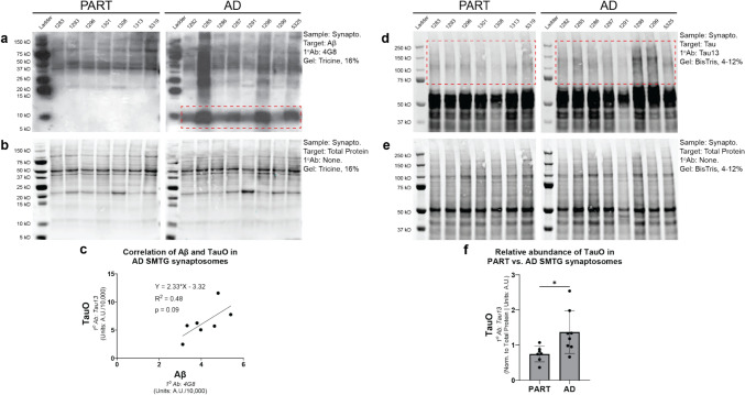
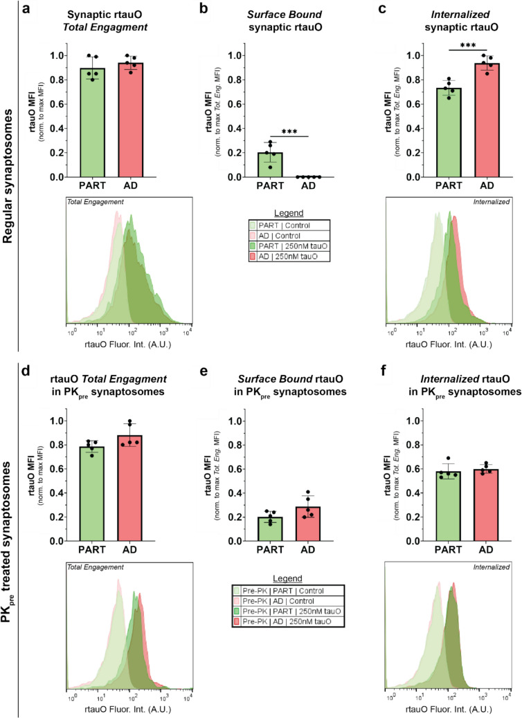
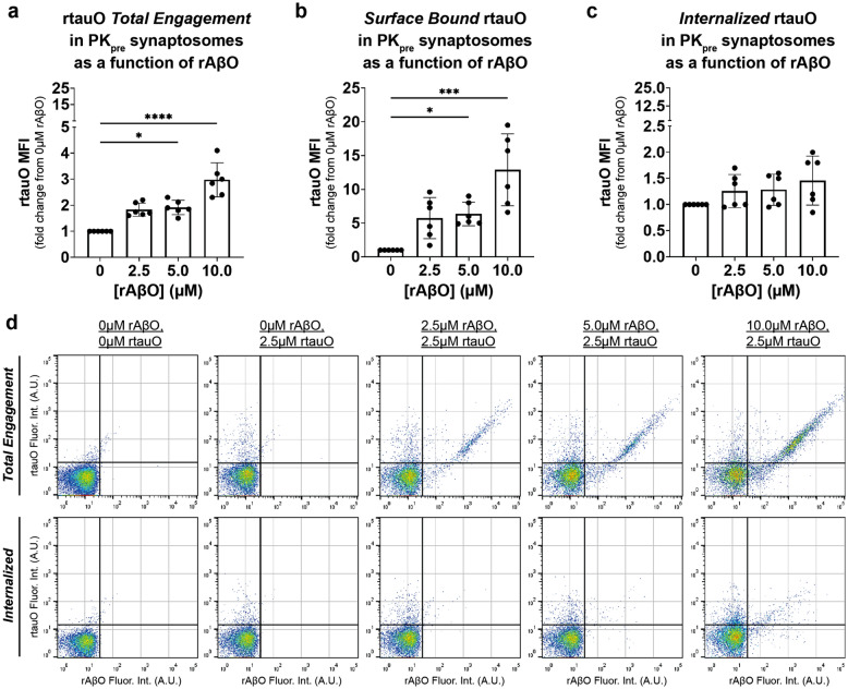
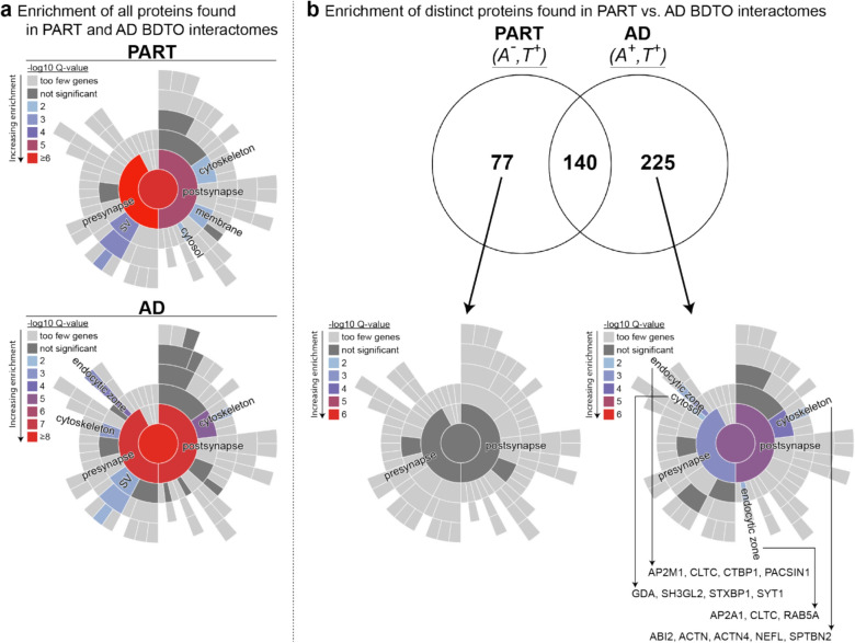
In Alzheimer's disease (AD), the propagation and spreading of CNS tau pathology closely correlates with cognitive decline, positioning tau as an attractive therapeutic target. Amyloid beta (Aβ) has been strongly implicated in driving tau spread, whereas primary tauopathies such as primary age-related tauopathy (PART)-which lack Aβ pathology-exhibit limited tau spread and minimal-to-no cognitive decline. Emerging evidence converges on a trans-synaptic mechanism of tau spread, facilitated by the transfer of misfolded tau aggregates (e.g. soluble oligomers). However, it is unclear whether Aβ oligomers modulate the binding and internalization of tau oligomers in human synapses. Our translationally focused paradigms utilize post-mortem brain specimens from Control, PART, and AD patients. Synaptosomes isolated from the temporal cortex of all three groups were incubated with preformed recombinant tauO (rtauO), ± preformed recombinant AβO (rAβO), and oligomer binding/internalization was quantified via flow cytometry following proteinase K (PK) digestion of surface-bound oligomers. TauO-synapse interactions were visualized using EM immunogold. Brain-derived tau oligomers (BDTO) from AD and PART PBS-soluble hippocampal fractions were co-immunoprecipitated and analyzed via mass spectrometry to compare synaptic tauO interactomes in primary and secondary tauopathies, thereby inferring the role of Aβ. AD synaptosomes, enriched in endogenous Aβ pathology, exhibited increased rtauO internalization compared to PART synaptosomes. This observation was mirrored in Control synaptosomes, where recombinant rAβO significantly increased rtauO binding and internalization. PK pre-treatment abolished this effect, implicating synaptic membrane proteins in AβO-mediated tauO internalization. While both PART and AD BDTO were broadly enriched in synaptic proteins, AD BDTO exhibited differential enrichment of endocytic proteins across pre- and post-synaptic compartments, whereas PART BDTO showed no significant synaptic enrichment. This study demonstrates that Aβ oligomers enhance tau oligomer binding and drive its internalization through synaptic membrane proteins. These findings offer novel mechanistic insights underlying pathological tau spreading directly within human synapses and emphasize the therapeutic potential of targeting Aβ-tau interactions.

Keywords: Aβ oligomers; Tau oligomers; Trans-synaptic tau spreading.


Conflict of interest statement

Declarations. Conflict of interest: The authors declare that they have no financial or non-financial competing interests to disclose. Ethical approval and consent to participate: Postmortem frozen brain tissues used in this study were obtained through established material transfer agreements with the Alzheimer’s Disease Research Center (ADRC) at Sanders-Brown Center on Aging, University of Kentucky, and the Layton Aging and Alzheimer’s Disease Center (ADC) at Oregon Health and Science University. All donors were enrolled in ongoing brain aging studies, with informed consent obtained prior to participation, and all protocols adhered to Institutional Review Board (IRB) guidelines at each institution. All donor subject samples were de-identified before being provided to the University of Texas Medical Branch (UTMB); therefore, no approval was required from the UTMB IRB per CFR §46.101(a). Animals (mice) used procure EM immunogold images were housed in adherence with USDA standards and sacrificed in compliance with UTMB’s Institutional Animal Care and Use Committee (IACUC), as well as UTMB IRB, approved protocols.

Figures

Fig. 1
Fig. 1
Schematic of experimental design and techniques implemented in the current study. This figure illustrates the integrated approach used to investigate the cellular (panel a) and molecular (panel b) mechanisms underlying the binding and internalization of tau oligomers in human synapses, and the modulating effects of Aβ oligomers. a Synaptosomes were isolated from SMTG human autopsy specimens and pooled to generate a representative experimental set. Pooled synaptosomes were treated with fluorescently labeled pre-formed rtauO and/or rAβO, followed by differential PK treatment to distinguish between oligomer surface binding and internalization (detailed PK experimental paradigm in Supp. Figure 1). Samples were subsequently analyzed using flow cytometry optimized for small particle detection. b BDTO isolated from the PBS-soluble fraction of autopsy hippocampal tissue from PART and AD patients via co-immunoprecipitation. BDTO and their interacting proteins were profiled via LC–MS/MS and subjected to SynGO enrichment analysis, facilitating the direct comparison of the synaptic BDTO interactome in PART (A, T+) vs. AD (A+, T+). A amyloid-β pathology, BDTO brain-derived tau oligomers, LC–MS/MS liquid-chromatography with tandem mass spectrometry, PK proteinase K, rAβO recombinant amyloid-β oligomers, rtauO recombinant tau oligomers, SMTG superior medial temporal gyrus, T tau pathology. Created in https://BioRender.com
Fig. 2
Fig. 2
Western blot analysis of endogenous Aβ and tau pathology in SMTG synaptosomes from PART and AD cases. Synaptosomes isolated from the SMTG of PART (n = 7) and AD (n = 8) post-mortem brain specimens, used in binding and internalization assays, were evaluated for endogenous Aβ and tau pathology. Each lane represents an individual patient sample, with identifiers indicated above each lane. Pertinent experimental details are provided on the right side of each blot. a and b Aβ detection with corresponding total protein staining for normalization, performed using the Tricine gel system. d and e tauO detection with corresponding total protein staining for normalization, performed using the Bis–Tris gel system. Red dashed boxes highlight quantified regions for each probe. c Linear regression analysis of Aβ and tauO levels in AD synaptosomes. f Quantification of tauO in PART versus AD synaptosomes. Statistical significance was assessed using an independent samples t-test (α = 0.05), with *p < 0.05 indicating significance
Fig. 3
Fig. 3
rtauO internalization is increased in AD synaptosomes, as compared to PART, and is modulated by synaptic membrane proteins. The binding and internalization of rtauO was assessed by flow cytometry in human synaptosomes isolated from the SMTG of PART and AD cases. a Total engagement, b surface-bound, and c internalized rtauO were measured and normalized to the maximum total engagement MFI within each group. df To evaluate the role of synaptic membrane proteins, the same parameters were assessed in synaptosomes pre-treated with proteinase K (PKpre). Representative flow cytometry histograms display fluorescence intensity distributions for total engagement and internalized tauO. Biological replicates, nPART = 7, and nAD = 8; independent experiments, n = 5. Statistical analysis was performed using an independent samples t-test (α = 0.05). Significance levels are indicated as follows: ***p < 0.001
Fig. 4
Fig. 4
rAβO increase the binding and internalization of rtauO in human synaptosomes. The effect of rAβO on the binding and internalization of rtauO in standardized human synaptosomes, isolated from the SMTG of Control cases, was assessed by flow cytometry. a Total engagement of rtauO (2.5 µM) as a function of increasing rAβO concentrations (0–10.0 µM) is shown. b Surface-bound rtauO and c internalized rtauO per synapse were measured under the same conditions. Biological replicates, n = 8; independent experiments, n = 8. Statistical analysis was performed using Kruskal–Wallis (α = 0.05) with Dunn’s post-hoc test. Significance levels are indicated as follows: ****p < 0.0001, **p < 0.01, *p < 0.05. d Representative flow cytometry dot plots displaying the fluorescence intensity of total engaged and internalized rtauO in the presence of varying rAβO concentrations
Fig. 5
Fig. 5
Synaptic membrane proteins modulate the binding and internalization of rtauO in human synaptosomes. The influence of synaptic surface proteins on the binding and internalization of rtauO (2.5 µM) was assessed via flow cytometry in Control human SMTG synaptosomes pre-treated with PK (PKpre, 1 mg/mL). a Total engagement, b surface bound, and c internalized rtauO, were measured as a function of rAβO (0–10.0 µM). Data are expressed as fold change from baseline (0 µM rAβO). Biological replicates, n = 8; independent experiments, n = 4. Statistical analysis was performed using Kruskal–Wallis (α = 0.05) with Dunn’s post-hoc test. Significance levels are indicated as follows: ****p < 0.0001, ***p < 0.001, **p < 0.01, *p < 0.05. d Representative flow cytometry dot plots display the fluorescence intensity of total engaged and internalized rtauO in the presence of varying rAβO concentrations
Fig. 6
Fig. 6
Synaptic protein interactome of PART and AD brain-derived tau oligomers (BDTO). BDTO were co-immunoprecipitated (antibody: T18) from the PBS-soluble fractions of PART and AD human hippocampal autopsy tissue. Pooled PART (n = 4) and AD (n = 4) BDTO samples were subjected to proteomic analysis via LC–MS/MS and subsequent Synaptic GO (SynGO) enrichment analysis with evidence filters set to maximum stringency. SynGO results are displayed as sunburst plots, where color-coded segments denote significantly enriched synaptic compartments or pathways. a Enrichment of synaptic compartments associated with PART (top panel) and AD (bottom panel) BDTO. b Venn diagram showing the number of shared and unique proteins between PART and AD BDTO. Unique proteins for each condition were further analyzed via SynGO enrichment. Individual proteins comprising enriched terms are listed below the respective sunburst plots

References

    1. Adams JN, Harrison TM, Maass A, Baker SL, Jagust WJ (2021) Distinct factors drive the spatiotemporal progression of tau pathology in older adults. J Neurosci. 10.1523/JNEUROSCI.1601-21.2021 - PMC - PubMed
    1. Amro Z, Yool AJ, Collins-Praino LE (2021) The potential role of glial cells in driving the prion-like transcellular propagation of tau in tauopathies. Brain Behav Immunity Health 14:100242. 10.1016/j.bbih.2021.100242 - PMC - PubMed
    1. Asai H, Ikezu S, Tsunoda S, Medalla M, Luebke J, Haydar T et al (2015) Depletion of microglia and inhibition of exosome synthesis halt tau propagation. Nat Neurosci 18:1584–1593. 10.1038/nn.4132 - PMC - PubMed
    1. Askenazi M, Kavanagh T, Pires G, Ueberheide B, Wisniewski T, Drummond E (2023) Compilation of reported protein changes in the brain in Alzheimer’s disease. Nat Commun 14:4466. 10.1038/s41467-023-40208-x - PMC - PubMed
    1. Ayers JI, Giasson BI, Borchelt DR (2018) Prion-like spreading in tauopathies. Biol Psychiat 83:337–346. 10.1016/j.biopsych.2017.04.003 - PMC - PubMed

Publication types

LinkOut - more resources