. 2024 Dec 17;19(12):e0313444.
doi: 10.1371/journal.pone.0313444. eCollection 2024.

Quantitative analysis of septin Cdc10 & Cdc3-associated proteome during stress response in the fungal pathogen Cryptococcus neoformans

Affiliations

Quantitative analysis of septin Cdc10 & Cdc3-associated proteome during stress response in the fungal pathogen Cryptococcus neoformans

Stephani Martinez Barrera et al. PLoS One. .

Abstract

Cryptococcus neoformans is a pathogenic basidiomycetous yeast that primarily infects immunocompromised individuals. Fatal outcome of cryptococcosis depends on the ability of C. neoformans to sense and adapt to 37°C. A complex of conserved filament forming GTPases, called septins, composed of Cdc3, Cdc10, Cdc11, and Cdc12, assembles at the mother-bud neck in C. neoformans. Septins Cdc3 and Cdc12 are essential for proliferation of C. neoformans at 37°C and for virulence in the Galleria mellonella model of infection, presumably due to their requirement for septin complex formation, and the involvement in cytokinesis. However, how exactly Cdc3, and Cdc12 contribute to C. neoformans growth at 37°C remains unknown. Based on studies investigating roles of septins in Saccharomyces cerevisiae, septin complex at the mother-bud neck of C. neoformans is predicted to interact with proteins involved in cell cycle control, morphogenesis, and cytokinesis, but the septin-associated proteome in C. neoformans has not been investigated. Here, we utilized tandem mass spectrometry to define C. neoformans proteins that associate with either Cdc3 or Cdc10 at ∼25°C or after the shift to 37°C. Our findings unveil a diverse array of septin-associated proteins, highlighting potential roles of septins in cell division, and stress response. Two proteins, identified as associated with both Cdc3 and Cdc10, the actin-binding protein profilin, which was detected at both temperatures, and ATP-binding multi-drug transporter Afr1, which was detected exclusively at 37°C, were further confirmed by co-immunoprecipitation. We also confirmed that association of Cdc3 with Afr1 was enhanced at 37°C. Upon shift to 37°C, septins Cdc3 and Cdc10 exhibited altered localization and Cdc3 partially co-localized with Afr1. In addition, we also investigated changes to levels of individual C. neoformans proteins upon shift from ∼25 to 37°C in exponentially grown culture and when cells entered stationary phase at ∼25°C. Our study reveals changes to C. neoformans proteome associated with heat and nutrient deprivation stresses and provides a landscape of septin-associated C. neoformans proteome, which will facilitate elucidating the biology of septins and mechanisms of stress response in this fungal pathogen.


Conflict of interest statement

The authors have declared that no competing interests exist.

Figures

Fig 1
Fig 1. Schematic representation of the GeLC-MS/MS workflow.
(A) C. neoformans cells were grown for 24 h in YPD rich media at ∼25°C. Subsequently, the cell culture was split, and half of the culture was further grown in the same media for an additional 48 h at ∼25°C (to ensure it approached the stationary phase of growth (SP)), while the other half was refreshed in YPD media and grown for 3 h at ∼25°C (to assure it was at an exponential phase of growth (EP)). Each EP culture was then divided into two cultures and grown either at ∼25 or at 37°C for 2 h before harvesting the cells. (B) Cell lysis and immunoprecipitation of bait protein using RFP-TRAP resin. Bound proteins were eluted and resolved on SDS-PAGE gel. (C) Each lane of the SDS-PAGE gel was excised into twelve fragments and subjected to in-gel trypsin digestion and analyzed by in-gel liquid chromatography-tandem mass spectrometry (GeLC-MS/MS) on an Orbitrap Fusion Tribrid mass spectrometer (Thermo). Raw data was processed with Scaffold DIA.
Fig 2
Fig 2. Schematic representation of samples that were analyzed via label-free quantitative mass spectrometry.
The bulk protein cell lysate and subsequent co-immunoprecipitation elution fractions from Cdc3-mCherry and Cdc10-mCherry pulldowns of the following strains were analyzed via mass spec: H99 (WT), Cdc10-mCherry, and Cdc3-mCherry. The experiment was conducted under three independent conditions (N = 5): [1] exponential growth phase at ∼25°C (Ambient Temperature), [2] stationary growth phase at ∼25°C (Nutrient deprivation stress), and [3] exponential growth phase at 37°C (Heat Stress).
Fig 3
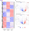
Fig 3. Heat stress and starvation stress associated proteome changes in C. neoformans revealed by label-free quantitative proteomics.
(A) Heat map representation of differentially expressed proteins across the following conditions: ∼25°C during the exponential growth phase (control), ∼25°C during the stationary growth phase (starvation stress), and 37°C during the exponential growth phase (heat stress). The Log2 Cyclic Loess Normalized Exclusive Intensities of each protein per sample were transformed to Z-scores. Clustering was performed both on proteins and samples using an average linkage method and Kendall’s Tau distance measurement method. Upregulated and downregulated proteins are represented with red and blue colors, respectively. (B) Top: Volcano plot displaying results of differential expression analysis comparing proteome of control (∼25°C during exponential growth phase), and nutrient starvation stress (∼25°C during stationary growth phase) proteome. Bottom: Volcano plot displaying results of differential expression analysis comparing proteome of control (∼25°C during exponential growth phase), and heat stress (37°C during exponential growth phase) proteome. The dashed vertical lines differentiate the upregulated and downregulated proteins (absolute value FC > 2). The proteins above the dashed horizontal line represent the statistically significant proteins (BH-adjusted p < 0.001). Proteins highlighted in red are significantly upregulated, and proteins highlighted in blue are significantly downregulated.
Fig 4
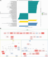
Fig 4. Gene set enrichment analysis (GSEA) of differentially expressed proteome in stationary growth phase versus exponential growth phase.
(A) Bar plot for significantly enriched gene ontology (GO) categories with p-value < 0.05 and FDR < 0.25 from GSEA. To avoid overlooking potentially significant gene ontology terms, an FDR of 0.25 rather than 0.05 was used for GSEA. (B) Directed acyclic graph of enriched GO terms with GSEA. The GO terms with positive enrichment are highlighted in red; while the GO terms with negative enrichment are highlighted in blue.
Fig 5
Fig 5. Gene set enrichment analysis (GSEA) of differentially expressed proteome at 37°C (heat stress) vs. ∼25°C (ambient temperature).
(A) Bar plot illustrating significantly enriched gene ontology (GO) categories with p-value < 0.05 and FDR < 0.25 from GSEA. To account for the potential overlook of significant gene ontology terms, an FDR of 0.25 rather than 0.05 was used for GSEA. (B) Directed acyclic graph of enriched GO terms from GSEA, with positive enrichment are highlighted in red, while the GO terms with negative enrichment are highlighted in blue.
Fig 6
Fig 6. Characterization of Cdc10-mCherry interactome at ∼25°C and 37°C (heat stress).
(A) Volcano plots of protein interactome results show proteins that are significantly enriched by Cdc10-mCherry co-immunoprecipitation from samples grown at ∼25 and 37°C, respectively. Significant interacting partners were determined by statistical t-test using logFC >2 and FDR of 0.05. The most significant candidates (highlighted) were considered those with a logFC >2 and FDR of 0.001. (B) Dot plots of functional enrichment analysis of septin Cdc10 protein interactome obtained from both temperature conditions using Fisher’s Exact Test. Gene count refers to the number of genes enriched in a GO term, and gene ratio is the percentage of total differential expressed genes in the given GO term.
Fig 7
Fig 7. Characterization of Cdc3-mCherry interactome at ∼25°C and 37°C (heat stress).
(A) Volcano plots of protein interactome results showing proteins that are significantly enriched by Cdc3-mCherry co-immunoprecipitation during ambient temperature and heat stress, respectively. Significant interacting partners were determined by statistical t-test using logFC >2 and FDR of 0.05. The most significant candidates (highlighted) were considered those with a logFC >2 and FDR of 0.001. (B) Dot plots of functional enrichment analysis of septin Cdc3 protein interactome during ambient temperature and heat stress, respectively, using Fisher’s Exact Test. Gene count refers to the number of genes enriched in a GO term, and gene ratio is the percentage of total differential expressed genes in the given GO term.
Fig 8
Fig 8. Overlap between Cdc10 and Cdc3 protein interactome.
Significant interacting partners were determined by statistical t-test using logFC >2 and adjusted p-value of 0.05. Venn Diagram showing the overlap of significantly enriched proteins for co-immunoprecipitation of both Cdc3-mCherry and Cdc10-mCherry between ambient temperature (∼25°C) and heat stress (37°C).
Fig 9
Fig 9. Septin Cdc3 and Cdc10 shared protein enrichment analysis.
(A) Dot plots of functional enrichment analysis of septin Cdc10 and Cdc3 shared protein interactome during ambient temperature (∼25°C) and heat stress (37°C) using Fisher’s Exact Test. (B) Dot plots of KEGG pathway functional analysis of septin Cdc10 and Cdc3 shared protein interactome during ambient temperature and heat stress. Gene count refers to the number of genes enriched in a GO term, and gene ratio is the percentage of total differential expressed genes in the given GO term.
Fig 10
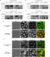
Fig 10. Co-immunoprecipitation and fluorescence microscopy of septins Cdc3 and Cdc10 and their interacting partners profilin and AFR1.
A co-IP of the Cdc10-mCherry using the GFP-Trap resin and a co-IP of the GFP-profilin using the RFP-Trap resin, at (A) 25°C and (B) heat stress (37°C). A co-IP of the Cdc3-mCherry using the GFP-Trap resin and a co-IP of the GFP-AFR1 using the RFP-Trap resin, at (C) 25°C and (D) heat stress (37°C). Strain that expresses only Cdc10-mCherry or Cdc3-mcherry, and strains that express only GFP-tagged interacting proteins were used as negative controls. The membranes were initially probed with an anti-GFP antibody and imaged and subsequently membranes were stripped and probed with an anti-RFP antibody to detect the precipitated GFP-tagged and mCherry-tagged proteins, respectively. (E) Cells expressing GFP-profilin and Cdc10-mCherry were visualized via live fluorescent microscopy from a culture grown at ∼25°C and after 2 hours of incubation at 37°C. GFP-profilin doesn’t show co-localization with septin Cdc10-mCherry. (F) Cells expressing GFP-Afr1 and Cdc3-mCherry were visualized via live fluorescent microscopy from a culture grown at ∼25°C and after 2 hours of incubation at 37°C. GFP-AFR1 does not colocalize with Cdc3-mCherry at 25°C; however, it co-localizes partially during high temperature stress.

Update of

Similar articles

Cited by

References

    1. Rajasingham R, Govender NP, Jordan A, Loyse A, Shroufi A, Denning DW, et al.. The global burden of HIV-associated cryptococcal infection in adults in 2020: a modelling analysis. Lancet Infect Dis. 2022. Dec;22(12):1748–55. - PMC - PubMed
    1. Zhao Y, Ye L, Zhao F, Zhang L, Lu Z, Chu T, et al.. Cryptococcus neoformans, a global threat to human health. Infect Dis Poverty. 2023. Mar 17;12(1):20. - PMC - PubMed
    1. Idnurm A, Bahn YS, Nielsen K, Lin X, Fraser JA, Heitman J. Deciphering the model pathogenic fungus Cryptococcus neoformans. Nat Rev Microbiol. 2005;3(10):753–64. - PubMed
    1. Bojarczuk A, Miller KA, Hotham R, Lewis A, Ogryzko N V., Kamuyango AA, et al.. Cryptococcus neoformans Intracellular Proliferation and Capsule Size Determines Early Macrophage Control of Infection. Sci Rep. 2016;6(January):1–15. - PMC - PubMed
    1. Mednick AJ, Nosanchuk JD, Casadevall A. Melanization of Cryptococcus neoformans affects lung inflammatory responses during cryptococcal infection. Infect Immun. 2005;73(4):2012–9. - PMC - PubMed

MeSH terms

LinkOut - more resources