. 2025 Apr;292(7):1696-1709.
doi: 10.1111/febs.17349. Epub 2024 Dec 17.

Porphyromonas gingivalis outer membrane vesicles increase vascular permeability by inducing stress fiber formation and degrading vascular endothelial-cadherin in endothelial cells

Affiliations

Porphyromonas gingivalis outer membrane vesicles increase vascular permeability by inducing stress fiber formation and degrading vascular endothelial-cadherin in endothelial cells

Mana Mekata et al. FEBS J. 2025 Apr.

Abstract

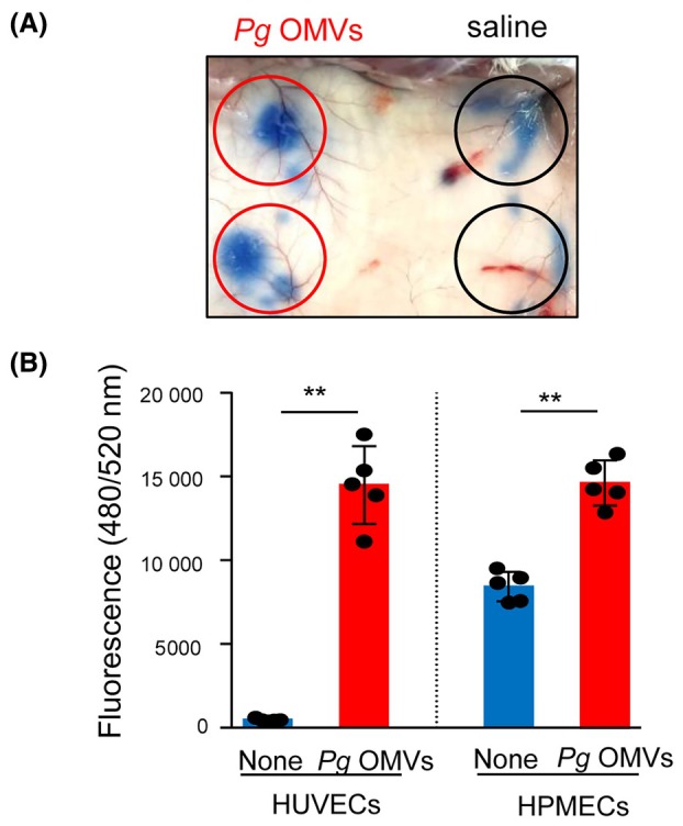
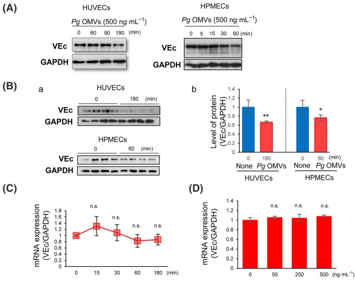
Porphyromonas gingivalis (Pg) is a keystone bacterium associated with systemic diseases, such as diabetes mellitus and Alzheimer's disease. Outer membrane vesicles (OMVs) released from Pg have been implicated in systemic diseases by delivering Pg virulence factors to host cells in distant organs and inducing cellular dysfunction. Pg OMVs also have the potential to enter distant organs via the bloodstream. However, the effects of Pg OMVs on the vascular function are poorly understood. Here, we showed that Pg OMVs increase vascular permeability by promoting stress fiber formation and lysosome/endosome-mediated vascular endothelial-cadherin (VEc) degradation in human umbilical vein endothelial cells (HUVECs) and human pulmonary microvascular endothelial cells (HPMECs). F-actin, visualized via fluorescein isothiocyanate-phalloidin, became thicker and longer, leading to the formation of radical stress fibers in response to Pg OMVs in HUVECs and HPMECs. Western blotting and quantitative real-time polymerase chain reaction analyses revealed that Pg OMVs decreased VEc protein levels in a gene-independent manner. Pg OMVs enhanced vesicular VEc accumulation in the cytoplasm around lysosome-associated membrane protein 1-positive structures during pretreatment with the lysosomal inhibitor chloroquine. This suggests that Pg OMVs decrease VEc protein levels by accelerating their internalization and degradation via lysosomes and endosomes. A27632 inhibition of Rho kinases impaired the Pg OMV-induced stress fiber formation and VEc degradation, resulting in the recovery of hyperpermeability. These findings provide new insights into the pathogenesis of systemic diseases that are associated with periodontal diseases.

Keywords: Porphyromonas gingivalis; outer membrane vesicles; periodontal diseases; vascular permeability.


Conflict of interest statement

The authors declare no conflict of interest.

Figures

Fig. 1
Fig. 1
Pg OMVs increase vascular permeability in vivo and in vitro. (A) Representative dorsal skin images of mice in the Miles assay. Porphyromonas gingivalis (Pg) outer membrane vesicles (OMVs) containing 15 μg of protein (right side of the skin) or saline (left side of the skin) were singly injected into dorsal skin of BALB/cAJc1 mice. Subsequently, 100 μL of 0.5% Evans blue was injected into the tail vein. After 10 min, the gross Evans blue dye level was visually observed on the skin surface in Pg OMVs‐injected area (red circles) and saline‐injected area (black circles). Four mice were used per group for each experiment and a typical image of visual discoloration is shown. (B) Human umbilical vein endothelial cells (HUVECs) and human pulmonary microvascular endothelial cells (HPMECs) were grown to confluence on the insert of a transwell system and treated with (n = 5) or without (none, n = 4) Pg OMVs (500 ng·mL−1) for 60 min. FITC‐labeled dextran (1 mg·mL−1) was then added to the insert. After 10 min, the FITC‐labeled dextran leakage from the apical compartment of the insert to the bottom well was quantified by measuring the fluorescence intensity. Student's t‐test was used for statistical analysis. Error bars represent standard deviation. **P < 0.01 compared with no‐treated cells (None).
Fig. 2
Fig. 2
Pg OMVs induce stress fiber formation in HUVECs and HPMECs. HUVECs (A, B) and HPMECs (C, D) were cultured in monolayers and treated with (B, D, F) or without (none; A, C, E) 500 ng·mL−1 of Porphyromonas gingivalis (Pg) outer membrane vesicles (OMVs) for 60 min. Cells were fixed and stained with vascular endothelial‐cadherin (VEc) to label adhesion junctions (AJs) (magenta) (A, B). F‐actin was labeled with FITC‐phalloidin (green) (C, D), and the nuclei were stained with Hoechst 33342 (white) and merged (E, F). High‐magnification merged images (2.5×) are shown in (B) and (D). Arrows indicate radical stress fibers. The arrowhead represents focal AJs connected to stress fibers. Scale bars indicate 20 μm. n = 4 in each group. Four samples were set up in each group (n = 4), and the typical photographs were shown from four independent experiments. (E). F‐actin was labeled with FITC‐phalloidin (green) in HUVECS and HPMECs treated with or without 500 ng·mL−1 of Pg OMVs for 60 min (n = 4 in each group), and green fluorescence intensity were measured. Student's t‐test was used for statistical analysis. Error bars represent standard deviation. **P < 0.01 compared with no‐treated cells (None).
Fig. 3
Fig. 3
Pg OMVs downregulate the level of VEc protein but not mRNA in HUVECs and HPMECs. (A) Human umbilical vein endothelial cells (HUVECs) and human pulmonary microvascular endothelial cells (HPMECs) were treated with 500 ng·mL−1 Porphyromonas gingivalis (Pg) outer membrane vesicles (OMVs). Subsequently, vascular endothelial‐cadherin (VEc) expression was analyzed via western blotting at indicated periods. (B) HUVECs and HPMECs were treated with Pg OMVs for 0 or 180 min, and 60 min, respectively. (A) The cell lysates were analyzed via western blotting using antibodies for VEc and GAPDH. (B) Densitometric analysis of each band was performed using imagej, and the VEc/GAPDH ratio in each group were presented and analyzed using Student's t‐test (n = 4 in each group). Values are shown as the fold change in expression levels compared to that in the 0 min group. Error bars represent standard deviation. *P < 0.05, **P < 0.01 compared with 0 min. (C) HUVECs were treated with 500 ng·mL−1 Pg OMVs for 0–180 min (n = 4). (D) HUVECs were treated with 0–500 ng·mL−1 Pg OMVs for 180 min (n = 4). VEc mRNA expression was analyzed using real‐time PCR. Values are shown as the fold change in expression levels compared to that in the 0 min group. Student's t‐test was used for statistical analysis (n = 4 in each group; n.s., not significant). Error bars represent standard deviation.
Fig. 4
Fig. 4
Pg OMVs accelerate VEc degradation through the lysosomal pathway in HUVECs. (A) Human umbilical vein endothelial cells (HUVECs) were pretreated with (+) (B, C, E, F) or without (−) (A, D) chloroquine for 30 min. Subsequently, cells were treated with (D–F) or without (A–C) 500 ng·mL−1 Porphyromonas gingivalis (Pg) outer membrane vesicles (OMVs) for 180 min before fixation and subjected to vascular endothelial‐cadherin (VEc) staining for immunofluorescence analysis. Nuclei were stained with Hoechst 33342. High‐magnification images (4×) of (B) and (E) are shown (C, F). Scale bar, 20 μm. Arrowheads indicate the accumulation of intracellular vesicular VEc. Four samples were set up in each group (n = 4), and the typical photographs were shown from three independent experiments. (B) Vesicular VEc was induced by same method shown as (A). VEc and lysosome‐associated membrane protein 1 (LAMP‐1) were co‐stained with specific antibodies for VEc (A, E) or LAMP‐1 (B, F). Nuclei were stained with Hoechst 33342 and the microscopic images of the same field were merged (C, G). High‐magnification images (4×) of the merge are shown (D, H). Scale bars indicate 20 μm. Arrowheads indicate vesicular VEc surrounded by LAMP‐1. (I) The merged white fluorescent intensity and green fluorescent intensity around nucleus were quantified. The ratio of merged (white)/LAMP‐1 (green) is shown as the co‐localization coefficients of VEc and LAMP‐1. Student's t‐test was used for statistical analysis (n = 4, samples; n = 8, field of view, in each group). Error bars represent standard deviation. **P < 0.01 compared with no‐treated cells (None). (C) The same samples used in (B) were subjected to confocal microscopy (A, C), and high‐magnification images (4×) are shown in (B, D). VEc and LAMP‐1 double‐staining revealed co‐localization in some vesicles induced by Pg OMVs (D, arrows).
Fig. 5
Fig. 5
Rho kinases are involved in Pg OMVs‐induced permeability in HUVECs. (A) The hyperpermeability signaling pathway promoted by actin filament rearrangement. (B) Human umbilical vein endothelial cells (HUVECs) were treated with 500 ng·mL−1 Porphyromonas gingivalis (Pg) outer membrane vesicles (OMVs) for the indicated periods and cofilin phosphorylation was assessed via western blotting. The immunoblots are representative figures of three independent experiments. (C) HUVECs were pretreated with 10 μm Y27632 for 30 min before Pg OMVs challenge for 60 min (C, F, I). Cells were treated, fixed, and stained as described in Fig. 2. Arrows indicate the radical stress fibers (E, H). The arrowhead indicates focal adhesion junctions (AJs) connected to stress fibers (E, H). Bars indicate 20 μm. The fluorescence intensity of F‐actin labeled with FITC‐phalloidin (green, in D, E and F) was quantified (J). Student's t‐test was used for statistical analysis (n = 4 in each group). Error bars represent standard deviation. **P < 0.01 compared with no‐treated cells (None). (D) HUVECs were pretreated with 10 μm Y27632 for 30 min before Pg OMVs challenge for 180 min. Vascular endothelial‐cadherin (VEc) protein levels were analyzed via western blotting. WT, wild‐type; KDP136, gingipain deficient. (E) HUVECs were grown on Transwell inserts and pretreated with 10 μm Y27632 for 30 min before Pg OMVs challenge for 60 min. Vascular permeability was assessed as shown in Fig. 1B. Student's t‐test was used for statistical analysis (n = 4). Error bars represent standard deviation. **P < 0.01.
Fig. 6
Fig. 6
Graphical summary. In healthy individuals, the cell–cell junction is constructed with linear adhesion junctions (AJs) supported by circumferential actin bundles. Therefore, the endothelial barrier was strong and had normal vascular permeability. Porphyromonas gingivalis (Pg) outer membrane vesicles (OMVs) released during periodontal disease may induce Rho activation. Activated Rho kinases increase stress fiber formation, thereby weakening the endothelial barrier by forming focal AJs. Pg OMVs‐activated Rho kinases also promote vascular endothelial‐cadherin (VEc) internalization via endocytosis and subsequent degradation via the endosomal and lysosomal pathways, leading to a decrease in VEc levels.

Similar articles

Cited by

References

    1. Kanagasingam S, Chukkapalli SS, Welbury R & Singhrao SK (2020) Porphyromonas gingivalis is a strong risk factor for Alzheimer's disease. J Alzheimers Dis Rep 4, 501–511. - PMC - PubMed
    1. Watanabe K, Katagiri S, Takahashi H, Sasaki N, Maekawa S, Komazaki R, Hatasa M, Kitajima Y, Maruyama Y, Shiba T et al. (2021) Porphyromonas gingivalis impairs glucose uptake in skeletal muscle associated with altering gut microbiota. FASEB J 35, e21171. - PubMed
    1. Bielaszewska M, Daniel O, Nyč O & Mellmann A (2021) In vivo secretion of β‐lactamase‐carrying outer membrane vesicles as a mechanism of β‐lactam therapy failure. Membranes 11, 806. - PMC - PubMed
    1. Ishikawa M, Yoshida K, Okamura H, Ochiai K, Takamura H, Fujiwara N & Ozaki K (2013) Oral Porphyromonas gingivalis translocates to the liver and regulates hepatic glycogen synthesis through the Akt/GSK‐3beta signaling pathway. Biochim Biophys Acta 1832, 2035–2043. - PubMed
    1. Veith PD, Chen YY, Gorasia DG, Chen D, Glew MD, O'Brien‐Simpson NM, Cecil JD, Holden JA & Reynolds EC (2014) Porphyromonas gingivalis outer membrane vesicles exclusively contain outer membrane and periplasmic proteins and carry a cargo enriched with virulence factors. J Proteome Res 13, 2420–2432. - PubMed

MeSH terms

LinkOut - more resources