. 2024 Dec 18;9(1):352.
doi: 10.1038/s41392-024-02042-5.

Macrophages promote pre-metastatic niche formation of breast cancer through aryl hydrocarbon receptor activity

Affiliations

Macrophages promote pre-metastatic niche formation of breast cancer through aryl hydrocarbon receptor activity

Xu Jiang et al. Signal Transduct Target Ther. .

Abstract

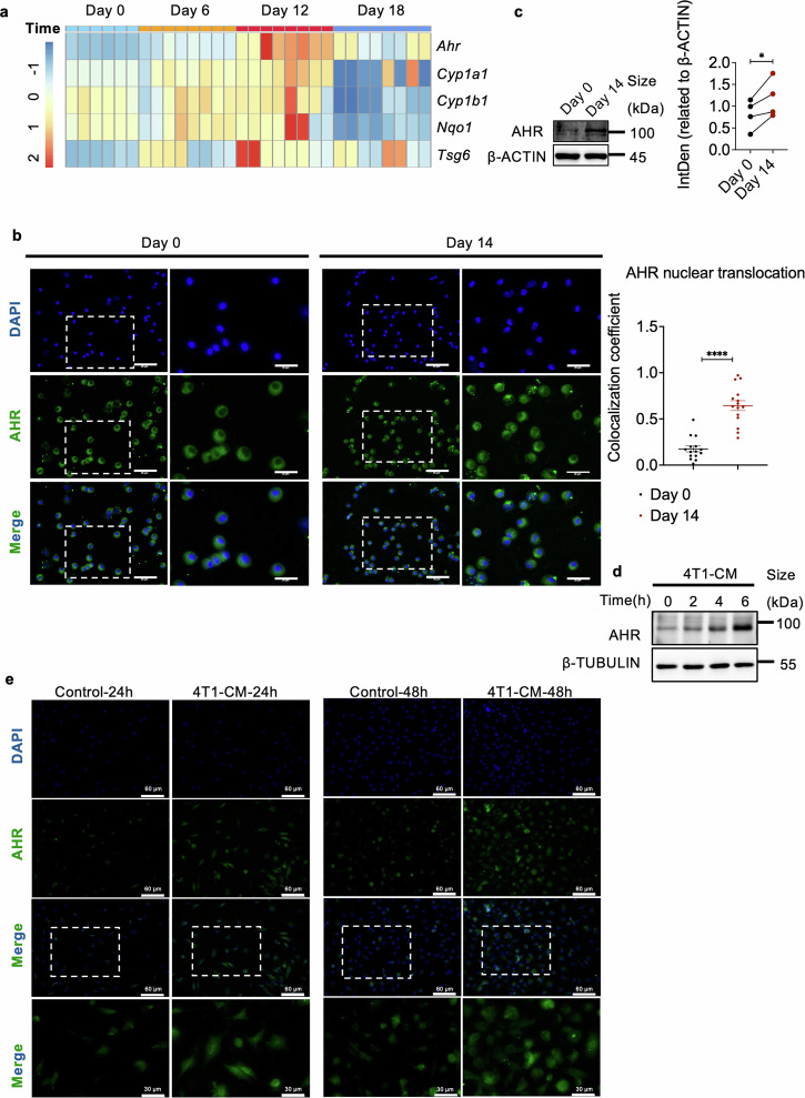
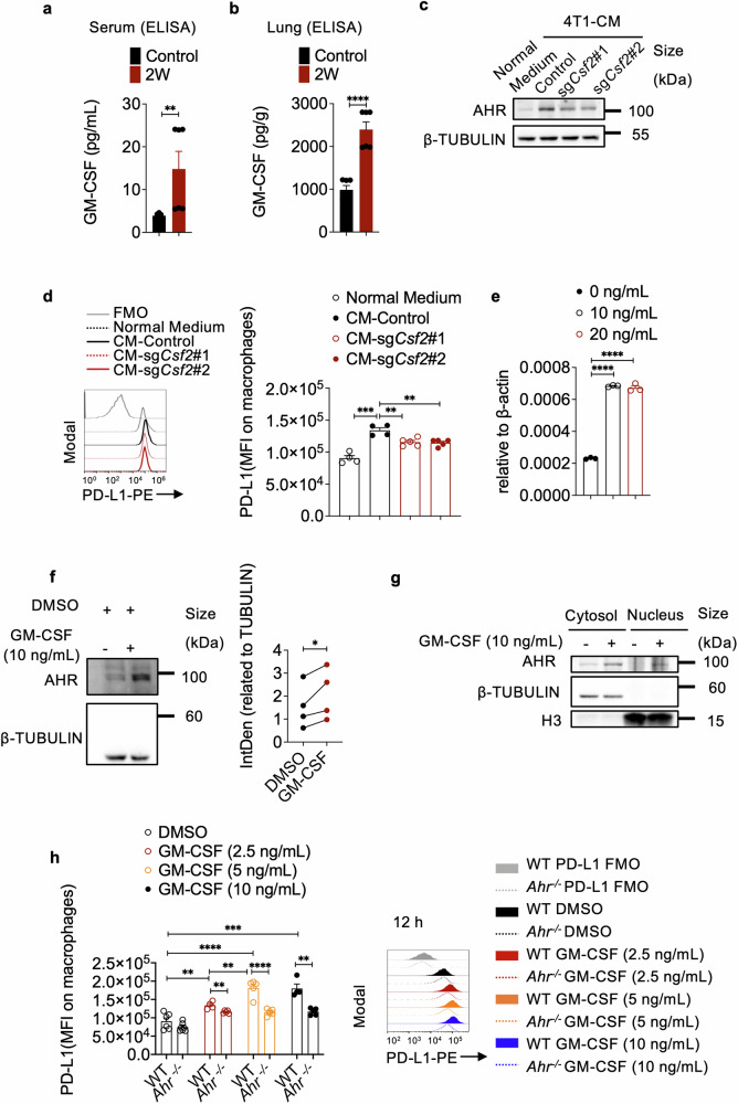
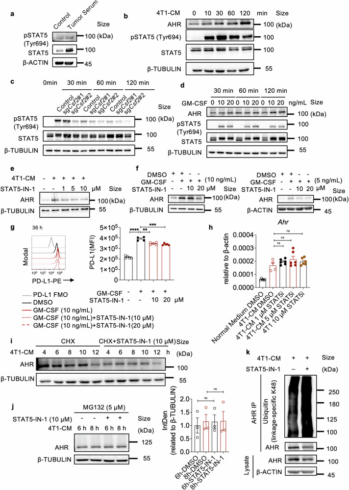
Macrophages that acquire an immunosuppressive phenotype play a crucial role in establishing the pre-metastatic niche (PMN), which is essential for facilitating breast cancer metastasis to distant organs. Our study showed that increased activity of the aryl hydrocarbon receptor (AHR) in lung macrophages plays a crucial role in establishing the immunosuppressive PMN in breast cancer. Specifically, AHR activation led to high expression of PD-L1 on macrophages by directly binding to the promoter of Pdl1. This upregulation of PD-L1 promoted the differentiation of regulatory T cells (Tregs) within the PMN, further enhancing immunosuppressive conditions. Mice with Ahr conditional deletion in macrophages had reduced lung metastasis of breast cancer. The elevated AHR levels in PMN macrophages were induced by GM-CSF, which was secreted by breast cancer cells. Mechanistically, the activated STAT5 signaling pathway induced by GM-CSF prevented AHR from being ubiquitinated, thereby sustaining its activity in macrophages. In breast cancer patients, the expression of AHR and PD-L1 was correlated with increased Treg cell infiltration, and higher levels of AHR were associated with a poor prognosis. These findings reveal that the crosstalk of breast cancer cells, lung macrophages, and Treg cells via the GM-CSF-STAT5-AHR-PD-L1 cascade modulates the lung pre-metastatic niche during breast cancer progression.


Conflict of interest statement

Competing interests: The authors declare no competing interests.

Figures

Fig. 1
Fig. 1
Changes in AHR activity in lung macrophages prior to breast cancer metastasis. a Heatmap showing AHR and its downstream gene expression in lung macrophages at indicated timepoints post primary 4T1 tumor establishment. b Representative immunofluorescent staining of AHR nuclear translocation in CD45+ CD11b CD11c+ SiglecF+ macrophages from the lungs of mice bearing 4T1 tumors for 14 days (n = 15 for each group, repeated twice). The scale bars, 60 µm. The scale bars in the enlarged images, 30 µm. The column bars show the coefficient of AHR nuclear translocation in macrophages. c Western blotting analysis and quantification of AHR levels in peritoneal macrophages treated with serum from mice bearing 4T1 tumors for 14 days (repeated four times). d Western blotting analysis of AHR levels in peritoneal macrophages treated with 4T1-conditioned medium (CM) for indicated times. e Representative immunofluorescent staining of AHR nuclear translocation in peritoneal macrophages with or without 4T1-CM treatment. Scale bars, 60 µm. Scale bars in the enlarged images, 30 µm. Data are analyzed by unpaired two-tailed t-test (b) or paired two-tailed t-test (c) and presented as mean ± SEM (b) or symbols & lines (c). *p < 0.05, ****p < 0.0001
Fig. 2
Fig. 2
AHR in macrophages influences breast cancer metastasis and Treg cell differentiation. ac Wild-type (Control) and AhrflflLyz2Cre+/− mice were inoculated with 4T1 cells in the mammary gland fat pad for 35 days. Tumor weight (n = 7 for each group) (a), the number of macroscopic metastases in the lungs (n = 7 for each group) (b), and picric acid-stained lungs (n = 3 for each group) (c) were assessed. d H&E staining of lung sections from AhrflflLyz2Cre−/− (Control) and AhrflflLyz2Cre+/− tumor-bearing mice(n ≥ 6). Scale bars, 400 µm. e Survival of 4T1 tumor-bearing wild-type (Control) and AhrflflLyz2Cre+/− mice (n = 8 for each group). f Flow cytometry analysis on Treg cells in the lungs of AhrflflLyz2Cre−/− (Control) and AhrflflLyz2Cre+/− mice bearing tumor for 14 days (n = 3 for each group, repeated three times). g Flow cytometry analysis on the impact of AHR in macrophages on Treg cell differentiation. BMDMs from wild-type (Control) or Ahr−/− mice were pretreated with normal medium (Control medium) or 4T1-CM for 2 days. Naïve CD4+ T cells were co-cultured with pretreated BMDMs for 3 days under the Treg differentiation conditions (n = 4 for each group, repeated four times). Data are analyzed by unpaired two-tailed t-test (a, b, f, g) or Mann Whitney test (d), or Log-rank (Mantel-Cox) test (e) and presented as mean ± SEM. *p < 0.05, **p < 0.01, ***p < 0.001, ****p < 0.0001, ns, no significance
Fig. 3
Fig. 3
Promotion of Treg cells by AHR activated macrophages through PD-L1. a Representative immunofluorescent images showing F4/80 and PD-L1 staining in the lungs of AhrflflLyz2Cre−/− (Control) and AhrflflLyz2Cre+/− mice inoculated with 4T1 cells for 14 days (n ≥ 15 for each group). Scale bar, 60 μm. b Schematic of lung macrophage isolation from mice bearing 4T1 tumors at indicated timepoints (created with BioRender.com). Correlation analysis between Pdl1 and Ahr mRNA expressions in F4/80+ cells from the PMN (n = 15 for each group). c Pdl1 mRNA expression in peritoneal macrophages, with or without addition of serum from 4T1 tumor bearing mice (n ≥ 3 for each group, repeated twice). d PD-L1 expression on peritoneal macrophages of wild-type (WT) and Ahr−/− mice, with or without 4T1-CM treatment for 12 h (n ≥ 3 for each group). e PD-L1 expression on peritoneal macrophages from AhrflflLyz2Cre−/− (Control) and AhrflflLyz2Cre+/− mice, with or without 4T1-CM (n ≥ 3 for each group, repeated twice). f Flow cytometry analysis of Treg cell differentiation when co-cultured with normal medium or 4T1-CM pretreated macrophages from WT mice and Ahr−/− mice in the presence of isotype control or anti-PD-L1. BMDMs from WT mice (Control) or Ahr−/− mice were pretreated with normal medium (Control) or 4T1-CM for 2 days. Naïve CD4+ T cells were co-cultured with pretreated BMDMs for 3 days under the Treg cell differentiation conditions, with the addition of PD-L1 neutralizing antibody or Isotype control. g Bar graph showing quantification of Treg cell differentiation (n = 4 for each group). h ChIP analysis of AHR recruitment to the Pdl1 promoter in peritoneal macrophages stimulated with 4T1-CM (n = 4 for each group). Data are analyzed by unpaired two-tailed t-test (a, c, d, e, g, h) or Pearson correlation analysis (b) and presented as mean ± SEM. *p < 0.05, **p < 0.01, ***p < 0.001, ****p < 0.0001, ns, no significance
Fig. 4
Fig. 4
GM-CSF released from breast cancer cells promotes AHR expression in macrophages. Quantification of GM-CSF levels in both serum (a) and lung tissue (b) of mice inoculated with 4T1 cells in the mammary gland fat pad for 2 weeks using ELISA (n = 6 for each group). c AHR expression in peritoneal macrophages cultured in normal medium, or CM from control 4T1 cells, or sgCsf2 transfected 4T1 cells for 12 h (repeated twice). d Histogram showing PD-L1 expression on peritoneal macrophages cultured in normal medium, CM from control 4T1 cells, or CM from sgCsf2-transfected 4T1 cells for 24 h (n ≥ 4 for each group, repeated three times). The bar graph shows MFI of PD-L1 on each group of macrophages. e mRNA expression of Ahr in peritoneal macrophages treated with GM-CSF at indicated concentrations for 24 h (n = 3 for each group, repeated twice). f Western blotting analysis and quantification of AHR expression in peritoneal macrophages treated with GM-CSF (10 ng/mL) for 24 h. g Western blotting analysis of AHR expression in the cytoplasm and nucleus of macrophages treated with or without GM-CSF. h Histogram showing PD-L1 expression on WT or Ahr−/− peritoneal macrophages treated with or without GM-CSF (2.5, 5, or 10 ng/mL) for 12 h. The bar graph shows MFI of PD-L1 on each group of macrophages (n ≥ 4 for each group). Data are analyzed by unpaired two-tailed t-test (b, d, e, h) or Mann Whitney test (a) or paired two-tailed t-test (f) and presented as mean ± SEM (a, b, d, e, h) or symbols & lines (f). *p < 0.05, **p < 0.01, ***p < 0.001, ****p < 0.0001
Fig. 5
Fig. 5
GM-CSF-initiated STAT5 signaling enhances AHR stability by inhibiting AHR ubiquitination. a Western blotting analysis of pSTAT5 and STAT5 expressions in peritoneal macrophages treated with serum from mice inoculated with or without 4T1 cells for 14 days (repeated twice). b Western blotting analysis of AHR, pSTAT5, and STAT5 levels in peritoneal macrophages treated with 4T1-CM at indicated time points (repeated three times). c Western blotting analysis of pSTAT5 and STAT5 expressions in peritoneal macrophages treated with CM from control 4T1 cells or Csf2 knockout 4T1 cells at indicated time points. d Western blotting analysis of AHR, pSTAT5, and STAT5 expressions in peritoneal macrophages treated with GM-CSF (0, 10, 20 ng/mL) for 30 min, 60 min, and 120 min. e Western blotting analysis of AHR expression in STAT5-IN-1-treated peritoneal macrophages, with or without 4T1-CM treatment for 24 h (repeated twice). f Western blotting analysis of AHR expression in peritoneal macrophages treated with GM-CSF (10 ng/mL or 5 ng/mL), with or without the addition of STAT5-IN-1 (10 or 20 μM) (repeated twice). g Histogram of PD-L1 expression on peritoneal macrophages treated with DMSO or GM-CSF (10 ng/mL) for 36 h, with or without the addition of STAT5-IN-1 (10 or 20 μM) (n ≥ 4 for each group, repeated twice). The bar graph shows MFI of PD-L1 on each group of macrophages. h mRNA levels of Ahr in peritoneal macrophages treated with or without 4T1-CM, in the presence or absence of STAT5-IN-1 (1, 5, or 10 μM) for 24 h (n ≥ 4 for each group, repeated three times). i The protein levels of AHR in peritoneal macrophages treated with 4T1-CM and CHX (5 µg/ml), with or without the addition of STAT5-IN-1 (10 μM) (repeated three times). j The protein levels and quantification of AHR in peritoneal macrophages treated with 4T1-CM and MG132 (5 μM) for 6 or 8 h, with or without the addition of STAT5-IN-1 (10 μM). k Western blotting analysis of ubiquitin in macrophages treated with 4T1-CM and STAT5-IN-1. Peritoneal macrophages cultured in 4T1-CM were treated with DMSO or STAT5-IN-1 (10 μM) for 12 h. The cell lysates were immunoprecipitated with an anti-AHR antibody and immunoblotted with an anti-ubiquitin antibody (repeated three times). Data are analyzed by unpaired two-tailed t-test (g, h, j) and presented as mean ± SEM. **p < 0.01, ***p < 0.001, ****p < 0.0001, ns no significance
Fig. 6
Fig. 6
The prognosis value of the AHR-PD-L1-Tregs axis in breast cancer patients. a Representative immunofluorescent images showing F4/80 and AHR staining in the lungs of patients and control. Scale bars, 60 µm. The bar graph shows the percentage of AHR⁺ F4/80⁺ cells among AHR⁺ cells (n ≥ 35 for each group). b Overall survival of breast cancer patients with high and low AHR expression. c Overall survival of TNBC patients with high and low AHR expression. AHR expression and breast cancer patient survival analysis using the Kaplan-Meier Plotter online tool (http://kmplot.com/analysis/). d The correlation between AHR and PD-L1 (CD274) expression in breast cancer and its subtype, basal-like breast cancer. e The correlation between Treg cell infiltration and PD-L1 expression in breast cancer. f The correlation between Treg cell infiltration and AHR expression in breast cancer. The correlation analysis among AHR, PD-L1, and Treg cells was analyzed using the TIMER2.0 online tool (http://timer.cistrome.org/). Data are analyzed by unpaired two-tailed t-test (a) and presented as mean ± SEM. *p < 0.05

References

    1. Weigelt, B., Peterse, J. L. & van ‘t Veer, L. J. Breast cancer metastasis: markers and models. Nat. Rev. Cancer5, 591–602 (2005). - PubMed
    1. Valastyan, S. & Weinberg, R. A. Tumor metastasis: molecular insights and evolving paradigms. Cell147, 275–292 (2011). - PMC - PubMed
    1. Peinado, H. et al. Pre-metastatic niches: organ-specific homes for metastases. Nat. Rev. Cancer17, 302–317 (2017). - PubMed
    1. Huang, Y. et al. Pulmonary vascular destabilization in the premetastatic phase facilitates lung metastasis. Cancer Res.69, 7529–7537 (2009). - PubMed
    1. Medeiros, B. et al. Triple-negative primary breast tumors induce supportive premetastatic changes in the extracellular matrix and soluble components of the lung microenvironment. Cancers12, 172 (2020). - PMC - PubMed

Publication types

MeSH terms

Substances