. 2025 May;480(5):3147-3160.
doi: 10.1007/s11010-024-05179-7. Epub 2024 Dec 17.

Interleukin-22 promotes endometrial carcinoma cell proliferation and cycle progression via ERK1/2 and p38 activation

Affiliations

Interleukin-22 promotes endometrial carcinoma cell proliferation and cycle progression via ERK1/2 and p38 activation

Shiqi Liu et al. Mol Cell Biochem. 2025 May.

Abstract

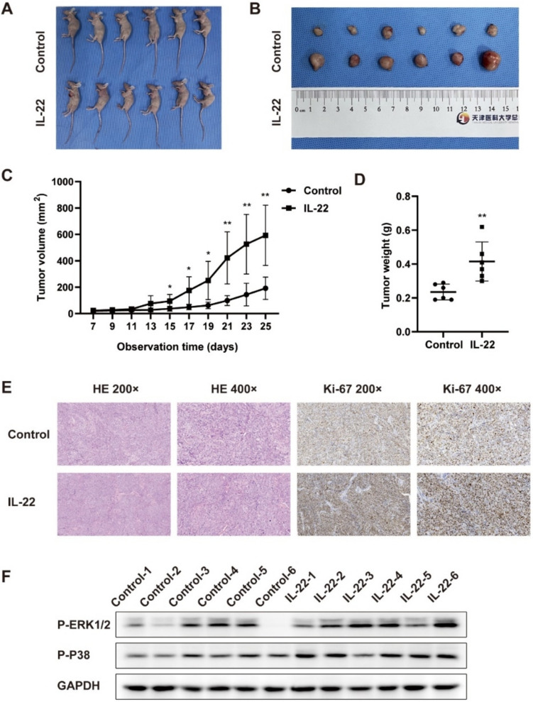
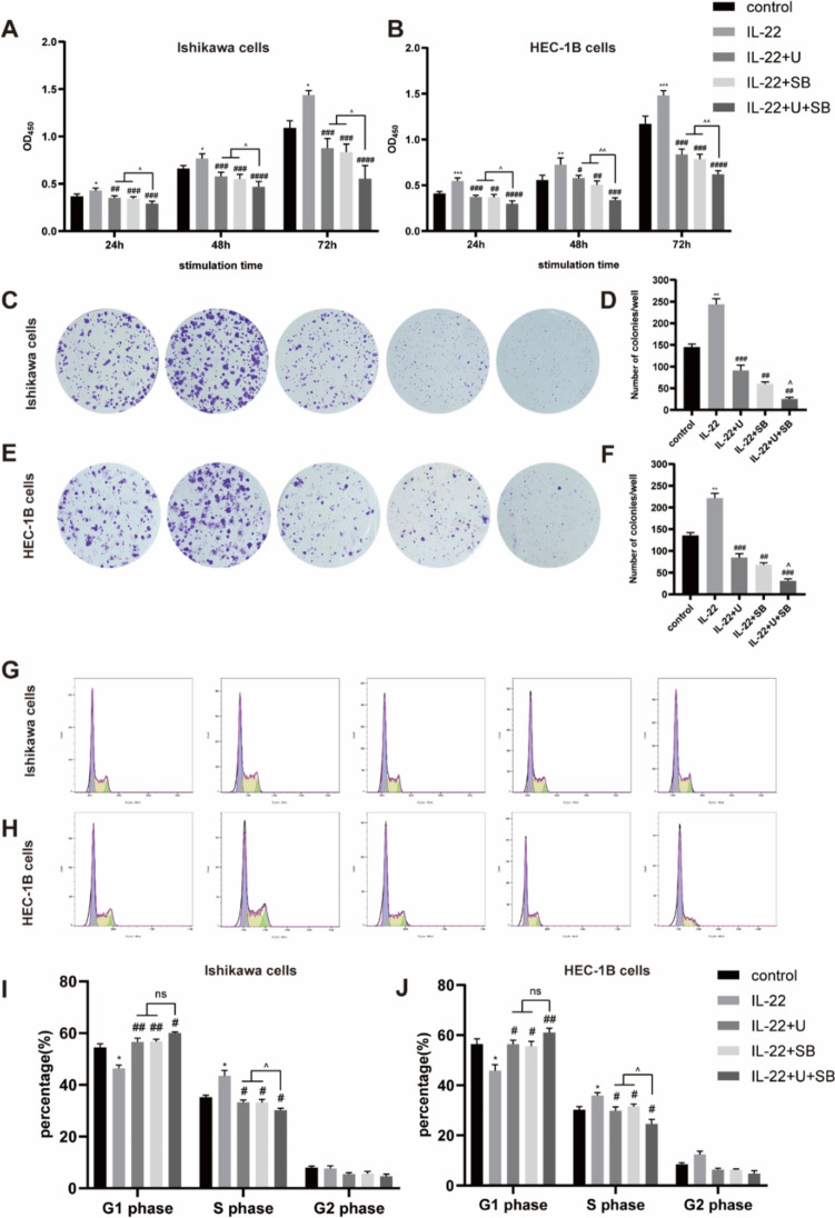
Endometrial carcinoma (EC) is one of the most common gynecological malignant tumors, but its underlying pathogenic mechanisms are largely obscure. Interleukin-22 (IL-22), one cytokine in the tumor immune microenvironment, was reported to be associated with carcinoma progression. Here, we aimed to investigate the regulation of IL-22 in endometrial carcinoma. Enzyme-linked immunosorbent assay (ELISA) analysis of IL-22 was done in 27 controls and 51 patients with EC. We examined the proliferative potential, cycle progression, and signaling pathways modulated by IL-22 in EC cells. Western blot analysis was performed to investigate the expression of proliferative and cycle-related proteins in EC cells. The effect of IL-22 mediated by interleukin-22 receptor alpha 1 (IL-22RA1) was examined using cell transfection with small interfering RNA (siRNA). In addition, a xenograft tumor model was performed to assess the effect of IL-22 in vivo. We demonstrated significant up-regulation of serum IL-22 concentrations in EC patients (42.59 ± 23.72 pg/mL) compared to the control group (27.47 ± 8.29 pg/mL). High levels of IL-22 concentrations appear to correlate with malignant clinicopathological features of EC. Treatment with IL-22 promoted cell proliferation and G1/S phase progression in Ishikawa and HEC-1B cells. Western blot analysis revealed that c-Myc, cyclin E1, cyclin-dependent kinase (CDK)2, cyclin D1, CDK4, CDK6, p-extracellular signal-regulated kinase1/2 (p-ERK1/2), and p-p38 were highly expressed in EC cells exposed to IL-22. Moreover, in the EC mice model, we found that giving exogenous IL-22 increased tumor volume and weight. Immunohistochemistry showed that intra-tumor Ki-67 expression was up-regulated upon IL-22 treatment. The IL-22-mediated changes in cell proliferation, cycle progression, and protein expression can be effectively inhibited by the ERK1/2 inhibitor U0126 and the p38 inhibitor SB202190. In addition, the role of IL-22 in EC is receptor-dependent. Our findings suggest that IL-22 promotes endometrial carcinoma cell proliferation and G1/S phase progression by activating ERK1/2 and p38 signaling. Therefore, IL-22 may represent a potential therapeutic target for the treatment of endometrial carcinoma.

Keywords: Cycle progression; Endometrial carcinoma; Interleukin-22; MAPKs; Proliferation.


Conflict of interest statement

Declarations. Conflict of interest: Conflict of interest the authors declare no conflict of interest. Ethical approval: The study was conducted in accordance with the Declaration of Helsinki, and approved by the Institutional Review Board of Tianjin Medical University General Hospital (protocol code IRB2021-KY-356; approved December 24, 2021). The animal study protocol was approved by the Institutional Review Board of Tianjin Medical University General Hospital (protocol code IRB2021-DWFL-422; approved December 24, 2021). Consent to participate: Informed consent was obtained from all individual participants included in the study. Consent to publication: The authors affirm that human research participants provided informed consent for publication of Tables 1 and 2.

Figures

Fig. 1
Fig. 1
IL-22 promotes endometrial carcinoma cell proliferation and G1/S phase progression. A, B Ishikawa (A) and HEC-1B (B) cells were treated with indicated concentrations of IL-22 for 24, 48, and 72 h to analyze cell viability by CCK-8 assay. C, E The colony formation assay was performed to further verify the effect of IL-22 on cell proliferation. D, F Quantitative analysis of colonies’ number per well in control and IL-22 groups. G, I Ishikawa (G) and HEC-1B (I) cell cycle in response to IL-22 were assessed by flow cytometry. H, J Results are expressed as the percentage of cells in the G1, S, and G2 phases. *p < 0.05, **p < 0.01, ***p < 0.001, ****p < 0.0001 vs control, respectively
Fig. 2
Fig. 2
IL-22 upregulates the proliferative and cycle-related proteins of endometrial carcinoma cells. A, C Western blot analysis of c-Myc, cyclin E1, CDK2, cyclin D1, CDK4, and CDK6 in Ishikawa (A) and HEC-1B (C) cells treated with IL-22 at different times. B, D Quantitative analysis of c-Myc, cyclin E1, CDK2, cyclin D1, CDK4, and CDK6 protein expression in Ishikawa (B) and HEC-1B (D) cells. The values represent the relative density of each band normalized to GAPDH. *p < 0.05, **p < 0.01, ***p < 0.001 vs control, respectively. ns: no significance
Fig. 3
Fig. 3
IL-22 activates ERK1/2 and p38 expression in endometrial carcinoma cells. A, E Western blot analysis of p-ERK, ERK, p-p38, p38, p-JNK, and JNK in Ishikawa (A) and HEC-1B (E) cells treated with IL-22 at different times. B, F Quantitative analysis of p-ERK protein expression in Ishikawa (B) and HEC-1B (F) cells. C, G Quantitative analysis of p-p38 protein expression in Ishikawa (C) and HEC-1BG) cells. D, H Quantitative analysis of p-JNK protein expression in Ishikawa (D) and HEC-1B (H) cells. The values represent the relative density of each band normalized to GAPDH. *p < 0.05, **p < 0.01, ***p < 0.001, ****p < 0.0001 vs control, respectively. ns: no significance
Fig. 4
Fig. 4
IL-22 stimulates endometrial carcinoma tumor growth and activates ERK1/2 and p38 expression in vivo. A, B Images of mice (A) and transplanted tumor (B) of control (n = 6) and IL-22 (n = 6) groups. C Changes in tumor volume over the 25 days of observation. Tumor volume was calculated according to the formula tumor volume (mm3) = (length × width2)/2. D Tumor weight of mice in control and IL-22 groups. E H&E staining and immunohistochemical (anti-Ki-67) staining of xenograft tissues from mice treated with or without IL-22. F Western blot analysis of p-ERK and p-p38 protein expression in xenograft tissues. *p < 0.05, **p < 0.01 vs control, respectively
Fig. 5
Fig. 5
Inhibition of ERK1/2 and p38 antagonizes the effects of IL-22 on endometrial carcinoma cell proliferation and G1/S phase progression. A, B Ishikawa (A) and HEC-1B (B) cells were pretreated with ERK1/2 inhibitor U0126 or p38 inhibitor SB202190 prior to IL-22 treatment for 24, 48, and 72 h to analyze cell viability by CCK-8 assay. C, E The colony formation assay was performed to further verify the effects of U0126 and SB202190 on the EC cell proliferation. D, F Quantitative analysis of colonies’ number per well in different groups. G, H Ishikawa (G) and HEC-1B (I) cell cycle in response to U0126 or SB202190 were assessed by flow cytometry. I, J Results are expressed as the percentage of cells in the G1, S, and G2 phases. U: U0126. SB: SB202190. *p < 0.05, **p < 0.01, ***p < 0.001 vs control, #p < 0.05, ##p < 0.01, ###p < 0.001, ####p < 0.0001 vs IL-22, ^p < 0.05, ^^p < 0.01 vs IL-22 + U or IL-22 + SB. ns: no significance
Fig. 6
Fig. 6
Inhibition of ERK1/2 and p38 antagonizes the effects of IL-22 on proliferative and cycle-related proteins of endometrial carcinoma cells A, B Western blot analysis of p-ERK1/2, p-p38, c-Myc, cyclin E1, CDK2, cyclin D1, CDK4, and CDK6 in Ishikawa (A) and HEC-1B (B) cells pretreated with ERK1/2 inhibitor U0126 or p38 inhibitor SB202190 prior to IL-22 treatment. C, D The mRNA (C) and protein (D) levels of IL-22RA1 were detected in EC cells transfected with negative control siRNA or IL-22RA1 siRNA. E, F Western blot analysis of p-ERK1/2, p-p38, c-Myc, cyclin E1, CDK2, cyclin D1, CDK4, and CDK6 in different groups of Ishikawa (E) and HEC-1B (F) cells. U: U0126. SB: SB202190

References

    1. Siegel RL, Giaquinto AN, Jemal A (2024) Cancer statistics, 2024. CA Cancer J Clin 74(1):12–49 - PubMed
    1. Crosbie EJ, Kitson SJ, McAlpine JN, Mukhopadhyay A, Powell ME, Singh N (2022) Endometrial cancer. Lancet 399(10333):1412–1428 - PubMed
    1. Lv B, Wang Y, Ma D, Cheng W, Liu J, Yong T et al (2022) Immunotherapy: reshape the tumor immune microenvironment. Front Immunol 13:844142 - PMC - PubMed
    1. Li J, Huang L, Zhao H, Yan Y, Lu J (2020) The role of interleukins in colorectal cancer. Int J Biol Sci 16(13):2323–2339 - PMC - PubMed
    1. Zang Y, Li H, Liu S, Zhao R, Zhang K, Zang Y et al (2022) The roles and clinical applications of interleukins in endometrial carcinoma. Front Oncol 12:1001693 - PMC - PubMed

MeSH terms

LinkOut - more resources