. 2025 Apr 22;121(2):311-323.
doi: 10.1093/cvr/cvae258.

Meis transcription factors regulate cardiac conduction system development and adult function

Affiliations

Meis transcription factors regulate cardiac conduction system development and adult function

Noelia Muñoz-Martín et al. Cardiovasc Res. .

Abstract

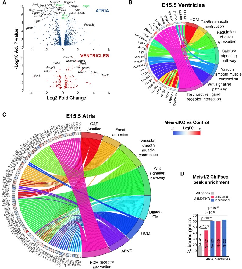
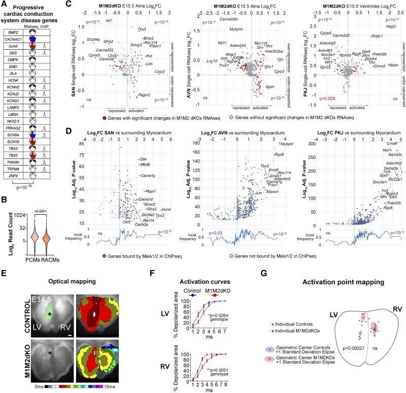
Aims: The cardiac conduction system (CCS) is progressively specified during development by interactions among a discrete number of transcription factors (TFs) that ensure its proper patterning and the emergence of its functional properties. Meis genes encode homeodomain TFs with multiple roles in mammalian development. In humans, Meis genes associate with congenital cardiac malformations and alterations of cardiac electrical activity; however, the basis for these alterations has not been established. Here, we studied the role of Meis TFs in cardiomyocyte development and function during mouse development and adult life.

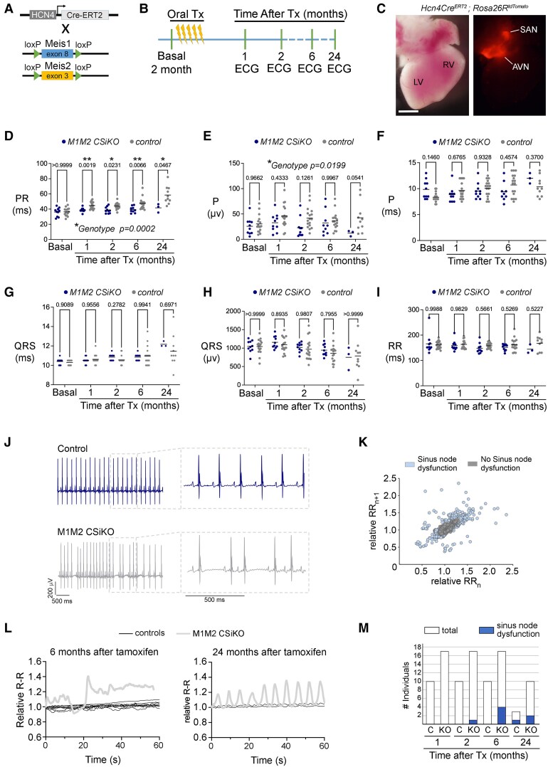
Methods and results: We studied Meis1 and Meis2 conditional deletion mouse models that allowed cardiomyocyte-specific elimination of Meis function during development and inducible elimination of Meis function in cardiomyocytes of the adult CCS. We studied cardiac anatomy, contractility, and conduction. We report that Meis factors are global regulators of cardiac conduction, with a predominant role in the CCS. While constitutive Meis deletion in cardiomyocytes led to congenital malformations of the arterial pole and atria, as well as defects in ventricular conduction, Meis elimination in cardiomyocytes of the adult CCS produced sinus node dysfunction and delayed atrio-ventricular conduction. Molecular analyses unravelled Meis-controlled molecular pathways associated with these defects. Finally, we studied in transgenic mice the activity of a Meis1 human enhancer related to an single-nucleotide polymorphism (SNP) associated by Genome-wide association studies (GWAS) to PR (P and R waves of the electrocardiogram) elongation and found that the transgene drives expression in components of the atrio-ventricular conduction system.

Conclusion: Our study identifies Meis TFs as essential regulators of the establishment of cardiac conduction function during development and its maintenance during adult life. In addition, we generated animal models and identified molecular alterations that will ease the study of Meis-associated conduction defects and congenital malformations in humans.

Keywords: Cardiac development; Mouse targeted mutation; PR elongation; Sinus node dysfunction; Transcription factor.


Conflict of interest statement

Conflict of interest: none declared.

Figures

Figure 1
Figure 1
Expression of Meis1 and Meis2 in the developing and adult heart. (A) Meis1 and Meis2 mRNA in situ hybridization showing expression in the second heart field, pericardium, endocardium (Ec), and epicardium (Ep) at E10.5. Boxed regions indicate magnifications shown in the panels to the right side. (B) Confocal images of ventricles at the indicated embryonic days showing anti-Meisa immunofluorescence. Boxed areas are magnified in the panels below. (CE′) Confocal images from sections of E16.5 hearts with the Meis1ECFP line combined with anti-Meisa. (FH′) Confocal images from sections of E16.5 hearts showing anti-Meis2 and anti-cTNT immunofluorescence. Boxes in (C) and (F) indicate magnified regions in (D and E′) and (G and H′). (I–L) Confocal images showing the distribution of ECFP in the CCS and atrial myocardium of Meis1ECFP adult hearts. (M–P) Anti-Meisa and anti-Hcn4 immunofluorescence in the SAN (M) and AVN (N), RA (O), and ventricular CMs (P). Dotted lines indicate magnified areas, and arrowheads within show anti-Meisa-positive CMs. (Q and Q′) Whole-mount brightfield and fluorescent confocal images of Meis1CreER; R26RTdTomato adult hearts showing strong recombination in the SAN following tamoxifen administration. Tamoxifen was administered by oral gavage to 10 weeks old mice at a dose of 1 mg/day for five consecutive days. Hearts were harvested 2 days after tamoxifen administration. (R and S) Two hundred micrometer-thick sections of the same heart, stained with DAPI and acquired by confocal microscopy (maximum projection). (R) Shows expression in RA and SAN and (S) expression in the BBs. Scale bars: 100 μm, except in (Q) (1 mm) and (R) (300 µm). CM, ventricular compact myocardium; TM, trabecular myocardium; MS, membranous septum; V, ventricle; M, myocardium; LA, left atrium; RBB, right BB; LBB, left BB.
Figure 2
Figure 2
Anatomical and functional consequences of the elimination of Meis1 and Meis2 function in cardiomyocytes during embryonic development. (A) Model for simultaneous constitutive deletion of Meis1 and Meis2 in CMs. (B) Confocal images of control and M1M2DKO embryos showing the loss of Meis expression in mutant cardiomyocytes by immunofluorescence with anti-Meisa and cTNT. Boxes indicate the magnified regions shown in the panels on the right side. Arrowheads point to cardiomyocyte nuclei with or without Meis expression. Scale bars: 100 µm. (C) Expected and observed frequencies of M1M2DKO foetuses at different embryonic days. E.14.5 n = 72; E16.5 n = 66; E18.5 n = 130; P1 n = 27. One-tailed Fisher’s test. (D) Four-chamber view of E16.5 control and mutant embryonic heart sections stained with H&E. Arrowheads point to VSD and RA morphology. Scale bar: 200 µm. (E) Ventral view of representative whole-mount E18.5 hearts from control and mutant littermates. Arrowhead points to finger-like projections in the left atrium. Scale bar: 500 µm. Panels on the right side show magnification of left atria for better appreciation of the altered morphology. Scale bar: 200 µm. (F) Three-dimensional reconstruction of whole-mount confocal images from E18.5 control and mutant atria stained with WGA. Arrowheads indicate finger-like projections in the mutant atrium. Panels on the right side show individual confocal sections the reconstructed specimens. Scale bar: 400 μm. (G) Classification of M1M2DKO fetuses at E16.5 according to the presence of VSD, n = 12. (H) Results from transuterine echocardiography of control and mutant foetuses at 18.5. n = 15 control and 9 mutant specimens for all graphs except for bmp, in which 26 control and 18 mutant specimens were used. LV and RV masses were corrected. Unpaired two-tailed Mann–Whitney test. Lines show the mean and dots, individual measurements on different specimens.
Figure 3
Figure 3
Transcriptomic analysis of mutant hearts with cardiomyocyte-specific Meis1 and Meis2 deletion. (A) Volcano plots showing transcriptome changes in atria (above) and ventricles (below) of E15.5 M1M2DKO hearts. n = 4 control and 4 mutant specimens. Some genes potentially relevant in cardiac biology are highlighted. For the analysis in atria, genes of the Gene Ontology class ‘Wnt signalling pathway’ are shown. (B and C) Gene ontology plots summarizing results from gene set enrichment analysis in E15.5 M1M2DKO ventricles (B) and atria (C). Genes and fold changes are represented on the left side and the associated disease categories on the right side. (D) Graph showing the over-representation of genes bound by Meis1/2 (ChIP-seq peaks from 3 kb upstream to1 kb downstream; data from) within the genes activated (≥1.5-fold) or repressed (≤−1.5-fold) in the RNA-seq analysis in E15.5 atria and ventricles. χ2 test with two-tailed P-values.
Figure 4
Figure 4
Elimination of Meis function affects the CCS during heart development. (A) Graphic table shows the incidence of Meis regulation and binding to genes associated to progressive cardiac conduction disease. Blue filling indicates repression in M1M2DKO and red filling activation in M1M2DKO of the indicated gene in the coloured heart region. The presence of a Meis1/2 ChIP-seq peak from 3 kb upstream to 1 kb downstream of the transcription unit is represented by a ChIP-seq ‘peak’ icon. χ2 test with Yate’s correction and two-tailed P-values. (B) Violin plot representing the distribution of reads/peak in ATAC-seq peaks coincident with Meis ChIP-seq-binding sites in RA CMs and pacemaker CMs. The dotted lines show the median and quartiles. Wilcoxon matched-pairs signed rank test. Two-tailed P-value <0.0001. (C) Plots correlating single-cell RNA-seq fold-change between CCS cardiomyocytes and nearby non-CCS cardiomyocytes from, with RNA-seq fold-change upon Meis1/2 elimination in cardiomyocytes. n = 4 control and 4 mutant specimens for RNA-seq of Meis mutants. Dots highlighted in red indicate genes that change significantly in the Meis1/2 mutant hearts. From left to right, graphs show the comparison of Meis1/2 mutant atria with the SAN region, Meis1/2 mutant atria with the AVN region, and Meis1/2 mutant ventricles with the VCS region. (D) Volcano plots showing the distribution of single-cell RNA-seq fold-change between CCS cardiomyocytes and nearby non-CCS cardiomyocytes from with highlight of the genes bound by Meis1/2 in ChIP-seq adult heart experiments from. Line graphs below indicate the local enrichment in Meis-bound genes according to the fold enrichment in the single-cell RNA-seq analyses. Data are shown from left to right for the SAN, AVN, and VCS regions. χ2 test with Yate’s correction and two-sided P-values. (E) Representative optical maps of ventricular depolarization (dorsal side) in control and M1M2DKO hearts at E14.5. *The area where the first signal appears. Colour bar shows the temporal scale (each colour = 1 ms). Scale bar: 200 µm. (F) LV and RV activation curves showing depolarized area percentage per millisecond obtained from the maps (n = 8/group). Graphs show the mean ± SEM. Two-way ANOVA with Sidak’s correction for multiple measurements and two-sided P-value. (G) Map of the ventricular conduction activation points (or breakthrough point), indicated with an asterisk for each control and mutant hearts, including representation of the geometric centre and a standard deviation ellipse (dorsal view). n = 6 control and 7 mutant specimens. Type II multivariate analysis of variance (MANOVA) test with Pillai statistics was applied to the orthogonal co-ordinates defining the position of each activation point.
Figure 5
Figure 5
Conditional elimination of Meis function in the adult CCS produces progressive dysfunction of sinus rhythm function and PR elongation. (A) Model for double deletion of Meis1 and Meis2 in the conduction system using Hcn4CreERT2. (B) Experimental timeline showing tamoxifen treatment and the schedule for the electrocardiographic analyses. (C) Dorsal views in brightfield and epifluorescence of a tamoxifen-induced newborn heart in which Tomato reports the sites of Cre activity provided by the Hcn4CreERT2 allele. Scale bar: 1 mm. (D–I) Representation of the values for different parameters of the ECG analyses in control and mutant M1M2 CSiKO mice before tamoxifen administration (basal) and at different times after tamoxifen administration. Each dot represents the average value for a single specimen. n = 10 control and 16 mutant in basal, 10 control and 16 mutant at 1 and 2 months, 9 control and 17 mutant at 6 months, and 3 control and 10 mutant at 24 months. Mixed model ANOVA with Sidak’s correction for multiple measurements and two-tailed P-values. Adjusted P-values are shown for each individual comparison. The P-value for the global analysis of the ‘genotype’ variable is shown only in case of significance. (J) Examples of ECGs of control and M1M2 CSiKO showing sinus rhythm dysfunction. (K) Poincaré plot of the RRn to RRn + 1 correlation in relative terms for all analysed data in (DI) (right). The domain for ‘no sinus node dysfunction’ was determined from control animals of up to 6 months of age, whereas all points outside this domain correspond to mutants up to 6 months of age. (L) Example of sinus rhythm alteration in the same mutant specimen 6 months and 2 years after tamoxifen administration. (M) Bar plot showing the incidence of sinus node dysfunction in control and mutant M1M2 CSiKO mice at different times after tamoxifen administration for all analysed data in (DI).
Figure 6
Figure 6
A Meis1 human enhancer in a GWAS-identified intron associated with PR elongation drives expression in CCS cardiomyocytes. (A) The Meis1 genomic region showing SNPs in different degrees of LD with the lead SNP associated with PR elongation (rs10865355). SNP linkage data were retrieved from LD information from the European ancestry (EUR) dataset of the 1000 Genomes Phase 3 project for rs10865355 using the LDproxy Tool provided by LDLink (https://ldlink.nci.nih.gov/?tab=ldproxy). The full list of SNPs is provided in Supplementary material online, Dataset S9. Below, a zoom-in to Intron 8, where the PR elongation–associated SNPs are located. Potential regulatory elements are indicated by epigenetic marks and detection of open chromatin by ATAC-seq in pacemaker-like CMs and ventricular-like CMs derived from human IPSCs (hIPSCs) and the developing human heart. The location of the 617-HCRE enhancer, previously characterized in the context of the RLS syndrome, is also shown. (B) Scheme showing the transgene carrying LacZ gene under the control of 617-HCRE and brightfield images of five different E17.5 transgenic hearts stained for LacZ. Arrowheads indicate the areas of LacZ expression. Scale bars: 500 µm. (CE) Confocal images of co-immunofluorescence of β-galactosidase and Hcn4 in sections from transgenic hearts at E17.5. Arrowheads indicate the areas of co-expression at the SAN, AVN, and AVB. (C′–E′) Single channel images from (CE) with β-galactosidase expression. Ao, aorta; Pa, pulmonary artery; SV, sinus venosus; AVVs, atrio-ventricular valves.

References

    1. Longobardi E, Penkov D, Mateos D, De Florian G, Torres M, Blasi F. Biochemistry of the tale transcription factors PREP, MEIS, and PBX in vertebrates. Dev Dyn 2014;243:59–75. - PMC - PubMed
    1. Moscow JA, Huang H, Carter C, Hines K, Zujewski J, Cusack G, Chow C, Venzon D, Sorrentino B, Chiang Y, Goldspiel B, Leitman S, Read EJ, Abati A, Gottesman MM, Pastan I, Sellers S, Dunbar C, Cowan KH. Engraftment of MDR1 and NeoR gene-transduced hematopoietic cells after breast cancer chemotherapy. Blood 1999;94:52–61. - PubMed
    1. Oulad-Abdelghani M, Chazaud C, Bouillet P, Sapin V, Chambon P, Dollé P. Meis2, a novel mouse Pbx-related homeobox gene induced by retinoic acid during differentiation of P19 embryonal carcinoma cells. Dev Dyn 1997;210:173–183. - PubMed
    1. Nakamura T, Jenkins NA, Copeland NG. Identification of a new family of Pbx-related homeobox genes. Oncogene 1996;13:2235–2242. - PubMed
    1. Delgado I, Giovinazzo G, Temiño S, Gauthier Y, Balsalobre A, Drouin J, Torres M. Control of mouse limb initiation and antero-posterior patterning by Meis transcription factors. Nat Commun 2021;12:3086. - PMC - PubMed

MeSH terms

Substances