Review
. 2024 Dec 17;14(53):39653-39691.
doi: 10.1039/d4ra05497k. eCollection 2024 Dec 10.

A review of the known MTA-cooperative PRMT5 inhibitors

Affiliations
Review

A review of the known MTA-cooperative PRMT5 inhibitors

Mei Hu et al. RSC Adv. .

Abstract

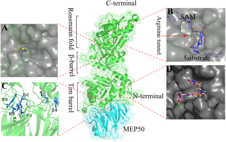
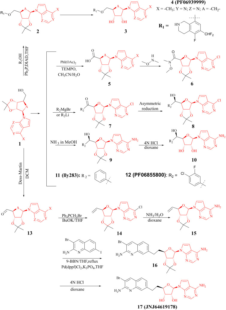
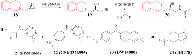
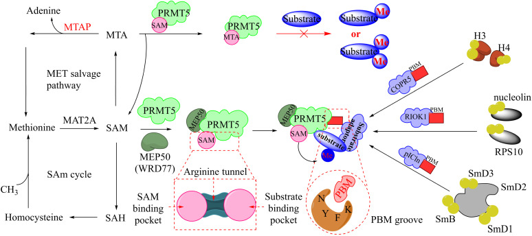
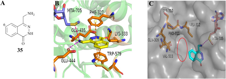
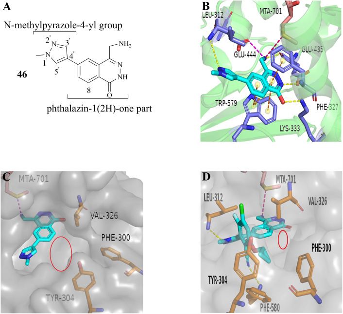
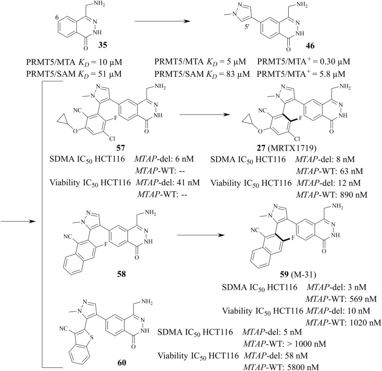
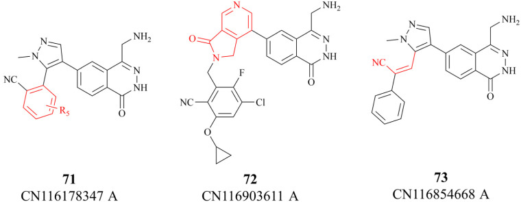
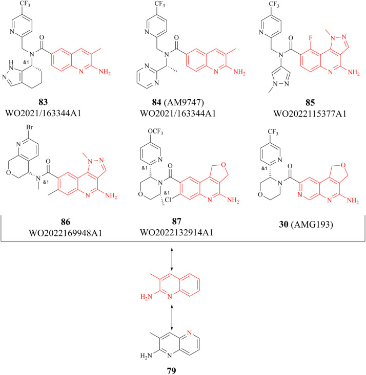
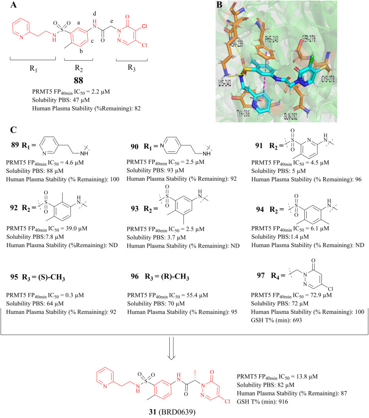
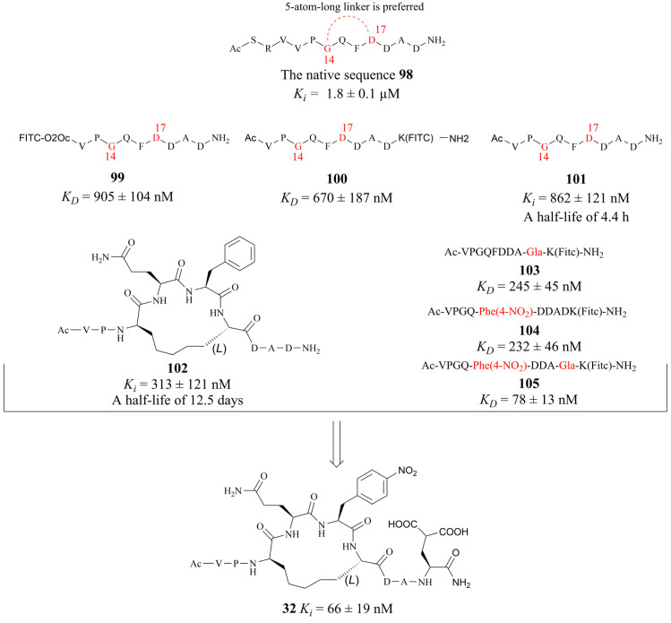
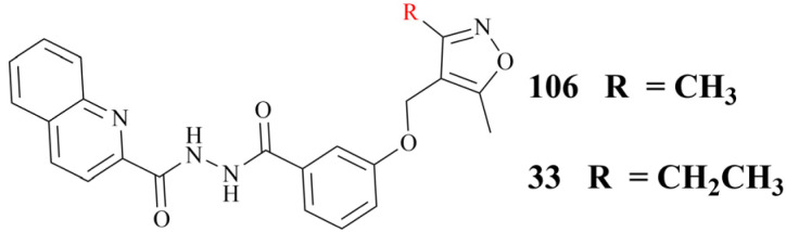
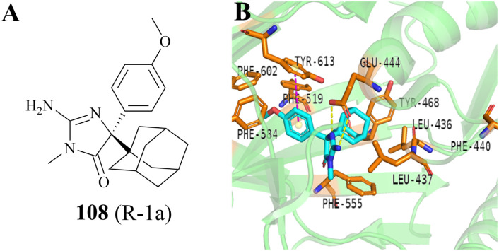
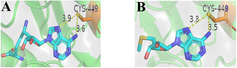
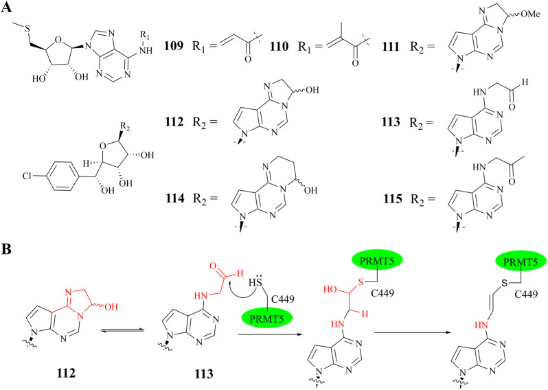
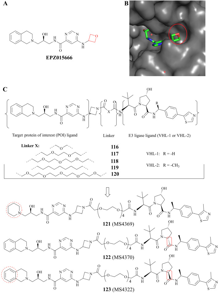
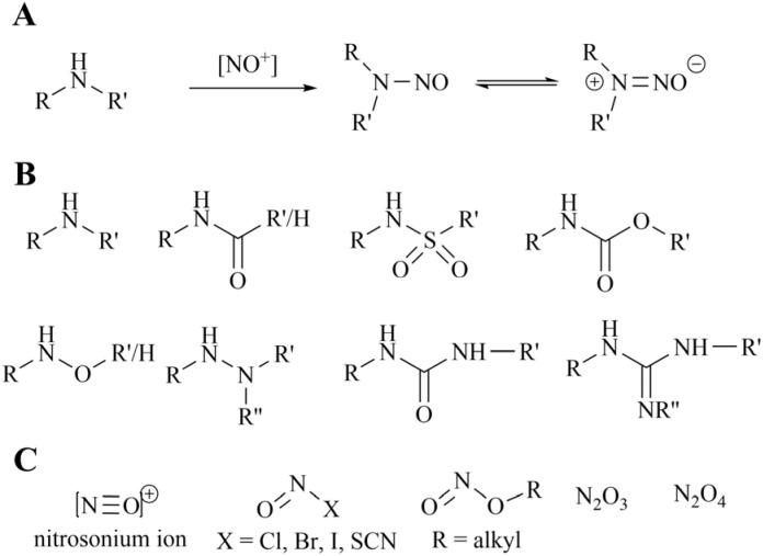
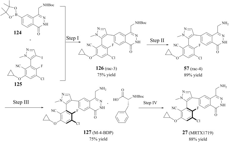
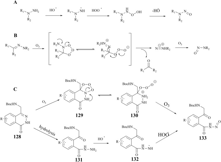
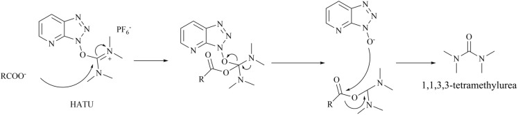
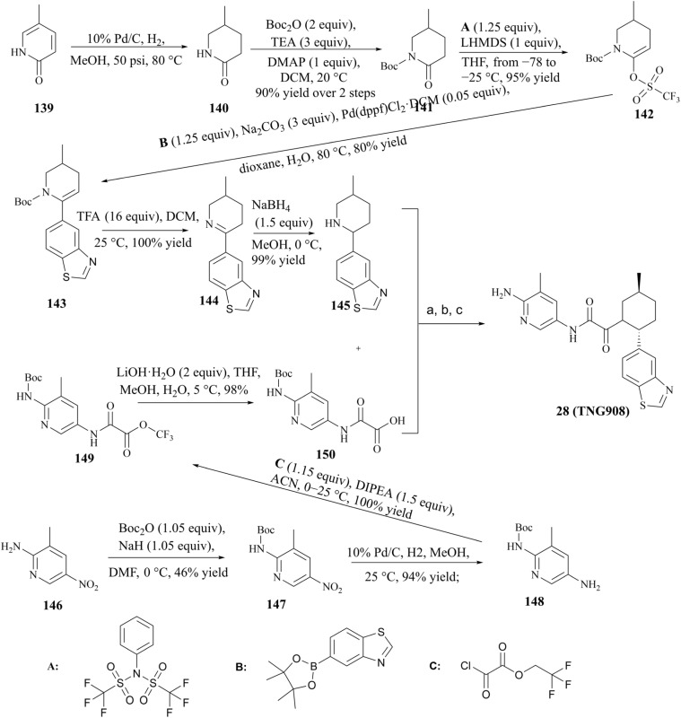
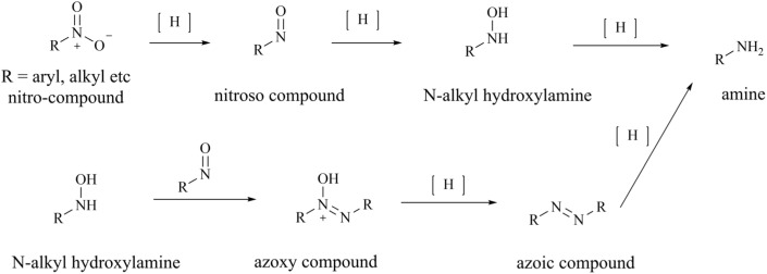
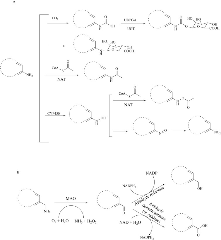
Protein arginine methyltransferase 5 (PRMT5), an epigenetic target with significant clinical potential, is closely associated with the occurrence and development of a range of tumours and has attracted considerable interest from the pharmaceutical industry and academic research communities. According to incomplete statistics, more than 10 PRMT5 inhibitors for cancer therapy have entered clinical trials in recent years. Among them, the second-generation PRMT5 inhibitors developed based on the synthetic lethal strategy demonstrate considerable clinical application value. This suggests that, following the precedent of poly ADP ribose polymerase (PARP), PRMT5 has the potential to become the next clinically applicable synthetic lethal target. However, due to the inherent dose-limiting toxicity of epigenetic target inhibitors, none of these PRMT5 inhibitors has been approved for marketing to date. In light of this, we have conducted a review of the design thoughts and the structure-activity relationship (SAR) of known methylthioadenosine (MTA)-cooperative PRMT5 inhibitors. Additionally, we have analysed the clinical safety of representative first- and second-generation PRMT5 inhibitors. This paper discusses the in vivo vulnerability of the aromatic amine moiety of the second-generation PRMT5 inhibitor based on its structure. It also considers the potential nitrosamine risk factors associated with the preparation process.


Conflict of interest statement

The authors declare that they have no known competing financial interests or personal relationships that could have appeared to influence the work reported in this paper.

Figures

Fig. 1
Fig. 1. Binding sites where PRMT5 can be used for the development of pharmaceutical agents. (A) Allosteric binding site (PDB: 6uxx). (B) SAM binding site and substrate binding site. The substrate binding site and SAM-binding pocket are closely connected by the arginine tunnel (PDB: 4X61), which can be exploited by certain inhibitors, such as JNJ-64619178, to block both the SAM-binding site and substrate binding pocket simultaneously. (C) The PRMT5: MEP50 protein–protein surface interaction (PDB: 4gqb). (D) The PBM groove (PDB: 6U0P). The substrate adaptor proteins (SAPs) contains a highly conserved linear peptide sequence, termed the PRMT5 binding motif (PBM). The PBM peptide and PRMT5 occurs within a shallow groove (the “PBM groove”) of the TIM barrel
Fig. 2
Fig. 2. Synthesis of the SAM mimetic using the adenosine scaffold as its structural core. The adenosine scaffold is highlighted in red.
Fig. 3
Fig. 3. Synthesis of compounds of formula 20.
Fig. 4
Fig. 4. The upstream and downstream molecular regulatory mechanisms of PRMT5. The substrate binding site and SAM-binding pocket are closely connected by the arginine tunnel, which can be exploited by certain inhibitors, such as 17 (JNJ-64619178), to block both the SAM-binding site and substrate binding pocket simultaneously. Furthermore, PRMT5's activity and substrate selectivity are regulated via the formation of multisubunit protein complexes. The SAPs, including Rio domain-containing protein (RioK1), cooperator protein of PRMT5 (COPR5), and chloride channel nucleotide-sensitive protein 1A (pICln), form the multisubunit complexes with PRMT5 by the interaction between the PBM and the PBM-binding groove.
Fig. 5
Fig. 5. (A) Chemical structure of 35. (B) The co-crystal structure of fragment 35 with PRMT5/MTA complex (PDB ID: 7S0U). Fragment 35 forms a hydrogen bonding network with Glu435 and Lys333. Additionally, it is observed that there is an ionic interaction between the primary amine (–NH2) of 35 and Glu444, a π–stack interaction between 35 and the side chains of Phe327 and Trp579, and a van der Waals interaction between 35 and the S-atom of MTA. (C) To investigate the pocket formed by five amino acid residues including the residues Leu312, the C6-position of 35 is the most appropriate position for fragment growth.
Fig. 6
Fig. 6. (A) Structure of 46. (B) The co-crystal structure of 46 with PRMT5/MTA (PDB 7S1Q). A new H-bond was observed between the backbone N–H of Leu312 and the N2 of N-methylpyrazole. (C) The lipophilic pocket formed by the side chains of Phe300, Tyr304 and Val326 is observed and there is enough room to accommodate a lipophilic group. (D) The co-crystal structure of 27 (MRTX1719) with PRMT5/MTA (PDB 7S1S). Additionally, there is also a smaller cavity at the side of the 8-position in the phthalazin-1(2H)-one that could accommodate a group.
Fig. 7
Fig. 7. The fragment 1 growth route of 27 (MRTX1719) and its analogues.
Fig. 8
Fig. 8. 27 (MRTX1719) analogues.
Fig. 9
Fig. 9. Analogues of fragment 35.
Fig. 10
Fig. 10. The chimeric design enabled optimisation of fragment 74 to obtain 80.
Fig. 11
Fig. 11. The development strategy, which combined the HTS and SBDD methodologies, yielded the 28 (TNG908). (A) The development route of 28 (TNG908) and 29 (TNG462). (B) X-ray cocrystal structure of PRMT5/SAM/substrate complex. Glu435 rotates toward substrate side chain (PDB 4X61). (C) X-ray cocrystal structure of PRMT5/MTA complex. Glu435 rotates to fill space previously filled by SAM (PDB 8VEO). (D) X-ray cocrystal structure of 82R (yellow) bound to PRMT5/MTA (PDB 8VET). (E) X-ray cocrystal structure of 28 (TNG908) bound to PRMT5/MTA (PDB 8VEY).
Fig. 12
Fig. 12. The structural relationship between the 3-methylpyridin-2-amine pharmacophore and 74.
Fig. 13
Fig. 13. The heterocycles as PRMT5/MTA complex inhibitors.
Fig. 14
Fig. 14. (A) Lead compound 88. (B) The cocrystal structure of compound 88 bound to the PRMT5 complex (PDB: 7M05). (C) Further SAR exploration of 88 results in a PBM-competitive inhibitor 31 (BRD0639). The FP is a competition fluorescence polarization assay.
Fig. 15
Fig. 15. Macrocyclization of the RioK1-derived PBM sequence results in a PAPII 32.
Fig. 16
Fig. 16. Structures of compounds 106 and 33.
Fig. 17
Fig. 17. (A) The structure of 108. (B) The co-crystal structure of the PRMT5/MEP50 complex with 108 (PDB: 6UXX).
Fig. 18
Fig. 18. (A) The space length between C449 and C6–NH2 (and N7 atom) of the adenine ring of SAM is 3.9 (and 3.6) Å (PDB: 4GQB). (B) The space length between C449 and C6–NH2 (and N7 atom) of the adenine ring of MTA is 3.8 (and 3.5) Å (PDB: 5FA5). Note: the standard length of a carbon–carbon bond is approximately 1.54 Å.
Fig. 19
Fig. 19. (A) PRMT5 covalent inhibitors. (B) Proposed mechanism of vinyl-thio ether formation.
Fig. 20
Fig. 20. (A) EPZ015666 (the oxetane moiety is shown in red); (B) the co-crystal structure of PRMT5/MEP50 complex with EPZ015666 (PDB: 4X61). It can be observed that the oxetane moiety of EPZ015666 is solvent-exposed. (C) A brief study of the SAR of 123 (MS4322).
Fig. 21
Fig. 21. (A) Mechanism of formation of nitrosamines from secondary amines, (B) the various secondary amine structures that have the potential to form nitrosamines. (C) Common nitrosating reagents.
Fig. 22
Fig. 22. Current multikilogram GMP synthesis of 27 (MRTX1719). (Step I) 3.0 mol% Ad2nBuP-Pd-G3,3.0 eq., Cs2CO3, 12 vol. toluene, 4 vol. H2O, 55–60 °C, 18 h. (Step II) 4 M HCl/EtOAc (5 vol.), 20 vol. MeOH, 15–25 °C, 18 h; 7 M NH3/MeOH (1 vol.),10 vol. MeOH, 10–15 °C, 20 h. (Step III) 1.2 eq. Boc-D-Phe, 15 vol. EtOH/H2O 98 : 2, crystallizer: 20–25 °C, 16 h, racemizer: 160 °C, tR 2–4 min; 7 vol. EtOH/H2O 85 : 15, 15–25 °C, 16 h. (Step IV) 4.5 vol. THF, 1.5 vol. H2O, 20–25 °C; 39 vol. H2O, 1 vol. conc. aq. NH3, 20–25 °C, 6 h; 9 vol. H2O, 1 vol. IPA, 20–25 °C, 6 h.
Fig. 23
Fig. 23. (A) The reaction mechanism of the oxidation of hydrazine. (B) The reaction mechanism of the ozonolysis of hydrazine (C) compound 133 possible production routes.
Fig. 24
Fig. 24. Preparation of 30 (AMG193).
Fig. 25
Fig. 25. Reaction mechanism of HATU to generate 1,1,3,3-tetramethylurea.
Fig. 26
Fig. 26. 28 (TNG908) preparation route. (a) HATU (1 equiv.), TEA (6 equiv.), DMF, 25 °C; (b) 4 M HCl in dioxane, 25 °C, 59% yield over 2 steps for 28 (TNG908); (c) chiral HPLC separation of enantiomers, 48% yield for 28 (TNG908).
Fig. 27
Fig. 27. Nitrohydrogenation reduction mechanism.
Fig. 28
Fig. 28. Major metabolic pathways of primary amines in vivo. (A) Major metabolic pathways of aryl amines in vivo. (B) Major metabolic pathways of benzyl amines in vivo. The vulnerability of arylamine in vivo.

Similar articles

Cited by

  • PRMT5 activity sustains histone production to maintain genome integrity.
    Roth JS, DeAngelo JD, Young DL, Maron MI, Saha A, Pinto H, Gupta V, Jacobs N, Hegde S, Aguilan JT, Basken J, Azofeifa J, Query CC, Sidoli S, Skoultchi AI, Shechter D. Roth JS, et al. bioRxiv [Preprint]. 2025 Jul 7:2025.07.03.663002. doi: 10.1101/2025.07.03.663002. bioRxiv. 2025. PMID: 40672221 Free PMC article. Preprint.

References

    1. Blanc R. S. Richard S. P. Arginine methylation: the coming of age. Mol. Cell. 2017;65:8–24. doi: 10.1016/j.molcel.2016.11.003. - DOI - PubMed
    1. Sauter C. Simonet J. Guidez F. Dumétier B. Pernon B. Callanan M. Bastie J.-N. Aucagne R. Delva L. Protein arginine methyltransferases as therapeutic targets in hematological malignancies. Cancers. 2022;14:5443. doi: 10.3390/cancers14215443. - DOI - PMC - PubMed
    1. Wu Q. Schapira M. Arrowsmith C. H. Barsyte-Lovejoy D. Protein arginine methylation: from enigmatic functions to therapeutic targeting. Nat. Rev. Drug Discovery. 2021;20:509–530. doi: 10.1038/s41573-021-00159-8. - DOI - PubMed
    1. Xiao W. Chen X.-Q. Liu L. Shu Y.-S. Zhang M. Zhong Y.-C. Role of protein arginine methyltransferase 5 in human cancers. Biomed. Pharmacother. 2019;114:10879. doi: 10.1016/j.biopha.2019.108790. - DOI - PubMed
    1. Yang Y.-Z. Bedford M. T. Protein arginine methyltransferases and cancer. Nat. Rev. Cancer. 2013;13:37–50. doi: 10.1038/nrc3409. - DOI - PubMed

LinkOut - more resources