. 2024 Nov 14;5(6):1201-1214.
doi: 10.1002/jha2.1046. eCollection 2024 Dec.

Association between microenvironment-related genes and prognosis of primary central nervous system lymphoma

Affiliations

Association between microenvironment-related genes and prognosis of primary central nervous system lymphoma

Keiichiro Hattori et al. EJHaem. .

Abstract

Background: Primary central nervous system lymphoma (PCNSL) is a rare lymphoid malignancy. Systemic profiling of the PCNSL tumor microenvironment (TME) was previously conducted through gene expression analysis. We investigated the prognostic impact of TME on survival to establish novel prognostic biomarkers in PCNSL patients.

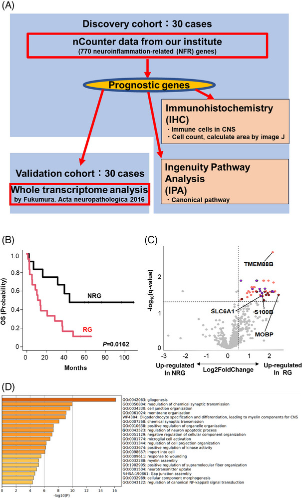
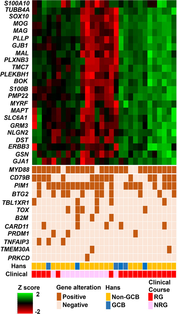
Methods: We analyzed expression levels of 770 neuroinflammation-related (NFR) genes via NanoString nCounter technology in tumor samples from 30 PCNSL patients. Genes related to the "recurrence group (RG)" or "non-recurrence group (NRG)" were identified and validated using whole transcriptomic analysis of an independent PCNSL cohort (n = 30).

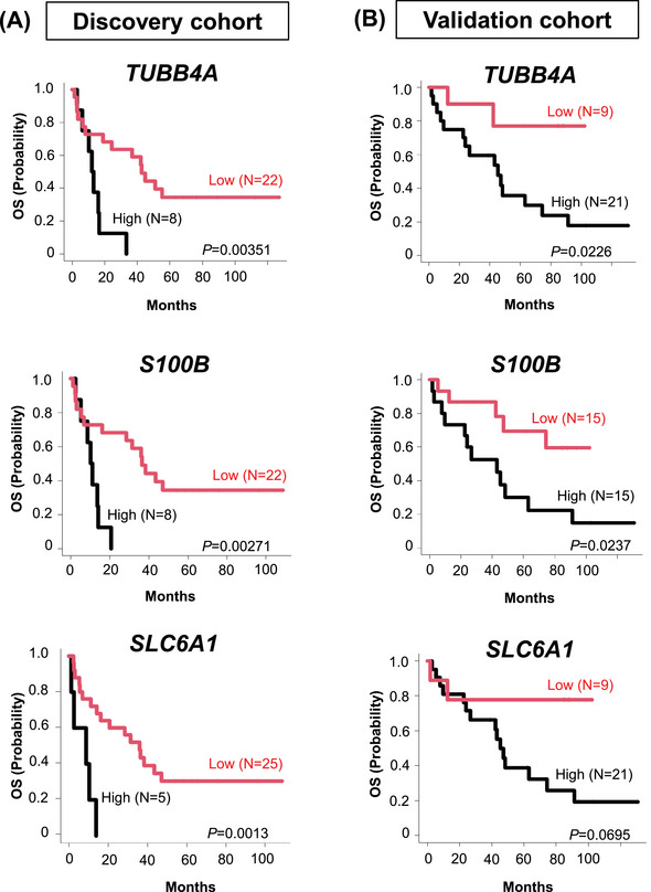
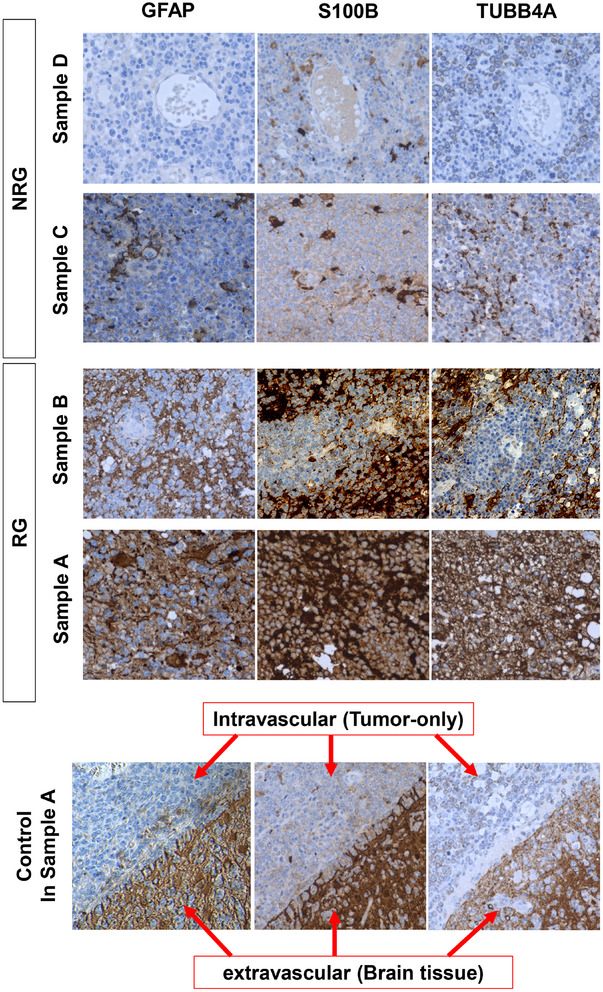
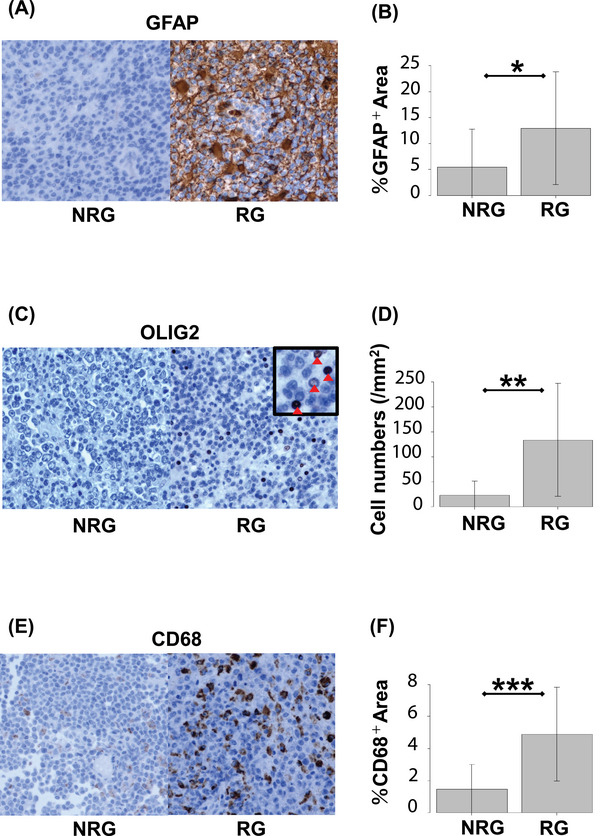
Results: Forty-five of 770 NFR genes were highly expressed in the RG (3-year overall survival (OS, 22.2%), compared with the NRG group (3-year OS 66.7%). Signatures related to glial cells were enriched in the RG-associated gene set. Multivariate analysis revealed that high expressions of TUBB4A (p = 0.028, HR: 3.88), S100B (p = 0.046, HR: 3.093), and SLC6A1 (p = 0.034, HR: 3.765) were significantly related to death. Expression levels of these three genes were also significantly associated with poor OS in the validation cohort. Immunohistochemical staining against TUBB4A, S100B, and proteins specific to glial cells (GFAP, OLIG2, and CD68) revealed significantly higher positivity in RG glial cells.

Conclusion: These data suggest that TME-related genes play a crucial role in the pathogenesis of PCNSL, complementing the well-known involvement of the NF-kB signaling pathway. TME targeting, especially glial cell-specific proteins, may thus open new and complementary avenues of therapy for all stages of PCNSL.

Keywords: Gliosis; Primary central nervous system lymphoma; Tumor microenvironment in central nervous system.


Conflict of interest statement

Lecture honoraria: Eiichi Ishikawa (Daiichi Sankyo, Eisai, Otuka Pharma); Yoshitaka Narita (Ono Pharma) Motoo Nagane (Ono Pharma); Research fund: Sakata‐Yanagimoto Mamiko (Eisai, Bristol Myers Squib, Otuska Pharma, Chugai Pharma), Shigeru Chiba (Thyas, Astellas, Kyowa Kirin); Eiichi Ishikawa (Mitsubishi Electric); Ryo Nishikawa (Ono Pharma) Motoo Nagane (Ono Pharma); Koichi Ichimura (Daiichi Sankyo, Riken Genesis, Gold Ribbon Network, Sumitomo Pharma); Scholarship grant: Shigeru Chiba (Bayer, Eisai, Chugai Pharma, Kyowa Kirin); Eiichi Ishikawa (Eisai) Ryo Nishikawa (Chugai Pharma); Yoshitaka Narita (Ono Pharma, Sumitomo Pharma, Eisai, Taiho Pharma, Ohara Pharma); Motoo Nagane (Chugai Pharma, Bayer, Eisai, Daiichi Sankyo); Endowed chair: Koichi Ichimura (Idorsia Pharmaceuticals).

Figures

FIGURE 1
FIGURE 1
Identification of predicting factors for primary central nervous system lymphoma (PCNSL) outcomes. (A) Schematic representation of overall study design. Thirty cases of newly diagnosed PCNSL were recruited for a discovery cohort using nCounter methods, followed by analysis of the validation cohort. Whole transcriptome analysis data (previously published) was included as a validation cohort. (B) The discovery cohort was divided into two groups according to prognoses. Relapsed PCNSL patients were classified into either the “recurrence group (RG)” or the “non‐recurrence group (NRG)”. (C) A volcano plot indicates differentially expressed genes highly expressed in RG (right) and NRG (left) of the discovery cohort. p‐value adjustment was performed using the Benjamini–Hochberg method. Forty‐five genes were significantly upregulated in RG (q‐value ≤ 0.05, log2 fold change > 0.5). Among these 45 genes, firebrick dots represent genes linked to gliogenesis (GO:0042063) (D), purple dots represent genes linked to modulation of chemical synaptic transmission (GO:0050804) (D) and orange dots represent other genes. (D) Gene ontology (GO) analysis of 45 prognostic genes from the differential expression analysis (C) was performed using the Metascape web application (http://metascape.org). Enriched GO terms are shown by ID and category. GO, gene ontology; NRG, nonrecurrence group; OS, overall survival; PCNSL, primary central nervous system lymphoma; RG, recurrence group; WTA, whole transcriptome analysis.
FIGURE 2
FIGURE 2
Clinical impact of extracted three prognostic genes in the discovery and validation cohort. Kaplan–Meier curves show the duration of OS based on expression levels of unfavorable candidate genes in the discovery (left) and the validation (right) cohorts. Statistical analysis was performed using the two‐sided log‐rank test. OS, overall survival.
FIGURE 3
FIGURE 3
Immunostaining of PCNSL for prognostic proteins. TUBB4A and S100B expression levels were examined by immunohistochemical (IHC) staining in the RG (samples A and B) and NRG (samples C and D) groups, respectively. In control in IHC staining of sample A, expression levels were compared between intravascular (only tumor cells inside the vessel) and extravascular (brain tissue infiltrated by tumor cells) space. Original magnification 400×. NRG, nonrecurrence group; PCNSL, primary central nervous system lymphoma; RG, recurrence group.
FIGURE 4
FIGURE 4
PCNSL tissues harbor tumor microenvironment immune cells. Tumor sections were stained with GFAP (A), OLIG2 (B), and CD68 (C). Representative data from NRG (left) and RG (right) are shown (panels A, C, and E). Quantifications of the immunoreactivity of immunohistochemical (IHC) markers shown in RG compared with NRG (panels B, D, and F). Numbers of OLIG2‐positive cells and percentage of GFAP‐ and CD68‐positive area were counted across sections using scanning software (see “Methods”; panels B, D, and F) *p < 0.05, **p < 0.01, ***p < 0.001. NRG, nonrecurrence group; PCNSL, primary central nervous system lymphoma; RG, recurrence group.
FIGURE 5
FIGURE 5
Integrated visualization of transcriptome data. The transcriptome data from nCounter analysis is shown as a heat map, with the 23 informative genes shown as rows and the 30 patient biopsies shown as columns. These were combined with genetic alterations, cell of origin (COO) classification, and prognosis. Gene expression levels are shown as a percentile of the normalized expression values for each gene. GCB, germinal center B; Hans, Hans criteria; NRG, nonrecurrence group; RG, recurrence group.

Similar articles

References

    1. Villano JL, Koshy M, Shaikh H, Dolecek TA, McCarthy BJ. Age, gender, and racial differences in incidence and survival in primary CNS lymphoma. Brit J Cancer. 2011;105(9):1414–1418. - PMC - PubMed
    1. Hattori K, Sakata‐Yanagimoto M, Okoshi Y, Goshima Y, Yanagimoto S, Nakamoto‐Matsubara R, et al. MYD88 (L265P) mutation is associated with an unfavourable outcome of primary central nervous system lymphoma. Brit J Haematol. 2017;177(3):492–494. - PubMed
    1. Lin CH, Yang CF, Yang HC, Fay LY, Yeh CM, Kuan AS, et al. Risk prediction for early mortality in patients with newly diagnosed primary CNS lymphoma. J Cancer. 2019;10(17):3958–3966. - PMC - PubMed
    1. Hattori K, Sakata‐Yanagimoto M, Kusakabe M, Nanmoku T, Suehara Y, Matsuoka R, et al. Genetic evidence implies that primary and relapsed tumors arise from common precursor cells in primary central nervous system lymphoma. Cancer Sci. 2019;110(1):401–407. - PMC - PubMed
    1. Nakamura T, Tateishi K, Niwa T, Matsushita Y, Tamura K, Kinoshita M, et al. Recurrent mutations of CD79B and MYD88 are the hallmark of primary central nervous system lymphomas. Neuropathol Appl Neurobiol. 2016;42(3):279–290. - PubMed

LinkOut - more resources