. 2024 Dec 3:15:1482898.
doi: 10.3389/fphar.2024.1482898. eCollection 2024.

In vitro hair growth-promoting effects of araliadiol via the p38/PPAR-γ signaling pathway in human hair follicle stem cells and dermal papilla cells

Affiliations

In vitro hair growth-promoting effects of araliadiol via the p38/PPAR-γ signaling pathway in human hair follicle stem cells and dermal papilla cells

Seokmuk Park et al. Front Pharmacol. .

Abstract

Background: Scalp hair plays a crucial role in social communication by expressing personal appearance and self-identity. Consequently, hair loss often leads to a perception of unattractiveness, negatively impacting an individual's life and mental health. Currently, the use of Food and Drug Administration (FDA)-approved drugs for hair loss is associated with several side effects, highlighting the need for identifying new drug candidates, such as plant-derived phytochemicals, to overcome these issues.

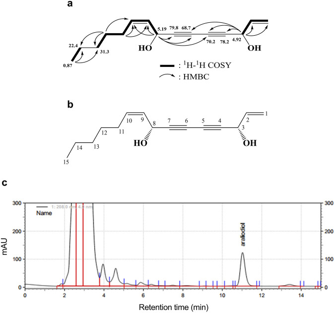
Objective: This study investigated the hair growth-promoting effects of araliadiol, a polyacetylene compound found in plants such as Centella asiatica.

Methods: We employed an in vitro model comprising human hair follicle stem cells (HHFSCs) and human dermal papilla cells (HDPCs) to evaluate the hair growth-promoting effects of araliadiol. The proliferation-stimulating effects of araliadiol were assessed using water-soluble tetrazolium salt assay, adenosine triphosphate content assay, and crystal violet staining assay. In addition, we performed luciferase reporter assay, polymerase chain reaction analysis, cell fractionation, Western blot analysis, and enzyme-linked immunosorbent assay (ELISA) to elucidate the mechanism underlying the hair growth-inductive effects of araliadiol.

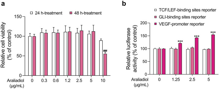
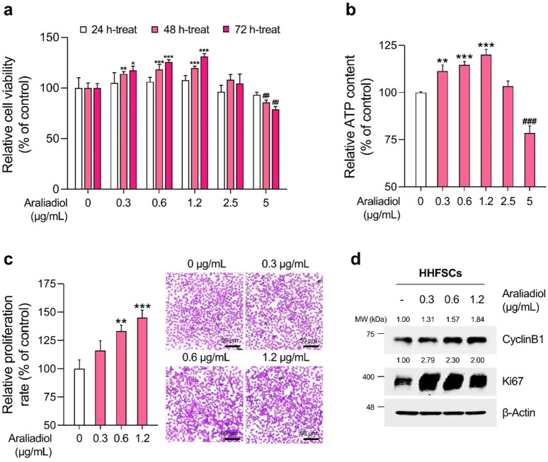
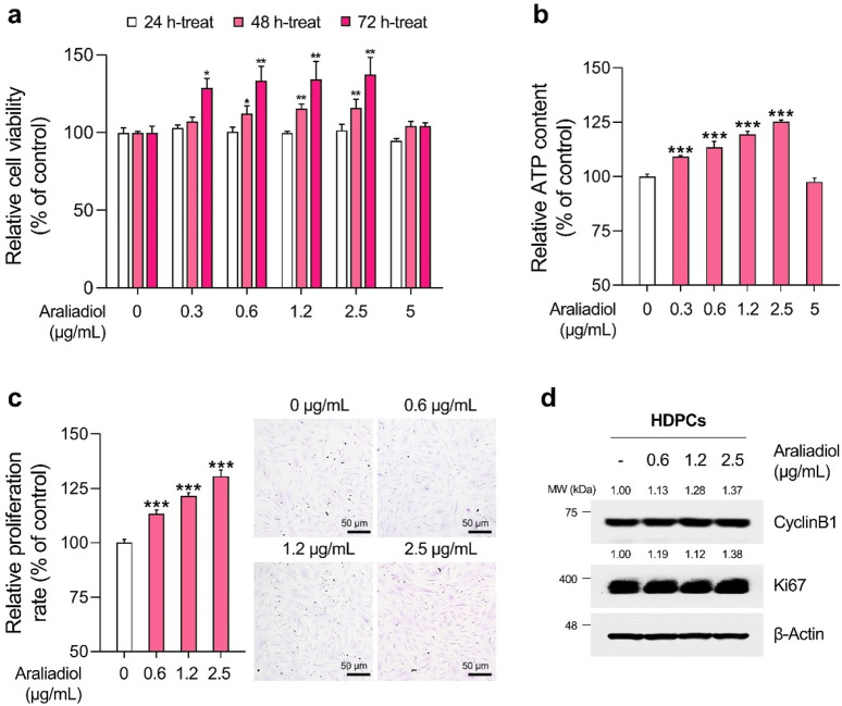
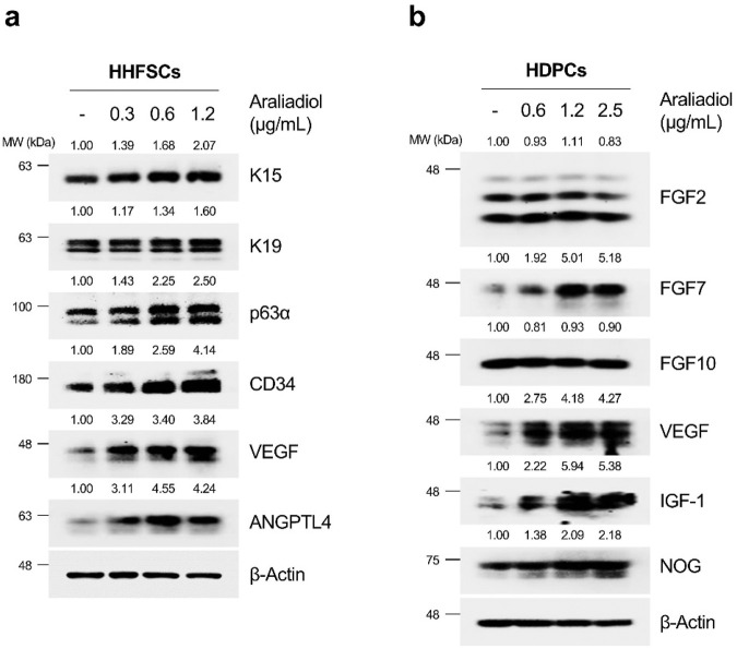
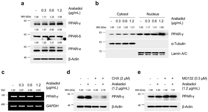
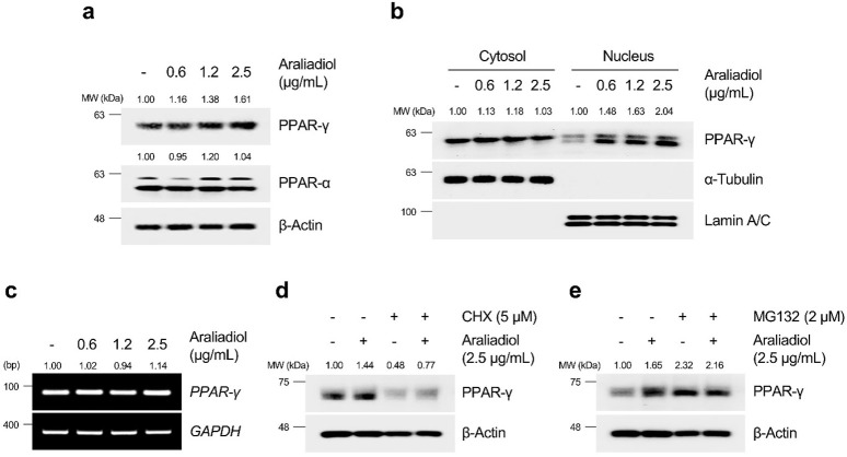
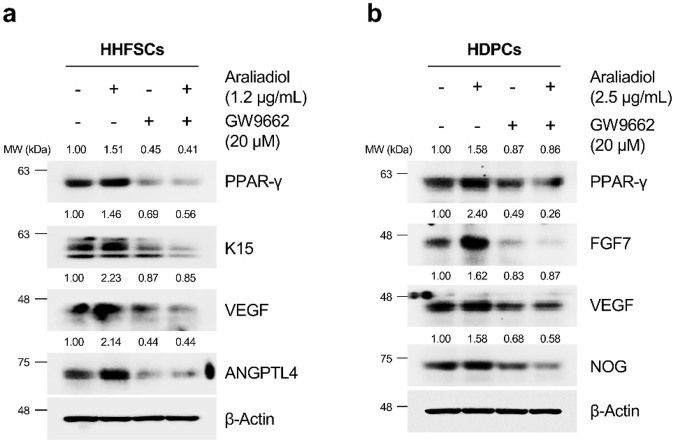
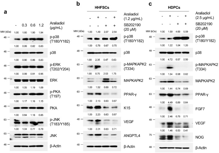
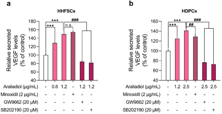
Results: Araliadiol exhibited both proliferation- and hair growth-promoting effects in HHFSCs and HDPCs. Specifically, it increased the protein expression of cyclin B1 and Ki67. In HHFSCs, it elevated the expression of hair growth-promoting factors, including CD34, vascular endothelial growth factor (VEGF), and angiopoietin-like 4. Similarly, araliadiol increased the expression of hair growth-inductive proteins such as fibroblast growth factor 7, VEGF, noggin, and insulin-like growth factor 1 in HDPCs. Subsequent Western blot analysis and ELISA using inhibitors such as GW9662 and SB202190 confirmed that these hair growth-promoting effects were dependent on the p38/PPAR-γ signaling in both HHFSCs and HDPCs.

Conclusion: Araliadiol promotes hair growth through the p38/PPAR-γ signaling pathway in human hair follicle cells. Therefore, araliadiol can be considered a novel drug candidate for the treatment of alopecia.

Keywords: PPAR-γ; alopecia; araliadiol; hair follicles; hair growth; hair loss; phytochemicals; polyacetylene.


Conflict of interest statement

Authors HP, DS, and DY were employed by ASK Company Co., Ltd. The remaining authors declare that the research was conducted in the absence of any commercial or financial relationships that could be construed as a potential conflict of interest.

Figures

FIGURE 1
FIGURE 1
Identification and chromatographic analysis of araliadiol. (A) 1H–1H COSY and HMBC correlations of the isolated compound from Centella asiatica extract. (B) Chemical structure of isolated compound, araliadiol. (C) HPLC chromatogram of Centella asiatica extract at 208 nm. Centella asiatica, Centella asiatica.
FIGURE 2
FIGURE 2
Effects of araliadiol on hair growth-related signaling pathways in 293T cells. (A) 293T cells were treated with varying concentrations of araliadiol (0–10 μg/mL) for up to 48 h, and cell viability was assessed using the WST-1 assay. (B) 293T cells were treated with araliadiol (0–5 μg/mL) for 48 h, and a luciferase reporter assay was conducted to evaluate the effects of araliadiol on hair growth-related signaling pathways. Luminescence values were normalized to β-galactosidase activities. Results are presented as the mean ± SD of three independent experiments and analyzed using a one-way analysis of variance followed by Tukey’s test. ###, *** p < 0.001 compared with the vehicle-treated group. WST-1, water soluble tetrazolium salt 1.
FIGURE 3
FIGURE 3
Araliadiol promotes proliferation in human hair follicle stem cells within non-toxic concentrations. (A) HHFSCs were treated with araliadiol (0–5 μg/mL) for up to 72 h, and cell viability was assessed using the WST-1 assay. (B, C) HHFSCs were treated with araliadiol (0–5 μg/mL) for 72 h, and their proliferative capacity was evaluated using an intracellular ATP detection assay (B) and crystal violet staining assay (C, D) Protein expression levels of cell cycle-related markers (CyclinB1 and Ki67) were analyzed by Western blotting and normalized to β-Actin levels. Results are presented as the mean ± SD of three independent experiments and analyzed using a one-way analysis of variance followed by Tukey’s test. * p < 0.05; ##,** p < 0.01; ###, *** p < 0.001 compared with the vehicle-treated group. HHFSCs, human hair follicle stem cells; ATP, adenosine triphosphate.
FIGURE 4
FIGURE 4
Araliadiol promotes proliferation in human dermal papilla cells within non-toxic concentrations. (A) HDPCs were treated with araliadiol (0–5 μg/mL) for up to 72 h, and cell viability was assessed using the WST-1 assay. (B, C) HDPCs were treated with araliadiol (0–5 μg/mL) for 72 h, and their proliferative capacity was evaluated using an intracellular ATP detection assay (B) and crystal violet staining assay (C, D) Protein expression levels of cell cycle-related markers (CyclinB1 and Ki67) were analyzed by Western blotting and normalized to β-Actin levels. Results are presented as the mean ± SD of three independent experiments and analyzed using a one-way analysis of variance followed by Tukey’s test. * p < 0.05; ** p < 0.01; *** p < 0.001 compared with the vehicle-treated group. HDPCs, human hair follicle dermal papilla cells.
FIGURE 5
FIGURE 5
Araliadiol enhances hair growth-promoting signals and anagen phase markers in human hair follicle cells. (A) HHFSCs were treated with different concentrations of araliadiol (0–1.2 μg/mL) for 48 h. Protein levels of hair follicle stem cell-specific markers (K15 and K19), anagen phase markers (CD34 and p63α), and hair growth-promoting proteins (VEGF and ANGPTL4) were analyzed by Western blotting, with β-Actin serving as a loading control. (B) HDPCs were stimulated with indicated concentrations of araliadiol (0–2.5 μg/mL) for 48 h. Protein levels of hair growth-promoting proteins (FGF2, FGF7, FGF10, VEGF, IGF-1, and NOG) were analyzed by Western blotting, with β-Actin serving as a loading control. The protein levels in all blots were quantified using ImageJ software version 1.53t. K15, cytokeratin 15; K19, cytokeratin 19; CD34, cluster of differentiation 34; p63α, tumor protein 63 alpha; VEGF, vascular endothelial growth factor; ANGPTL4, angiopoietin like 4; FGF2, fibroblast growth factor 2; FGF7, fibroblast growth factor 7; FGF10, fibroblast growth factor 10; IGF-1, insulin-like growth factor 1; NOG, noggin.
FIGURE 6
FIGURE 6
Araliadiol induces transcriptional activity of PPAR-γ by elevating protein stability rather than increasing mRNA expression in HHFSCs. (A) HHFSCs were treated with different concentrations of araliadiol (0–1.2 μg/mL) for 48 h. Protein levels of the PPAR family (PPAR-α, PPAR-δ, and PPAR-γ) were analyzed by Western blotting, with β-Actin as a loading control. (B) Nuclear translocation of PPAR-γ was analyzed by Western blotting. α-Tubulin and Lamin A/C served as loading controls for cytoplasmic and nuclear fractions, respectively. (C) mRNA expression of PPAR-γ was determined by PCR and normalized to GAPDH. (D) Protein stability of PPAR-γ was assessed after treatment with araliadiol (1.2 μg/mL) with or without CHX (2 μM) in HHFSCs for 48 h (E) HHFSCs were treated with araliadiol (0–1.2 μg/mL) with or without MG132 (0.3 μM) for 48 h, and protein levels of PPAR-γ were analyzed by Western blotting with β-actin as a loading control. mRNA and protein levels in all blots were quantified using ImageJ software version 1.53t. PCR, polymerase chain reaction; PPAR, peroxisome proliferator-activated receptor; GAPDH, glyceraldehyde 3-phosphate dehydrogenase.
FIGURE 7
FIGURE 7
Araliadiol induces transcriptional activity of PPAR-γ by elevating protein stability rather than increasing mRNA expression in HDPCs. (A) HDPCs were treated with different concentrations of araliadiol (0–2.5 μg/mL) for 48 h. Protein levels of PPAR-α and PPAR-γ were analyzed by Western blotting with β-Actin as a loading control. (B) Nuclear translocation of PPAR-γ was analyzed by Western blotting. α-Tubulin and Lamin A/C served as loading controls for the cytoplasmic and nuclear fractions, respectively. (C) mRNA expression of PPAR-γ was determined by PCR and normalized to GAPDH. (D) Protein stability of PPAR-γ was assessed after treatment with araliadiol (2.5 μg/mL) with or without CHX (5 μM) in HDPCs for 48 h (E) HDPCs were treated with araliadiol (0–2.5 μg/mL) with or without MG132 (2 μM) for 48 h, and protein levels of PPAR-γ were analyzed by Western blotting, with β-Actin as a loading control. mRNA and protein levels in all blots were quantified using ImageJ software version 1.53t.
FIGURE 8
FIGURE 8
Araliadiol elevates hair growth-promoting effects in human hair follicle cells by upregulating the PPAR-γ signaling pathway. (A) Protein levels of PPAR-γ and hair growth-inductive proteins (K15, VEGF, and ANGPTL4) were assessed after treatment with araliadiol (1.2 μg/mL) with or without GW9662 (20 μM) in HHFSCs for 48 h. (B) Protein levels of PPAR-γ and hair growth-inductive proteins (FGF7, VEGF, and NOG) were assessed after treatment with araliadiol (2.5 μg/mL) with or without GW9662 (20 μM) in HDPCs for 48 h. Protein levels in all blots were quantified using ImageJ software version 1.53t.
FIGURE 9
FIGURE 9
Araliadiol induces PPAR-γ/hair growth-promoting signaling by activating p38MAPK in human hair follicle cells. (A) Protein levels of PPAR-γ and its upstream kinases (p38, ERK, JNK, and PKA) were assessed after treatment with araliadiol (0–1.2 μg/mL) in HHFSCs for 48 h. The protein levels of the p38/PPAR-γ/hair growth-promoting signaling pathway were assessed after treatment with araliadiol with or without SB202190 (20 μM) in (B) HHFSCs and (C) HDPCs for 48 h. Protein levels in all blots were quantified using ImageJ software version 1.53t. p38, p38 mitogen-activated protein kinase; ERK, extracellular signal-regulated kinase; JNK, Jun N-terminal kinase; PKA, protein kinase (A).
FIGURE 10
FIGURE 10
Araliadiol enhances VEGF secretion via the p38/PPAR-γ signaling pathway in human hair follicle cells. (A, B) Human hair follicle cells were treated with araliadiol (0–2.5 μg/mL) for 48 h, either with or without SB202190 (20 μM) or GW9662 (20 μM). VEGF-A secretion levels were quantified using an ELISA on conditioned medium from the treated cells. Minoxidil (2 μg/mL) was included as a positive control. Results are presented as the mean ± SD of three independent experiments and analyzed using a one-way analysis of variance followed by Tukey’s test. *** p < 0.001 compared with the vehicle-treated group. ## p < 0.01; ### p < 0.001 compared with the araliadiol-treated group. ELISA, enzyme-linked immunosorbent assay.

Similar articles

Cited by

References

    1. Abdullah A. (2020). Comparative study of the online over-the-counter hair loss products. J. Dermatology Res. Ther. 6. 10.23937/2469-5750/1510077 - DOI
    1. Ahmadian M., Suh J. M., Hah N., Liddle C., Atkins A. R., Downes M., et al. (2013). PPARγ signaling and metabolism: the good, the bad and the future. Nat. Med. 19 (5), 557–566. 10.1038/nm.3159 - DOI - PMC - PubMed
    1. Ahmed M. H., Karkush S. I., Ali S. A., Mohammed A. A. (2024). Phytochemicals: a new arsenal in drug discovery. Int. J. Med. Sci. Dent. Health 10 (01), 29–44. 10.55640/ijmsdh-10-01-03 - DOI
    1. Ahmed N. S., Ghatak S., El Masry M. S., Gnyawali S. C., Roy S., Amer M., et al. (2017). Epidermal E-cadherin dependent β-catenin pathway is phytochemical inducible and accelerates anagen hair cycling. Mol. Ther. 25 (11), 2502–2512. 10.1016/j.ymthe.2017.07.010 - DOI - PMC - PubMed
    1. Asnaashari S., Javadzadeh Y. (2020). Herbal medicines for treatment of androgenic alopecia. Altern. Ther. Health and Med. 26 (4), 27–35. - PubMed

LinkOut - more resources