Anatomy of the aortic segmental arteries-the fundamentals of preventing spinal cord ischemia in aortic aneurysm repair
- PMID: 39691497
- PMCID: PMC11649645
- DOI: 10.3389/fcvm.2024.1475084
Anatomy of the aortic segmental arteries-the fundamentals of preventing spinal cord ischemia in aortic aneurysm repair
Abstract
Objective: Spinal cord ischemia due to damage or occlusion of the orifices of aortic segmental arteries (ASA) is a serious complication of open and endovascular aortic repair. Our study aims to provide detailed descriptions of the proximal course of the ASAs and metric information on their origins.
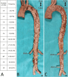
Materials and methods: Initially, 200 randomly selected, embalmed cadavers of human body donors were anatomically dissected and systematically examined. On macroscopic inspection, 47 showed severe pathologies and were excluded. Of the remaining 153, 73 were males and 80 females.
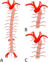
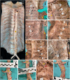
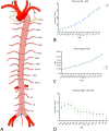
Results: In total, 69.9% of the aortae showed 26-28 ASA orifices. In 59.5% the most proximal ASA, at least unilaterally, was the third posterior intercostal artery, which originated from the descending aorta at approximately 10% of its length. In 56.2%, the left and right ASAs had a common origin in at least one body segment. This mainly affected the abdominal aorta and L4 in particular (54.2%). The ASAs of lumber segments 1-3 originated strictly segmentally. In contrast, in 80.4%, at least one posterior intercostal artery originated from a cranially or caudally located ipsilateral ASA. Such an arrangement was seen along the entire thoracic aorta. Further descriptions of variants and metric data on ASA orifices are presented.
Conclusion: Our large-scale study presents a detailed topographic map of ASAs. It underscores the value of preoperative CT councils and provides crucial information for interpreting the results. Furthermore, it aids in planning and conducting safe aortic intervention and assists in deciding on single- or two-staged stent graft procedures.
Keywords: ASA; CT; aorta; aortic aneurysm; aortic segmental arteries; prevention.
© 2024 Pruidze, Weninger, Didava, Schwendt, Geyer, Neumayer, Nanobachvili, Eilenberg, Czerny and Weninger.
Conflict of interest statement
The authors declare that they have no known competing financial interests or personal relationships that could have appeared to influence the work reported in this paper.
Figures


Similar articles
-
Extensive endovascular repair of thoracic aorta: observational analysis of the results and effects on spinal cord perfusion.J Cardiovasc Surg (Torino). 2013 Aug;54(4):523-30. Epub 2013 Feb 1. J Cardiovasc Surg (Torino). 2013. PMID: 23369947
-
Fenestrated endovascular grafts for the repair of juxtarenal aortic aneurysms: an evidence-based analysis.Ont Health Technol Assess Ser. 2009;9(4):1-51. Epub 2009 Jul 1. Ont Health Technol Assess Ser. 2009. PMID: 23074534 Free PMC article.
-
Endovascular repair of descending thoracic aortic aneurysm: an evidence-based analysis.Ont Health Technol Assess Ser. 2005;5(18):1-59. Epub 2005 Nov 1. Ont Health Technol Assess Ser. 2005. PMID: 23074469 Free PMC article.
-
Staged procedures for prevention of spinal cord ischemia in endovascular aortic surgery.Gefasschirurgie. 2018;23(Suppl 2):39-45. doi: 10.1007/s00772-018-0410-z. Epub 2018 Jul 2. Gefasschirurgie. 2018. PMID: 30147243 Free PMC article. Review.
-
Endovascular thoracic aortic repair and risk of spinal cord ischemia: the role of previous or concomitant treatment for aortic aneurysm.J Cardiovasc Surg (Torino). 2010 Apr;51(2):169-76. J Cardiovasc Surg (Torino). 2010. PMID: 20354486 Review.
Cited by
-
Intraspinal vascular perfusion territories of the descending thoracic aorta.Eur J Cardiothorac Surg. 2025 Jul 1;67(7):ezaf212. doi: 10.1093/ejcts/ezaf212. Eur J Cardiothorac Surg. 2025. PMID: 40576461 Free PMC article.
References
-
- Hanna L, Borsky K, Abdullah AA, Sounderajah V, Marshall DC, Salciccioli JD, et al. Trends in hospital admissions, operative approaches, and mortality related to abdominal aortic aneurysms in England between 1998 and 2020. Eur J Vasc Endovasc Surg. (2023) 66(1):68–76. 10.1016/j.ejvs.2023.03.015 - DOI - PubMed
-
- Isselbacher EM, Preventza O, Hamilton Black J, 3rd, Augoustides JG, Beck AW, Bolen MA, et al. 2022 ACC/AHA guideline for the diagnosis and management of aortic disease: a report of the American Heart Association/American College of Cardiology joint committee on clinical practice guidelines. Circulation. (2022) 146(24):e334–482. 10.1161/CIR.0000000000001106 - DOI - PMC - PubMed
LinkOut - more resources
Full Text Sources

