

A case of treating polypoidal choroidal vasculopathy subretinal fluid by subthreshold micropulse laser
- PMID: 39691633
- PMCID: PMC11650130
- DOI: 10.1016/j.ajoc.2024.102225
A case of treating polypoidal choroidal vasculopathy subretinal fluid by subthreshold micropulse laser
Abstract
Purpose: Assess the effectiveness of a subthreshold micropulse laser for treating a patient with polypoidal choroidal vasculopathy and subretinal fluid.
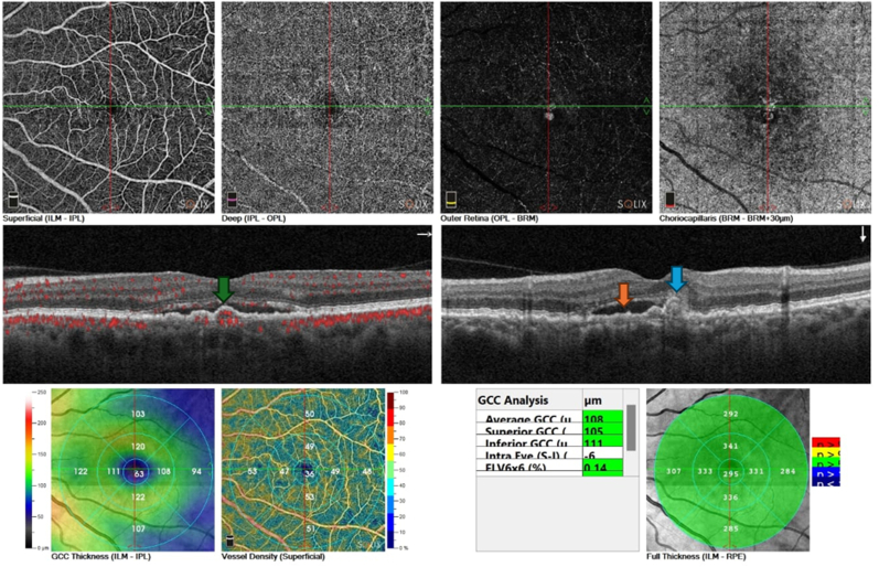
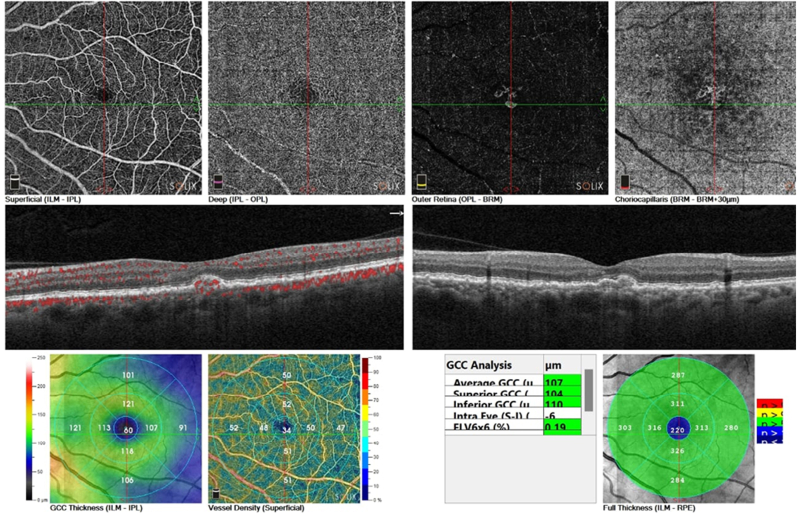
Observations: A 55-year-old female presented with left eye blurring vision and metamorphopsia, and her visual acuity was 20/60. Optical coherence tomography (OCT) and OCT angiography showed subretinal fluid and pigmented epithelium detachment with a small polyp under the retinal pigmented epithelium, which was diagnosed as polypoidal choroidal vasculopathy. She was treated with a subthreshold micropulse (577nm) laser and showed a dramatic response within 12 weeks with the disappearance of subretinal fluid and visual acuity improving to 20/25.
Conclusion and importance: A subthreshold micropulse laser might be an effective and safe option for treating patients with polypoidal choroidal vasculopathy and subretinal fluid.
Keywords: Micropulse laser; Polypoidal choroidal vasculopathy; Subretinal fluid.
© 2024 The Authors.
Conflict of interest statement
The authors declare that they have no known competing financial interests or personal relationships that could have appeared to influence the work reported in this paper.
Figures


References
-
- Yannuzzi L.A., Sorenson J., Spaide R.F., Lipson B. Idiopathic polypoidal choroidal vasculopathy (IPCV) Retina. 1990;10:1–8. - PubMed
-
- Imanmura Y., et al. Polypoidal choroidal vasculopathy: a review. Surv Ophthalmol. 2010;55:501–515. - PubMed
-
- Maruko I., Iida T., Saito M., et al. Clinical characteristics of exudative age-related macular degeneration in Japanese patients. Am J Ophthalmol. 2007;144:15–22. - PubMed
-
- Nakashizuka H., Mitsumata M., Okisaka S., et al. Clinicopathologic findings in polypoidal choroidal vasculopathy. Invest Ophthalmol Vis Sci. 2008;49:4729–4737. - PubMed
Publication types
LinkOut - more resources
Full Text Sources
