

Use of a double pigtail ureteral stent for surgical management of obstructive pancreatolithiasis in a feline patient
- PMID: 39691672
- PMCID: PMC11650493
- DOI: 10.1177/20551169241288217
Use of a double pigtail ureteral stent for surgical management of obstructive pancreatolithiasis in a feline patient
Abstract
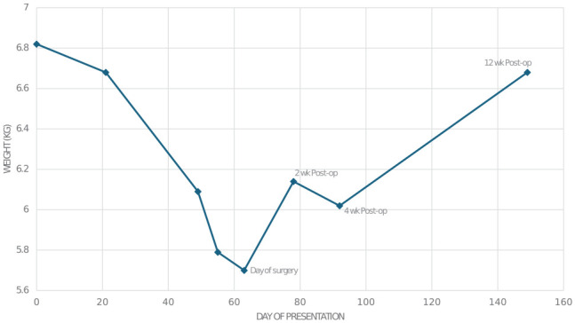
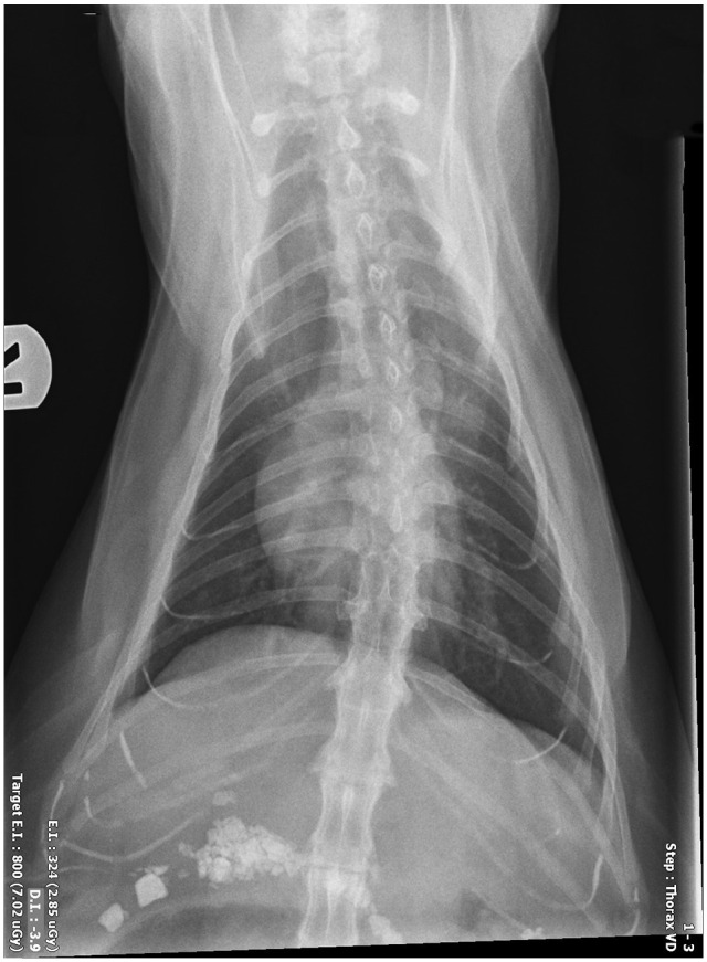
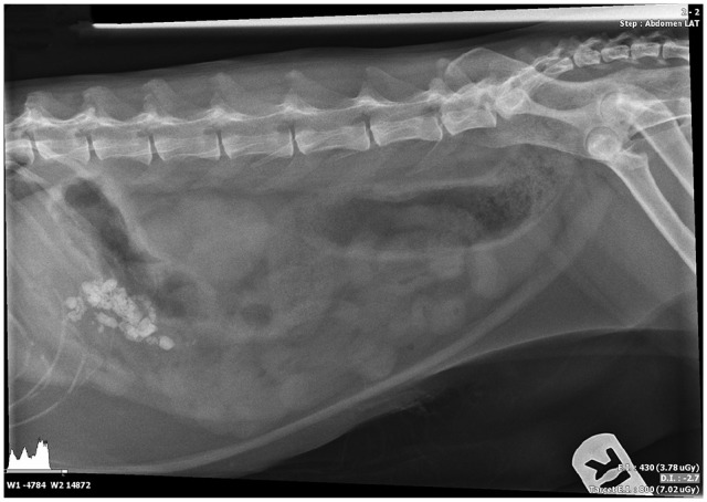
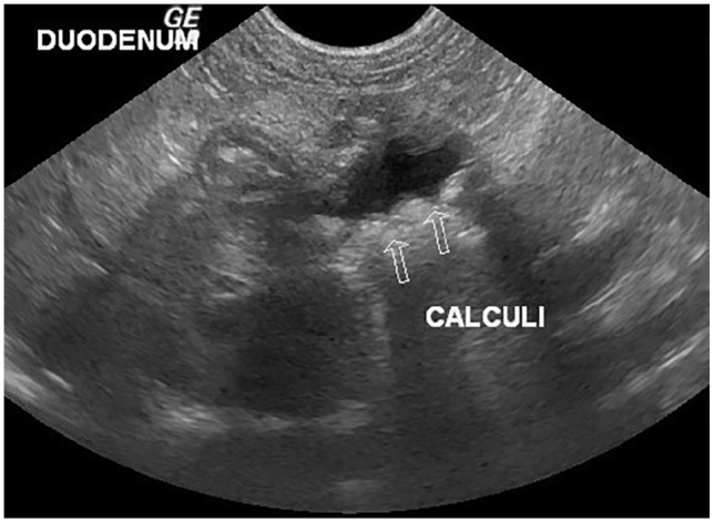
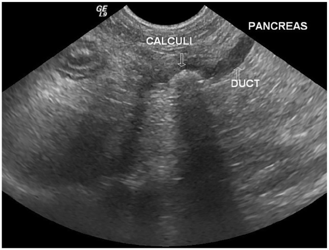
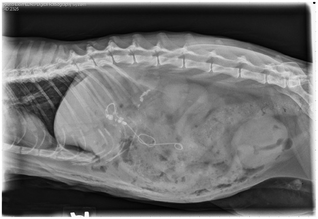
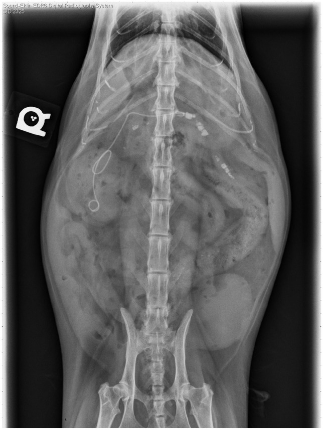
Case summary: A 7-year-old castrated male domestic shorthair cat was presented for surgical intervention for obstructive pancreatolithiasis. The patient had a history of chronic weight loss, vomiting and lethargy. Elevation of feline pancreas-specific lipase and a marked decrease in cobalamin were documented on blood biochemistry. Abdominal ultrasound revealed an enlarged right pancreatic limb with a dilated central duct and multiple pancreatoliths visualized within, consistent with partial pancreatic duct obstruction. The patient was successfully treated with a minor duodenal papilla construction using a 2.5 Fr double pigtail ureteral stent.
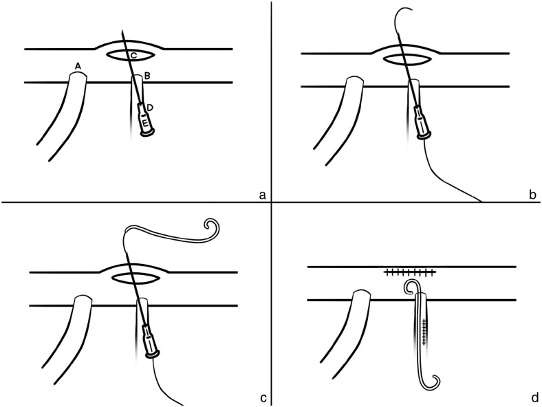
Relevance and novel information: Pancreatolithiasis is an exceptionally rare condition in veterinary medicine, particularly in cats, with only a few reported cases. Given its low incidence, there is no established consensus regarding the best therapeutic options in clinical practice. This case report outlines the successful application of a locking loop catheter to re-establish the patency of a pancreatic duct. Notably, this is the first documented use of this technique for the surgical management of obstructive pancreatolithiasis.
Keywords: Pancreatolith; chronic pancreatitis; double pigtail ureteral stent; feline pancreatitis; feline pancreatolithiasis; obstructive pancreatolithiasis; pancreatic stones.
© The Author(s) 2024.
Conflict of interest statement
The authors declared no potential conflicts of interest with respect to the research, authorship, and/or publication of this article.
Figures
References
-
- De Cock HE, Forman MA, Farver TB, et al. Prevalence and histopathologic characteristics of pancreatitis in cats. Vet Pathol 2007; 44: 39–49. - PubMed
-
- Sharzehi K. Management of pancreatic duct stones. Curr Gastroenterol Rep 2019; 21: 63. DOI: 10.1007/s11894-019-0727-0. - PubMed
-
- Farnbacher MJ, Schoen C, Rabenstein T, et al. Pancreatic duct stones in chronic pancreatitis: criteria for treatment intensity and success. Gastrointest Endosc 2002; 56: 501–506. - PubMed
-
- Inui K, Yoshino J, Miyoshi H, et al. New developments in diagnosis and non-surgical treatment of chronic pancreatitis. J Gastroenterol Hepatol 2013; 28 Suppl 4: 108–112. - PubMed
-
- Chandra A, Lesmana T. Pancreatolithiasis: a case report. Bali Med J 2023; 12: 131–134.
Publication types
LinkOut - more resources
Full Text Sources
Miscellaneous