. 2024 Nov 26:8:100327.
doi: 10.1016/j.crmicr.2024.100327. eCollection 2025.

Uncovering latent infections in kidneys: A novel molecular approach for differential Leptospira detection

Affiliations

Uncovering latent infections in kidneys: A novel molecular approach for differential Leptospira detection

Li-Fang Chou et al. Curr Res Microb Sci. .

Abstract

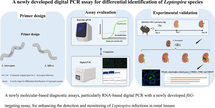
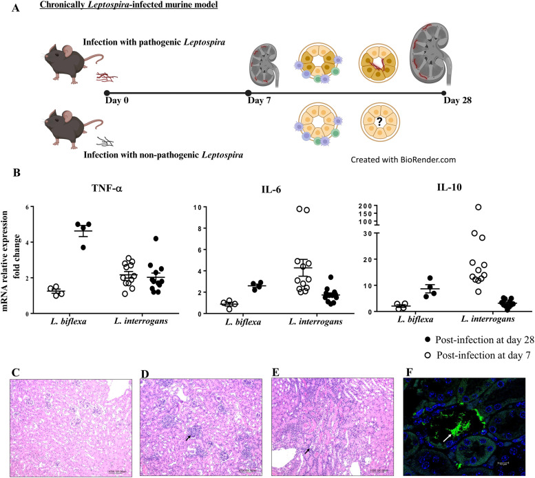
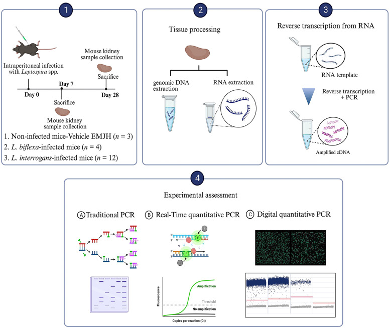
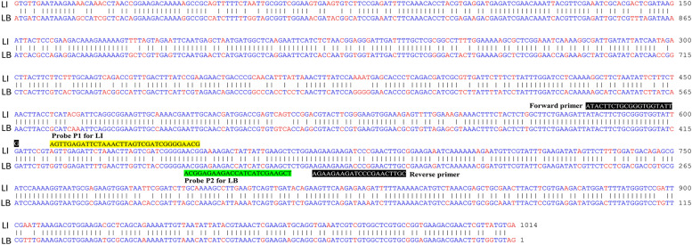
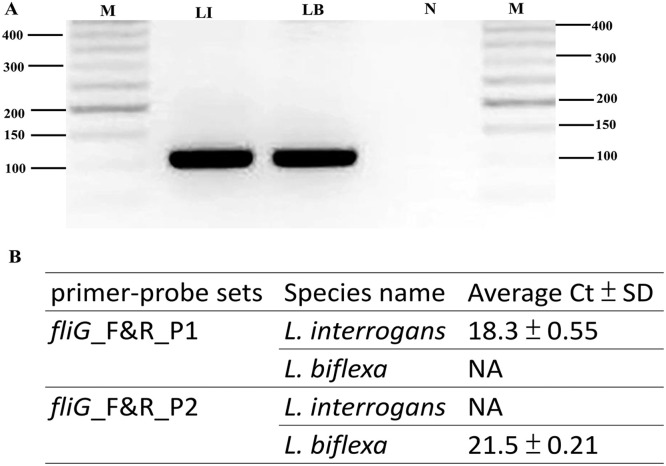
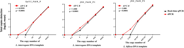
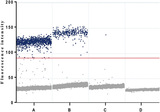
Leptospirosis, a re-emerging zoonotic disease caused by Leptospira spp., poses significant global health and veterinary challenges. Long-term colonization of renal tubules by Leptospira in asymptomatic hosts highlights the need for sensitive detection methods. This study evaluates the chronic or latent Leptospira infections in kidneys using a novel molecular approach to examine individual immune responses differences. Digital PCR strategies employing newly developed primer-probe sets targeting the flagellar fliG gene were used to assess the presence of trace Leptospira in infected murine kidneys and urine samples from laboratory-confirmed leptospirosis patients. RNA-based digital PCR detected leptospires in 58 % (targeting lipl32) and 83 % (targeting fliG) of infected kidneys, demonstrating that the digital PCR strategy targeting the fliG gene offers superior sensitivity. Notably, the newly developed fliG-targeting assay detected as low as 20 fg of Leptospira DNA, offering ten-fold greater sensitivity than traditional qPCR for trace detection. This allows for differential detection of Leptospira species and facilitates monitoring of extremely low bacterial loads with greater sensitivity than conventional methods. We also observed regenerating renal tubules with mitosis and elevated cytokine expression in kidneys with transcriptionally active Leptospira during chronic infection. This approach aids in identifying latent infections and offers insights into individual variations. Our research provides a powerful molecular tool for epidemiological studies and public health surveillance, contributing valuable insights into the prevalence and transmission dynamics of this pervasive zoonotic disease.

Keywords: Differential detection; Digital PCR; Latent infection; Leptospira spp.; Leptospirosis; Leptospirosis kidney disease.


Conflict of interest statement

L.-F. C., P.-Y. H. and C.-W. Y. are inventors (Kidney Research Center, Chang Gung Memorial Hospital) on a patent for “Innovative method for nucleic acid detection tracking minute quantities of Leptospira spp. in biological tissues and environmental samples” (US Provisional Patent Application 63/605,750, filed 4 December 2023). The remaining authors declare no competing interests.

Figures

Image, graphical abstract
Graphical abstract
Fig 1
Fig. 1
Experimental design for investigating the impact of Leptospira infection on renal immune responses. (A) Schematic view of the study of immune responses and bacterial colonization in the kidneys following infection with either pathogenic or nonpathogenic Leptospira spp. (B) The immune responses to Leptospira infection are characterized by the mRNA expression of pro-inflammatory cytokines (TNF-α and IL-6) and the anti-inflammatory cytokine IL-10. Representative renal histological findings in uninfected (C) and infected (D∼F) mice. These tissues were formalin-fixed and paraffin-embedded for histology and immunofluorescence study. Light microscopy of kidney tissues, stained with the hematoxylin and eosin (Magnification, 100 X). After infection at 28 days, inflammatory infiltration is observed in the nonpathogenic L. biflexa-infected (D) and pathogenic L. interrogans-infected (E) mice. (F) Bacterial adherence to the tubule lumen was found in the kidneys from mice infected with L. interrogans at 28 days post-infection. Confocal images of immunofluorescent staining (magnification of 400X). Anti-leptospiral LipL32 (green; white arrow) and DAPI staining (blue).
Fig 2
Fig. 2
The evaluation of digital PCR for the detection of pathogenic Leptospira infection in kidneys. Our study employed a digital PCR approach designed to target the lipl32 gene, which is specific only to pathogenic species of Leptospira. This methodology, combined with conventional and real-time qPCR, was utilized for the detection and quantification of pathogenic leptospires in the kidneys of mice subjected to experimental Leptospira infection. Negative control mice were used as a reference for noninfected status, while mice with nonpathogenic L. biflexa infection served as an infection control group. Figure was created with biorender.com (accessed on Jan 2024).
Fig 3
Fig. 3
Sequence alignment for each primer/probe pair using representative Leptospira species. Alignment of the DNA sequence of the fliG gene from L. interrogans serovar Copenhageni str. Fiocruz L1–130 (NCBI accession number: NC_005823.1) and the fliG2 gene from L. biflexa serovar Patoc strain 'Patoc 1 (Paris)' (NCBI accession number: CP000786.1) was performed. Alignment of the DNA sequence using the Martinez/Needlemen-Wunsch alignment method by MegAlign (DNASTAR, USA) revealed that the sequence similarity index was 78.3 %. Identical nucleotides are shown as blue letters, and different nucleotides aligned at the same position are shown as red letters. The positions of the forward and reverse primers are indicated as white letters on a black background. The nucleotides corresponding to the probe for the target gene from L. interrogans (LI) are highlighted in yellow, while those for the target gene from L. biflexa (LB) are highlighted in green.
Fig 4
Fig. 4
Validation of novel primer-probe sets for Leptospira species detection. (A) PCR amplification was performed using the primer pair fliG_F and fliG_R. Each reaction contained 25 ng of Leptospira genomic DNA under standard conditions. The amplification assay consisted of 30 cycles of 95 °C for 15 s, 60 °C for 75 s, and 72 °C for 75 s. Subsequently, the samples were maintained at 72 °C for 10 min, and the PCR products were separated via agarose gel electrophoresis and photographed. The genomic DNA from L. interrogans yielded a 110 bp fragment (Lane LI); genomic DNA from L. biflexa amplified a 110 bp fragment (Lane LB); negative control (water; Lane N); M, molecular size determined using a 50-bp ladder molecular marker. (B) RT‒PCR amplification was performed using the primer pairs fliG_F and fliG_R combined with different probes designed in this study. Duplicate Ct values were averaged, and the standard deviation (SD) of the Ct values was calculated. NA: samples with no amplification detected.
Fig 5
Fig. 5
Dynamic ranges and sensitivity for digital PCR assays targeting lipl32 and fliG genes. The total copy number of each sample was determined using a digital PCR (absolute total copies detection; log values) and estimated from the Ct values obtained from real-time qPCR (data are presented as means; detailed values are provided in Table 3). The X-axis represents the 10-fold dilution series of Leptospira genomic DNA, and the numbers of copies were theoretically calculated using the copy number calculator tool on the Technology Networks server (https://www.technologynetworks.com/tn/tools/copynumbercalculator). Correlation analysis between the theoretically calculated Leptospira copies (X-axis) and the copy number measured in the dilution series using both real-time qPCR and digital PCR (Y-axis, displayed as the mean values; n ≥ 3). Pearson's correlation coefficient (r) and p value are indicated. Statistical analyses were performed using Graph-Pad Prism 6.0 software (GraphPad Software, Inc.).
Fig 6
Fig. 6
Raw droplet scatter plot for the clinical validity of digital PCR assays targeting fliG genes in urine specimens from a laboratory-confirmed leptospirosis patient. Digital PCR for detection of Leptospira from A: positive control (cells infected with L. interrogans), B: urine specimens (before the initiation of antibiotic therapy), C: urine specimens (after the initiation of antibiotic therapy) and D: no template control. Positive droplets are obviously observed in the urine sample from before the initiation of antibiotic therapy. Blue dots represent the positive droplets, above the red horizontal threshold. Gray dots represent the negative droplets. The threshold for the detection of positive results was set at an amplitude of 90. Estimated copy numbers: Urine samples collected before the initiation of antibiotic therapy: 71.8 copy numbers per ml; Urine samples collected after the initiation of antibiotic therapy at day 10: 0.1 copy numbers per ml.

Similar articles

References

    1. Abela-Ridder B., Sikkema R., Hartskeerl R.A. Estimating the burden of human leptospirosis. Int. J. Antimicrob. Agents. 2010;36(Suppl 1):S5–S7. doi: 10.1016/j.ijantimicag.2010.06.012. - DOI - PubMed
    1. Bárbara Couto Roloff Padilha M.M.S., Daiane Drawanz Hartwig Molecular and serological diagnostic of leptospirosis: a review (2014–2020) Res, Soc Dev. 2022;11 doi: 10.33448/rsd-v11i2.25471. - DOI
    1. Bini Viotti J., Chan J.C., Rivera C., Tuda C. Sporadic leptospirosis case in Florida presenting as Weil;s disease. IDCases. 2020;19:e00686. doi: 10.1016/j.idcr.2019.e00686. - DOI - PMC - PubMed
    1. Bradley E.A., Lockaby G. Leptospirosis and the environment: a review and future directions. Pathogens. 2023;12 doi: 10.3390/pathogens12091167. - DOI - PMC - PubMed
    1. Carrillo-Larco R.M., Altez-Fernandez C., Acevedo-Rodriguez J.G., Ortiz-Acha K., Ugarte-Gil C. Leptospirosis as a risk factor for chronic kidney disease: a systematic review of observational studies. PLoS Negl. Trop. Dis. 2019;13 doi: 10.1371/journal.pntd.0007458. - DOI - PMC - PubMed

LinkOut - more resources