. 2024 Dec 4;18(4):e12055.
doi: 10.1002/ccs3.12055. eCollection 2024 Dec.

Cytokine expression and cytokine-mediated cell-cell communication during skeletal muscle regeneration revealed by integrative analysis of single-cell RNA sequencing data

Affiliations

Cytokine expression and cytokine-mediated cell-cell communication during skeletal muscle regeneration revealed by integrative analysis of single-cell RNA sequencing data

Pallob Barai et al. J Cell Commun Signal. .

Abstract

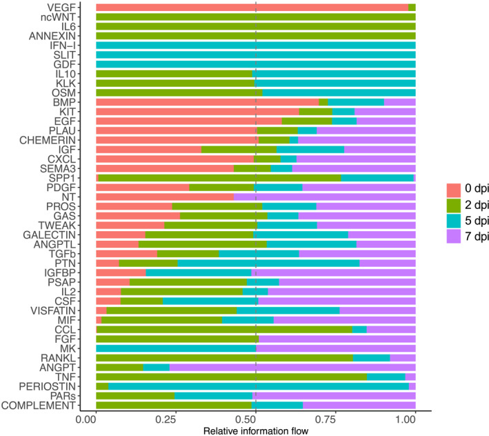
Skeletal muscles undergo self-repair upon injury, owing to the resident muscle stem cells and their extensive communication with the microenvironment of injured muscles. Cytokines play a critical role in orchestrating intercell communication to ensure successful regeneration. Immune cells as well as other types of cells in the injury site, including muscle stem cells, are known to secret cytokines. However, the extent to which various cell types express distinct cytokines and how the secreted cytokines are involved in intercell communication during regeneration are largely unknown. Here we integrated 15 publicly available single-cell RNA-sequencing (scRNA-seq) datasets of mouse skeletal muscles at early regeneration timepoints (0, 2, 5, and 7 days after injury). The resulting dataset was analyzed for the expression of 393 annotated mouse cytokines. We found widespread and dynamic cytokine expression by all cell types in the regenerating muscle. Interrogating the integrated dataset using CellChat revealed extensive, bidirectional cell-cell communications during regeneration. Our findings provide a comprehensive view of cytokine signaling in the regenerating muscle, which can guide future studies of ligand-receptor signaling and cell-cell interaction to achieve new mechanistic insights into the regulation of muscle regeneration.

Keywords: cell–cell communication; cytokines; muscle stem cells; scRNA‐seq; skeletal muscle regeneration.


Conflict of interest statement

The authors declare that they have no conflict of interest.

Figures

FIGURE 1
FIGURE 1
Integration of 15 scRNA‐seq datasets at various days post injury (dpi) of TA muscles and analysis of cell types during regeneration. (A) A schematic of scRNA‐seq data integration and downstream analysis. (B) Single‐cell atlas of 75,150 cells assembled from all the timepoints. Data are presented in UMAP to visualize the transcriptional differences between different cell clusters. (C) Cell type composition throughout the early phase of regeneration. (D) Single‐cell atlases at 0, 2, 5, and 7 dpi.
FIGURE 2
FIGURE 2
Regenerating muscles express a large array of cytokines. (A) A heatmap representing the number of cytokines detected in each cell type (diagonal axis) and number of overlapping cytokines between cell populations. Cell types are arranged in a descending order of number of cytokines detected. (B) A Venn diagram showing numbers of overlapping cytokines expressed by MuSCs, FAPs, macrophages, and endothelial cells. (C) Numbers of cytokines expressed in MuSCs, FAPs, macrophages, and tenocytes during regeneration. (D) A Venn diagram showing the numbers of upregulated cytokines in regenerating muscles (all cells combined) at 2, 5 and 7 dpi compared to 0 dpi. (E)–(G) Dot‐plots showing expression profiles of the most highly upregulated cytokines at 2, 5, and 7 dpi compared to 0 dpi in MuSCs (E), FAPs (F), and macrophages (G). Color gradient represents the average expression level and size of circle corresponds to percentage of cells in a cluster that expressed the gene.
FIGURE 3
FIGURE 3
Cell–cell communication during skeletal muscle regeneration. (A) Chord‐diagram visualization of cell–cell communication networks involving all cell types identified in the integrated data. Arrow indicates direction of communication and line thickness corresponds to number of interactions. (B) Total numbers of inferred interactions at 0, 2, 5, and 7 dpi. (C) Numbers of communications between MuSCs, FAPs, and macrophages at 0, 2, 5, and 7 dpi.
FIGURE 4
FIGURE 4
Contribution of cell types to sending and receiving cytokine signals during regeneration. (A)–(D) Scatter plots show signaling strength of various cell types in sending (outgoing, x‐axis) or receiving (incoming, y‐axis) cytokine signals at 0 dpi (A), 2 dpi (B), 5 dpi (C), and 7 dpi (D). Size of the circle represents total number of interactions (combining outgoing and incoming).
FIGURE 5
FIGURE 5
Overall information flow during regeneration. A paired Wilcoxon test was performed to identify signaling pathways with significant change over the course of regeneration. Relative strength of information flow is shown for 0, 2, 5, and 7 dpi.
FIGURE 6
FIGURE 6
Outgoing and incoming signaling in individual cell types during regeneration. (A)–(B) Heatmap graphs show the outgoing (A) and incoming (B) signaling patterns at 0, 2, 5, and 7 dpi. Green gradient bars denote relative signaling strengths. Colored bars at the top represent the overall signaling strength of a particular cell type from summing all signaling pathways in the heatmap. Gray bars at the right of each graph represent the total signaling strength of a pathway from all cell types. MuSC data are highlighted by red boxes.
FIGURE 7
FIGURE 7
Examples of signaling to and from MuSCs over the early course of regeneration. (A)–(B) PDGF (A) and TGFβ (B) signaling from MuSCs to other cell types at 0, 2, 5, and 7 dpi. (C)–(D) IGF (C) and TWEAK (D) signaling from other cell types to MuSCs at 0, 2, 5, and 7 dpi. Thickness of the lines corresponds to relative signaling strength and arrow indicates the direction of signaling.
FIGURE 8
FIGURE 8
Cytokine signaling to MuSCs upregulated in regenerating muscles. (A)–(C) Upregulated ligand–receptor signaling to MuSCs from macrophages, FAPs, tenocytes, and MuSCs at 2 dpi (A), 5 dpi (B), and 7 dpi (C) compared to 0 dpi. Relative communication probability is represented by color scheme as indicated. p‐value threshold was set to <0.01 to identify significantly upregulated signaling.

Similar articles

Cited by

References

    1. Yin, H. , Price F., and Rudnicki M. A.. 2013. “Satellite Cells and the Muscle Stem Cell Niche.” Physiological Reviews 93(1): 23–67. 10.1152/physrev.00043.2011. - DOI - PMC - PubMed
    1. Pannérec, A. , Marazzi G., and Sassoon D.. 2012. “Stem Cells in the Hood: The Skeletal Muscle Niche.” Trends in Molecular Medicine 18(10): 599–606. 10.1016/j.molmed.2012.07.004. - DOI - PubMed
    1. Joe, A. W. B. , Yi L., Natarajan A., Le Grand F., So L., Wang J., Rudnicki M. A., and Rossi F. M. V.. 2010. “Muscle Injury Activates Resident Fibro/adipogenic Progenitors that Facilitate Myogenesis.” Nature Cell Biology 12(2): 153–U144. 10.1038/ncb2015. - DOI - PMC - PubMed
    1. Kuang, S. , Gillespie M. A., and Rudnicki M. A.. 2008. “Niche Regulation of Muscle Satellite Cell Self‐Renewal and Differentiation.” Cell Stem Cell 2(1): 22–31. 10.1016/j.stem.2007.12.012. - DOI - PubMed
    1. Wosczyna, M. N. , and Rando T. A.. 2018. “A Muscle Stem Cell Support Group: Coordinated Cellular Responses in Muscle Regeneration.” Developmental Cell 46(2): 135–143. 10.1016/j.devcel.2018.06.018. - DOI - PMC - PubMed

LinkOut - more resources