

Giant Cell Myocarditis vs Cardiac Sarcoidosis: Reconsidering the Diagnosis With FDG PET Imaging
- PMID: 39691891
- PMCID: PMC11646884
- DOI: 10.1016/j.jaccas.2024.102738
Giant Cell Myocarditis vs Cardiac Sarcoidosis: Reconsidering the Diagnosis With FDG PET Imaging
Abstract
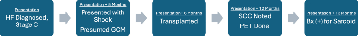
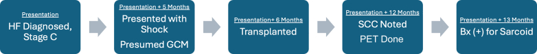
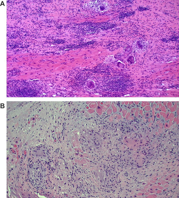
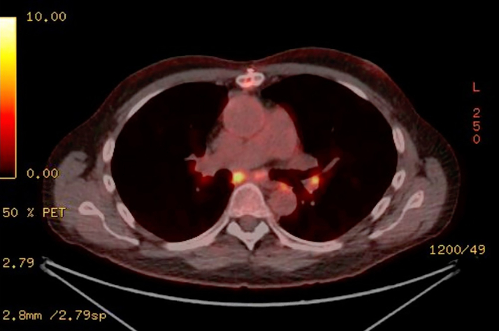
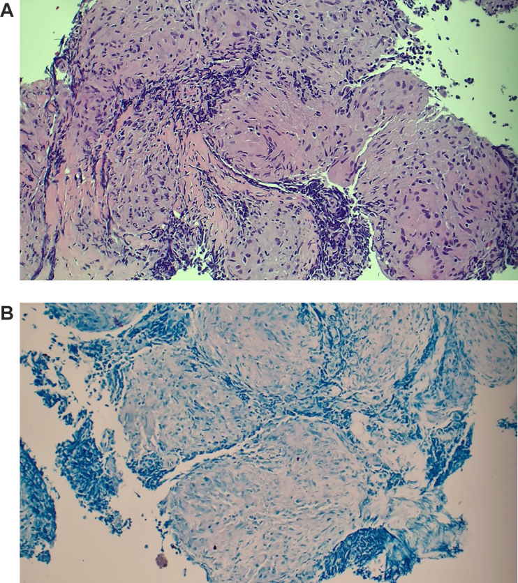
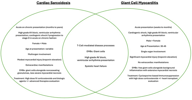
Giant cell myocarditis (GCM) and cardiac sarcoidosis share clinical and histologic features, but whether they represent separate processes or lie on an inflammatory cardiomyopathy spectrum is unclear. We present a case of cardiogenic shock thought to be secondary to biopsy-proven GCM with a subsequent post-transplant diagnosis of sarcoidosis through 18-fluorodeoxyglucose positron emission tomography and biopsy.
Keywords: cardiac sarcoidosis; giant cell myocarditis; inflammatory cardiomyopathy.
© 2024 The Authors.
Conflict of interest statement
Dr Sheikh has received institutional research support from Abbott, Alnylam, and Akcea; has received honoraria for educational presentations from Abbott; and has served as a consultant for Alnylam. Dr Gupta has served as a consultant for and has received honoraria for educational presentations from CVRx. All other authors have reported that they have no relationships relevant to the contents of this paper to disclose.
Figures
Similar articles
-
The Clinical and Histological Intersection of Cardiac Sarcoidosis and Giant Cell Myocarditis.Cureus. 2024 May 7;16(5):e59783. doi: 10.7759/cureus.59783. eCollection 2024 May. Cureus. 2024. PMID: 38846240 Free PMC article.
-
Fulminant cardiac sarcoidosis resembling giant cell myocarditis: a case report.Eur Heart J Case Rep. 2021 Mar 10;5(3):ytab042. doi: 10.1093/ehjcr/ytab042. eCollection 2021 Mar. Eur Heart J Case Rep. 2021. PMID: 33733047 Free PMC article.
-
Phenotyping of giant cell myocarditis versus cardiac sarcoidosis using cardiovascular magnetic resonance.Int J Cardiol. 2023 Sep 15;387:131143. doi: 10.1016/j.ijcard.2023.131143. Epub 2023 Jun 25. Int J Cardiol. 2023. PMID: 37364717
-
Idiopathic giant cell myocarditis and cardiac sarcoidosis.Heart Fail Rev. 2013 Nov;18(6):733-46. doi: 10.1007/s10741-012-9358-3. Heart Fail Rev. 2013. PMID: 23111533 Review.
-
[Inflammatory heart diseases--cardiac sarcoidosis and giant cell myocarditis].Duodecim. 2015;131(22):2127-33. Duodecim. 2015. PMID: 26749906 Review. Finnish.
References
-
- Bang V., Ganatra S., Shah S.P., et al. Management of patients with giant cell myocarditis. J Am Coll Cardiol. 2021;77:1122–1134. - PubMed
-
- Kociol R.D., Cooper L.T., Fang J.C., et al. Recognition and initial management of fulminant myocarditis: a scientific statement from the American Heart Association. Circulation. 2020;141(6):e69–e92. - PubMed
-
- Kouranos V., Sharma R. Cardiac sarcoidosis: state-of-the-art review. Heart. 2021;107:1591–1599. - PubMed
-
- Okura Y., Dec G.W., Hare J.M., et al. A clinical and histopathologic comparison of cardiac sarcoidosis and idiopathic giant cell myocarditis. J Am Coll Cardiol. 2003;41:322–329. - PubMed
Publication types
LinkOut - more resources
Full Text Sources
