

Case Reports
doi: 10.1016/j.jaccas.2024.102653.
Managing Left Ventricular Outflow Tract Obstruction in Combined Aortic Stenosis and Hypertrophic Cardiomyopathy
Affiliations
- PMID: 39691894
- PMCID: PMC11646875
- DOI: 10.1016/j.jaccas.2024.102653
Item in Clipboard
Case Reports
Managing Left Ventricular Outflow Tract Obstruction in Combined Aortic Stenosis and Hypertrophic Cardiomyopathy
JACC Case Rep.
.
Abstract
This study presents an elderly man with sequential hemodynamic obstructions caused by hypertrophic cardiomyopathy and aortic stenosis. Septal reduction therapy was performed to avoid outflow tract obstruction associated with potential future transcatheter aortic valve replacement. This case highlights the importance of resolving outflow tract obstruction during assessment of aortic valve disease.
Keywords: aortic stenosis; obstructive hypertrophic cardiomyopathy.
© 2024 The Authors.
Conflict of interest statement
The authors have reported that they have no relationships relevant to the contents of this paper to disclose.
Figures


Baseline Electrocardiogram Electrocardiogram demonstrated sinus bradycardia.

Diagnosis of Hypertrophic Cardiomyopathy by Echocardiography (A) Parasternal long-axis view demonstrated 2.0-cm basal septal hypertrophy (red arrow), along with calcification and restricted motion of the aortic valve leaflets (yellow arrow). (B) M-mode across the mitral valve demonstrated systolic anterior motion, with systolic displacement of the anterior mitral valve leaflet (red arrow) toward the left ventricular outflow tract.

Baseline Hemodynamics by Echocardiography (A) Continuous-wave Doppler across the left ventricular outflow tract (LVOT) and aortic valve (AV) identified a 4 m/s mid-systolic peak velocity and 34 mm Hg mean gradient. In an alternate examination, (B) continuous-wave Doppler detected a 5.3 m/s late-peaking velocity across the LVOT and AV and (C) pulse wave Doppler identified a 3 m/s late-peaking velocity at the LVOT.
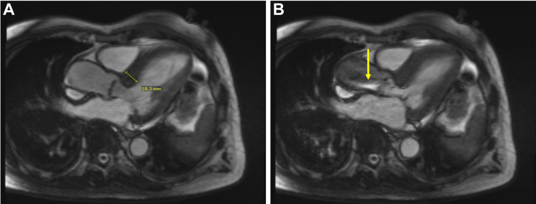
Baseline Measurements by Cardiac Magnetic Resonance Baseline cardiac magnetic resonance demonstrated (A) 1.8-cm basal septal hypertrophy and (B) severely restricted movement of the aortic valve leaflets with proton-dephasing across the left ventricular outflow tract and aortic valve (yellow arrow).

Baseline Hemodynamics by Cardiac Catheterization Representative tracing demonstrating peak-to-peak pressure differences between the left ventricle (yellow) and aorta (teal) before and after withdrawal of catheter from the left ventricle (LV) toward the cranial aspect of the left ventricular outflow tract (LVOT). Electrocardiogram leads II, aVL, and V1 are shown in black. Plethysmography tracing is shown in blue.

Follow-Up Echocardiography Four-month follow-up echocardiography demonstrated (A) 2.0-cm basal septal thickness and (B and C) moderate aortic stenosis with high flow.
References
Publication types
LinkOut - more resources
Full Text Sources
