The Median Nerve Displays Adaptive Characteristics When Exposed to Repeated Pinch Grip Efforts of Varying Rates of Force Development: An Ultrasonic Investigation
- PMID: 39692085
- PMCID: PMC11892086
- DOI: 10.1002/jum.16634
The Median Nerve Displays Adaptive Characteristics When Exposed to Repeated Pinch Grip Efforts of Varying Rates of Force Development: An Ultrasonic Investigation
Abstract
Objectives: Repeated gripping with high grip forces and high rates of grip force development are risk factors for carpal tunnel syndrome. As the nerve's adaptive ability is crucial to prevent disease progression, we investigated how these risk factors influence median nerve deformation and displacement over the time course of a repeated pinch grip task.
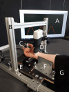
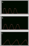
Methods: Seventeen healthy participants performed a repeated grip task against a load cell while their carpal tunnel was scanned with ultrasound. The grip task involved pulp-pinching three consecutive times from 0 to 40% maximal voluntary exertion (MVE), performed at three different rates of force development (RFD): 40% MVE/1 second; 2 seconds; and 5 seconds. Ultrasound images were analyzed at 10% MVE intervals. Nerve circularity, width, height, and cross-sectional area were measured to assess deformation. Median nerve displacement was assessed by its change in position relative to the flexor digitorum superficialis tendon of the third digit (FD) in both radioulnar and palmodorsal axes.
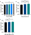
Results: Linear mixed modeling indicated that median nerve deformation increased, becoming more circular, with each repeated pulp-pinch (P < .01) and with grip force magnitude (P < .01). However, a faster RFD decreased nerve deformation (P < .01). Furthermore, the nerve displaced ulnarly during pulp-pinching, with greater displacement during the fastest (ie, 40% MVE/1 second) RFD (P < .01).
Conclusions: The median nerve deformed and displaced in response to pulp-pinching; however, faster rates of force development hindered this adaptive response. This likely reflects the viscoelastic properties of the healthy nerve and subsynovial connective tissue, highlighting the importance of tissue compliance in preventing nerve compression.
Keywords: carpal tunnel; connective tissue; ultrasound; viscoelastic substances.
© 2024 The Author(s). Journal of Ultrasound in Medicine published by Wiley Periodicals LLC on behalf of American Institute of Ultrasound in Medicine.
Figures


References
-
- Guimberteau JC, Delage JP, McGrouther DA, Wong JKF. The microvacuolar system: how connective tissue sliding works. J Hand Surg 2010; 35:614–622. - PubMed
-
- Hakim AJ, Cherkas L, El Zayat S, MacGregor AJ, Spector TD. The genetic contribution to carpal tunnel syndrome in women: a twin study. Arthritis Rheum 2002; 47:275–279. - PubMed
-
- Zyluk A. The role of genetic factors in carpal tunnel syndrome etiology: a review. Adv Clin Exp Med 2020; 29:623–628. - PubMed
MeSH terms
Grants and funding
LinkOut - more resources
Full Text Sources
Research Materials

