. 2025 Apr 3;148(4):1194-1211.
doi: 10.1093/brain/awae363.

Clinical and genetic characterization of a progressive RBL2-associated neurodevelopmental disorder

Affiliations

Clinical and genetic characterization of a progressive RBL2-associated neurodevelopmental disorder

Gabriel N Aughey et al. Brain. .

Abstract

Retinoblastoma (RB) proteins are highly conserved transcriptional regulators that play important roles during development by regulating cell-cycle gene expression. RBL2 dysfunction has been linked to a severe neurodevelopmental disorder. However, to date, clinical features have been described in only six individuals carrying five biallelic predicted loss-of-function (pLOF) variants. To define the phenotypic effects of RBL2 mutations in detail, we identified and clinically characterized a cohort of 35 patients from 20 families carrying pLOF variants in RBL2, including 15 new variants that substantially broaden the molecular spectrum. The clinical presentation of affected individuals is characterized by a range of neurological and developmental abnormalities. Global developmental delay and intellectual disability were observed uniformly, ranging from moderate to profound and involving lack of acquisition of key motor and speech milestones in most patients. Disrupted sleep was also evident in some patients. Frequent features included postnatal microcephaly, infantile hypotonia, aggressive behaviour, stereotypic movements, seizures and non-specific dysmorphic features. Neuroimaging features included cerebral atrophy, white matter volume loss, corpus callosum hypoplasia and cerebellar atrophy. In parallel, we used the fruit fly, Drosophila melanogaster, to investigate how disruption of the conserved RBL2 orthologue Rbf impacts nervous system function and development. We found that Drosophila Rbf LOF mutants recapitulate several features of patients harbouring RBL2 variants, including developmental delay, alterations in head and brain morphology, locomotor defects and perturbed sleep. Surprisingly, in addition to its known role in controlling tissue growth during development, we found that continued Rbf expression is also required in fully differentiated post-mitotic neurons for normal locomotion in Drosophila, and that adult-stage neuronal re-expression of Rbf is sufficient to rescue Rbf mutant locomotor defects. Taken together, our study provides a clinical and experimental basis to understand genotype-phenotype correlations in an RBL2-linked neurodevelopmental disorder and suggests that restoring RBL2 expression through gene therapy approaches might ameliorate some symptoms caused by RBL2 pLOF.

Keywords: Drosophila; RBL2; Rbf; cell cycle; neurodevelopmental disorder.


Conflict of interest statement

C.B. is an employee of Centogene GmbH. G.H.S. is an employee of 3billion. L.M. has received personal fees for ad hoc consultancy from Mendelian Ltd, a rare disease digital healthcare company. The remaining authors report no competing interests.

Figures

Figure 1
Figure 1
RBL2-related disorder is characterized by a range of neurological, behavioural and developmental abnormalities. (A) Representation of the most frequent clinical features observed in the RBL2 patients (y-axis, clinical features; x-axis, number of patients). (B) Time-line-style schematic diagram outlining the acquisition of key developmental milestones observed in the affected individuals. Most of the individuals did not attain independent sitting, walking or speech development (blue bar indicates range at last evaluation), and the others presented delayed acquisition (orange, line indicates median age). Normal range is indicated in green. (C) Schematic depiction of the degree of global developmental delay/intellectual disability (GDD/ID) observed in the patients (number of patients indicated at bottom). The spectrum ranged from moderate (left) to profound (right).
Figure 2
Figure 2
RBL2 patients present postnatal microcephaly and dysmorphic features, without a recognizable facial ‘gestalt’. (A) Left: Box plot showing the range of head circumference measurements in RBL2 predicted loss-of-function patients, expressed in standard deviations (SDs) from the mean of healthy controls. The box delineates the range between first and third quartile, the cross (×) represents the mean, and the line that divides the box indicates the median of the whole cohort. Head circumference was within normal ranges at birth and reduced at last examination. Right: Head circumference measurements with age at last follow-up across individual patients. (B) Facial features of the patients. P = patient.
Figure 3
Figure 3
Neuroimaging features of RBL2-related disorder. Sagittal T1-weighted image (left), axial T2-weighted or FLAIR image (middle) and coronal T1- or T2-weighted or FLAIR image (right). Most subjects have an enlargement of the cerebral CSF spaces, with an anteroposterior gradient associated with thinning of the corpus callosum (thick arrows), particularly in the anterior portions. There is additional cerebellar atrophy in P10, P12, P20, P22 and P30 (thin arrows). Bilateral mild-to-moderate signal changes are noted at the level of the forceps minor in P10, P19, P20, P21, P22 and P30 (arrowheads). Note the large prepontine lesion in P6 (curved arrow). FLAIR = fluid-attenuated inversion recovery; P = patient.
Figure 4
Figure 4
Molecular spectrum of loss-of-function variants in RBL2. (A) Schematic representation of the location of variants on the RBL2 gene. Top: Newly reported variants. Bottom: Previously reported variants. (B) Pedigrees of the newly reported patients. Filled black symbols = affected. Genotype, where indicated, represents the results of segregation. (C) Classification of variants according to type.
Figure 5
Figure 5
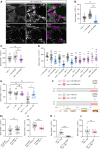
Drosophila Rbf regulates head and brain morphology. (A) Representative images of adult eyes in control (iso31) and Rbf120a hypomorphs. Scale bars: 0.3 mm. (B) Quantification of eye sizes in male Rbf120a hemizygotes (n = 16) compared with controls (n = 9). (C) Quantification of eye sizes in female Rbf allelic combinations (n = 5–8). (D) Representative images of adult brains in control and Rbf120a adult males. Scale bars: 50 μm. (EG) Measurements of brain morphology in control and Rbf120a hemizygotes adult males (n = 10 brains, 10 central brains and 20 optic lobes per genotype). (H) Quantification of apoptotic (DCP1+) cells in control and Rbf120a hemizygote adult male brains (n = 6 per genotype). (I) Representative images of DCP1-labelled control (n = 9) and Rbf120a hemizygote (n = 6) third instar larval nervous system. Nuclei are counterstained with DAPI. Scale bars = 20 μm. (J) Quantification of apoptosis in control and Rbf120a hemizygote third instar larval brains. Error bars are the standard error of the mean. *P < 0.05, **P < 0.005, ***P < 0.0005, ns = P > 0.05, unpaired t-test with Welch’s correction (B, E and F), one-way ANOVA with Dunnett’s post hoc test (C) or Mann–Whitney U-test (G, H and J).
Figure 6
Figure 6
Loss of Rbf disrupts movement and sleep in Drosophila. (A) Schematic diagram illustrating Drosophila life cycle and histogram of time to eclosion for iso31 controls (n = 201) and Rbf120a hemizygotes (n = 26). (B) Schematic representation of the Drosophila activity monitor (DAM) system. (C and D) DAM activity in Rbf120a hemizygotes (n = 38) and controls (iso31; n = 31) across a 24 h period (C) or during zeitgeber time (ZT) 0–1, a period of peak activity (D). (E) DAM activity in adult females harbouring trans-heterozygote or heterozygote Rbf allelic combinations and in wild-type iso31 controls during ZT0–1. n = 16–20. (F) Sleep traces of control (iso31) and Rbf120a hemizygote males showing the proportion of time spent asleep during 30 min windows across a 12 h light/12 h dark period. Loss of evening anticipation (left arrow) and reduced sleep during the first half of the night (right arrow), in Rbf120a males. (G and H) Proportion of time spent asleep during the hour before lights-off (G) and the first half of the night (H) in control and Rbf120a males. n = 31 iso31 males and 38 Rbf120a males. (I–K) Sleep traces (I), proportion of time spent asleep during the hour before lights-off (J) and the first half of the night (K), in control and Rbf120a homozygote females. Arrows in I again point to loss of evening anticipation (left arrow) and reduced sleep during the first half of the night (right arrow). n = 16 iso31 and 14 Rbf120a females. Error bars are the standard error of the mean. *P < 0.05, **P < 0.005, ***P < 0.0005, ns = P > 0.05, unpaired t-test with Welch’s correction (C and J), Mann–Whitney U-test (D, F, G and I) or one-way ANOVA with Dunnett’s post hoc test (E).
Figure 7
Figure 7
Adult-stage neuronal expression of Rbf rescues locomotor defects in Rbf hypomorphs. (A) Rbf-Gal4 driven nuclear mCherry expression in the adult central brain. Neurons and glia are counterstained with antibodies against ELAV and REPO, respectively. Scale bar: 20 µm. (B) Pan-neuronal post-mitotic knockdown of Rbf severely reduces peak locomotor activity during ZT0–1. n = 20–54. (C) Knockdown of Rbf in glial cells (using repo-Gal4 to express Rbf shRNA) does not significantly reduce peak locomotor activity during ZT0–1 compared with both driver and transgene alone controls (n = 18–24). (D) Knockdown of Rbf in cholinergic, glutamatergic and GABAergic neurons reduces peak activity during ZT0–1 in adult males. n = 11–41. Upper significance notation is relative to Rbf shRNA alone controls, lower significance notation is relative to Gal4 driver alone controls. (E) Effects of post-mitotic, neuron-specific Rbf expression on peak locomotor activity in either wild-type or Rbf120a hypomorph backgrounds. Data are from adult males. n = 15–21. (F) Experimental protocol for temperature-induced knockdown and rescue experiments shown in G and H. (G) Quantification of peak activity for: (i) control adult male flies kept at non-permissive temperature, mCherry (n = 33) or Rbf (n = 23) shRNA expression repressed; and (ii) experimental adult male flies maintained at a permissive temperature, mCherry (n = 24) or Rbf (n = 16) shRNA expression permitted. [H(i)] Constitutive suppression of neuronal RBF expression via tub-Gal80ts significantly decreases peak locomotor activity in Rbf120a; nsyb > Rbf adult males. Rbf120a, nsyb > Rbf: n = 13; Rbf120a, tub-Gal80ts, nsyb > Rbf: n = 10. [H(ii)] Peak activity in adult male flies that robust RBF expression solely permitted in adult-stage post-mitotic neurons is not significantly different from Rbf120a hypomorphs with constitutive post-mitotic neuronal expression of RBF. Rbf120a, nsyb > Rbf: n = 15; Rbf120a, tub-Gal80ts, nsyb > Rbf: n = 13. Error bars are the standard error of the mean. *P < 0.05, **P < 0.005, ***P < 0.0005, ns = P > 0.05, Kruskal–Wallis test with Dunn’s post hoc test (B and E), one-way ANOVA with Dunnett’s post hoc test (C and D) or unpaired t-test with Welch’s correction [G(i), G(ii), H(i) and H(ii)]. ZT = zeitgeber time.

References

    1. Harbour JW, Dean DC. Rb function in cell-cycle regulation and apoptosis. Nat Cell Biol. 2000;2:E65–E67. - PubMed
    1. Yao Y, Gu X, Xu X, Ge S, Jia R. Novel insights into RB1 mutation. Cancer Lett. 2022;547:215870. - PubMed
    1. Chai P, Luo Y, Yu J, et al. Clinical characteristics and germline mutation spectrum of RB1 in Chinese patients with retinoblastoma: A dual-center study of 145 patients. Exp Eye Res. 2021;205:108456. - PubMed
    1. Wadayama B, Toguchida J, Shimizu T, et al. Mutation spectrum of the retinoblastoma gene in osteosarcomas. Cancer Res. 1994;54:3042–3048. - PubMed
    1. Berge EO, Knappskog S, Geisler S, et al. Identification and characterization of retinoblastoma gene mutations disturbing apoptosis in human breast cancers. Mol Cancer. 2010;9:173. - PMC - PubMed

Substances