Review
. 2025 Jan 8;125(1):326-368.
doi: 10.1021/acs.chemrev.4c00570. Epub 2024 Dec 18.

Covalent Proximity Inducers

Affiliations
Review

Covalent Proximity Inducers

Nir London. Chem Rev. .

Abstract

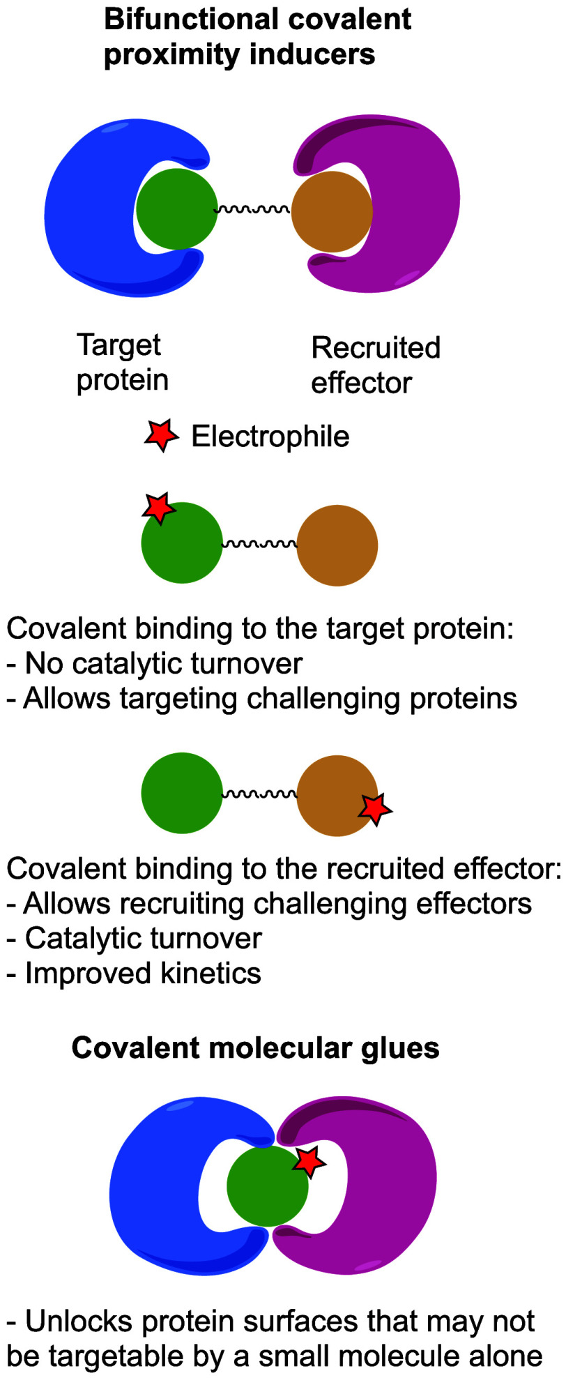
Molecules that are able to induce proximity between two proteins are finding ever increasing applications in chemical biology and drug discovery. The ability to introduce an electrophile and make such proximity inducers covalent can offer improved properties such as selectivity, potency, duration of action, and reduced molecular size. This concept has been heavily explored in the context of targeted degradation in particular for bivalent molecules, but recently, additional applications are reported in other contexts, as well as for monovalent molecular glues. This is a comprehensive review of reported covalent proximity inducers, aiming to identify common trends and current gaps in their discovery and application.


Conflict of interest statement

The author declares the following competing financial interest(s): N.L. serves on the SAB of Larkspur Biosciences, Tesseract Medicines and Eindura Therapeutics.

Figures

Figure 1
Figure 1
Classes of covalent proximity inducers. Covalent proximity inducers can be divided to bifunctional molecules and covalent molecular glues. Each with its advantages and disadvantages.
Figure 2
Figure 2
Covalent K-RasG12C PROTACs.
Figure 3
Figure 3
Covalent BTK PROTACs. (A) Chemical structures of various covalent BTK PROTACs. (B) Co-crystal structure (PDB 8DSO) of BTK (white) in ternary complex with covalent PROTAC BCCov (green) and E3 ligase cIAP1 (blue).
Figure 4
Figure 4
Covalent EGFR PROTACs.
Figure 5
Figure 5
Covalent SARS-CoV-2 Mpro PROTACs.
Figure 6
Figure 6
Chemical structures of HaloPROTACs
Figure 7
Figure 7
Covalent ligand directed PROTACs. (A) General scheme for covalent ligand directed PROTACs. After labeling the target protein with a degradation label, the guiding ligand acts as a leaving group and vacates the protein. (B) Chemical structure of CoLDR based BTK PROTAC 1n with a schematic mechanism of action. (C) Chemical structures of covalent ligand directed PROTACs, the reactive centers are indicated in red.
Figure 8
Figure 8
Structures of additional various covalent PROTACs.
Figure 9
Figure 9
DCAF16 recruiting covalent PROTACs.
Figure 10
Figure 10
RNF114 recruiting covalent PROTACs.
Figure 11
Figure 11
RNF4 recruiting covalent PROTAC.
Figure 12
Figure 12
Covalent, natural product based, Keap1-recruiting PROTACs.
Figure 13
Figure 13
DCAF11 recruiting covalent PROTACs.
Figure 14
Figure 14
FEM1B recruiting covalent PROTAC.
Figure 15
Figure 15
DCAF1 recruiting stereoselective covalent PROTAC.
Figure 16
Figure 16
Covalent recruiters and PROTACs for non-E3 ligase UPS components. (A) UBE2D, (B) DDB1, (C) SKP1.
Figure 17
Figure 17
CRBN and VHL covalent recruiters. (A) Structures of covalent CRBN recruiting ligands and PROTAC. (B) Structure of covalent VHL recruiting PROTAC.
Figure 18
Figure 18
FBXO22 recruiting covalent PROTACs.
Figure 19
Figure 19
Structures of OTUB1 recruiter and lumacaftor based CFTR stabilizer.
Figure 20
Figure 20
CoLDR based phosphorylation inducing chimeras (PHICs). (A) Schematic for the mechanism of action of covalent ligand directed release (CoLDR) based PHICs. (B) Chemical structures of 3 BTK targeting PHICs. While 17 and 19 recruited BTK to phosphorylate BRD4, 21 recruited ABL to phosphorylate BTK itself.
Figure 21
Figure 21
Imine based glues for the 14–3–3σ/p65 interaction. (A) Chemical structures of aldehyde glues. (B–D) Co-crystal structures of 14–3–3σ (white) with a p65 derived peptide (blue) and imine covalent glue (magenta). (B) TCF521; PDB 6YOW. (C) TCF521-123; PDB 6YPY. (D) TCF521-129; PDB 6YQ2.
Figure 22
Figure 22
Imine based glues for 14–3–3σ/Pin1 interaction. (A) Chemical structures of aldehyde glues. (B–D) Co-crystal structures of 14–3–3σ (white) with a Pin1 derived peptide (blue) and imine covalent glue (magenta). (B) L2; PDB 7AXN. (C) 13; PDB 7BDT. (D) 28; PDB 7BFW.
Figure 23
Figure 23
Disulfide 14–3–3 glues. (A) Chemical structures of disulfide stabilizers of 14–3–3 client peptides. (B–E) Co-crystal structures of 14–3–3σ (white) in complex with client peptides (blue) and disulfide glues (magenta). (B) ERα; C42–1; PDB 6HHP. (C) ERα; C38–1; PDB 8AFN. (D) C-RAF; C38-1; PDB 8AV0. (E) FOXO1; FOXO1-2 PDB 8A62. (F) C-RAF; CRAF5; PDB 8A68.
Figure 24
Figure 24
Dual covalent glues for 14–3–3 proteins. (A) Chemical structures of parent disulfide glue 1 and dual-covalent derivatives 5 and 10 that mediate an interaction between 14 and 3–3γ and ERRγ. (B,C) Co-crystal structures of the ternary complex between 14 and 3–3σ (white), ERRγ peptide (blue) and dual covalent glues (magenta) interacting with a cysteine on the client peptide and Lys122 on 14–3–3. (B) 5; PDB 8B4Q. (C) 10; PDB 8B5P.
Figure 25
Figure 25
Irreversible 14–3–3/peptide molecular glues. (A) Medicinal chemistry optimization campaign of chloroacetamide glues for 14–3–3σ to ERα client peptide. (B) Co-crystal structure of 14–3–3σ (white), ERα client peptide (blue), and compound 181 (magenta). (C) Structures of an irreversible fragment that binds to 14–3–3σ and stabilizes its interaction with a peptide from ERα, and a bifunctional that recruits BRD4 to 14–3–3σ and therefore sequesters it in the cytoplasm.
Figure 26
Figure 26
Covalent glues targeting K-RasG12C. (A) Chemical structure of compound 5. Despite being bifunctional, it labels K-RasG12C 4× better in the presence of FKBP12. (B,C) Chemical structures of covalent glues that mediated the interaction between CypA and K-RasG12C. (D,E) Co-crystal structures of the ternary complex between CypA (white), K-RasG12C (blue) in its GTP (nonhydrolizable analogue in green) bound form and a covalent glue (magenta). (D) Compound 1; PDB 8G9Q. (E) RMC-4998; PDB 8G9P.
Figure 27
Figure 27
Covalent BRD4/DCAF16 glues and related DCAF16/11 PROTACs. (A) A series of monovalent BRD4 degraders recruiting DCAF16 via template assisted covalent modification. (B). Schematic representation of the degradation mechanism of action of BRD4 degradation through template assisted covalent modification of DCAF16. (C) Additional glues and bifuncational degraders of BRD4 that recruit DCAF16 through alternative cysteines or DCAF11. (D) Structure of the ternary complex between BRD4 (white), MMH2 (magenta) and DCAF16 (blue; PDB 8G46).
Figure 28
Figure 28
RNF126 covalent recruitment handles. Chemical structures of covalent handles that can be installed on ligands to turn them into recruiters of RNF126 E3 ligase. In this example, installation on ribociclib turned it into a CDK4 degrader.
Figure 29
Figure 29
Various examples of covalent molecular glues. (A) A natural product covalent glue of UBR7 and p53. (B) A natural product molecular glue degrader of E2F2 recruiting ZPF91. (C) A covalent glue that stabilizes DNA interaction with the transcription factor FOXA1.
Figure 30
Figure 30
Strategies for the discovery of covalent proximity inducers.

References

    1. Schreiber S. L. The Rise of Molecular Glues. Cell 2021, 184 (1), 3–9. 10.1016/j.cell.2020.12.020. - DOI - PubMed
    1. Liu X.; Ciulli A. Proximity-Based Modalities for Biology and Medicine. ACS Cent Sci. 2023, 9 (7), 1269–1284. 10.1021/acscentsci.3c00395. - DOI - PMC - PubMed
    1. Chirnomas D.; Hornberger K. R.; Crews C. M. Protein Degraders Enter the Clinic - a New Approach to Cancer Therapy. Nat. Rev. Clin. Oncol. 2023, 20 (4), 265–278. 10.1038/s41571-023-00736-3. - DOI - PMC - PubMed
    1. Schreiber S. L.; Crabtree G. R. The Mechanism of Action of Cyclosporin A and FK506. Immunol. Today 1992, 13 (4), 136–142. 10.1016/0167-5699(92)90111-J. - DOI - PubMed
    1. Ito T.; Ando H.; Suzuki T.; Ogura T.; Hotta K.; Imamura Y.; Yamaguchi Y.; Handa H. Identification of a Primary Target of Thalidomide Teratogenicity. Science 2010, 327 (5971), 1345–1350. 10.1126/science.1177319. - DOI - PubMed

LinkOut - more resources