. 2024 Dec 18;15(1):797.
doi: 10.1007/s12672-024-01701-x.

Impact of prolactin treatment on enhancing the cellular responses of MCF7 breast cancer cells to tamoxifen treatment

Affiliations

Impact of prolactin treatment on enhancing the cellular responses of MCF7 breast cancer cells to tamoxifen treatment

Anwar Shams. Discov Oncol. .

Abstract

Breast cancer remains one of the most challenging diseases to treat due to its heterogeneity, propensity to recur, capacity to spread to distant vital organs, and, ultimately, patient death. Estrogen receptor-positive illness comprises the most common breast cancer subtype. Preclinical progress is hampered by the scarcity of medication-naïve estrogen receptor-positive tumour models that recapitulate metastatic development and treatment resistance. It is becoming increasingly clear that loss of differentiation and increased cellular stemness and plasticity are important causes of cancer evolution, heterogeneity, recurrence, metastasis, and treatment failure. Therefore, it has been suggested that reprogramming cancer cell differentiation could offer an effective method of reversing cancer through terminal differentiation and maturation. In this context, the hormone prolactin is well recognized for its pivotal involvement in the development of the mammary glands lobuloalveolar tissue and the terminal differentiation that drives the production of the milk protein gene and lactation. Additionally, numerous studies have examined the engagement of prolactin in breast cancer as a differentiation player that resulted in the ablation of tumour growth and progression. Here, we showed that a pre-treatment of the estrogen-positive breast cancer cell line with prolactin led to a considerable improvement in the sensitivity of this cancer cell to Tamoxifen endocrine therapy. We also showed a favourable prognostic value of prolactin receptors/estrogen receptors 1 (or alpha) co-expression on breast cancer patients outcomes, and this co-expression is highly correlated with the well-differentiated breast tumour type. Our results revealed a fruitful aspect of the effects of prolactin in improving the responses of breast cancer cells to conventional endocrine therapy. Moreover, these findings further validated the ability of prolactin as a persuader of a more differentiated and less aggressive breast cancer phenotype. Hence, it suggested a potential implication of prolactin as a therapeutic candidate.

Keywords: Breast cancer; Differentiation; Epithelial markers; Prolactin; Sensitivity; Tamoxifen.


Conflict of interest statement

Declarations. Ethics approval and consent to participate: Not applicable. Consent for publication: Not applicable. Competing interests: The authors declare no competing interests.

Figures
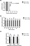
Fig. 1
Fig. 1
Prolactin treatment of MCF-7 breast cancer cells induced a more differentiated phenotype: MCF-7 cells were treated or left untreated with PRL (250 ng/ml) for 72 h. The mRNA expression levels of epithelial marker panel (A), EMT transcription markers panel (B), and proliferative & stemness markers panel (C) were measured using q-RT-PCR in both MCF-7/-PRL (ctrl) and MCF-7/ + PRL (tested) cells. Results are expressed as relative expression of duplicates of three independent experiments. ER-alpha (*p = 0.011), CK-18 (**p = 0.0076), CD-24 (**p = 0.0098), PRLR (***p = 0.00005), MUC-1 (**p = 0.0010), ECAD (p = 0.422), HER-2 (p = 0.085), PR (p = 0.422), SNAI (**p = 0.0076), SLUG (*p = 0.013), ZEB-1 (*p = 0.025), FN1 (p = 0.214), NCAD (p = 0.138), VIM (p = 0.353), ZEB-2 (p = 0.095), TWIST (p = 0.420), Ki-67 (*p = 0.025), CD-44 (*p = 0.030), and PCNA (p = 0.423), ns: not significant (multiple unpaired t-test)
Fig. 2
Fig. 2
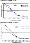
Prolactin treatment of MCF-7 breast cancer cells enhanced the cellular responses to the Tamoxifen treatment: MCF-7 cells were treated or left untreated with PRL (250 ng/ml) for 72 h panel (A) or 21 days panel (B). This was followed by treatment with TAM with different concentrations (0–100) mM for 72 h. Response to varying doses of TAM was measured using SRB assay at the OD of 490 nm. The IC50 values of TAM after 72 h of treatment were determined in both the control (MCF-7/-PRL + TAM) and the tested (MCF-7/ + PRL + TAM) breast cancer cells. Results are expressed as a percentage of cell viability of six replicates of three independent experiments, ****p < 0.0001(two-way ANOVA), panels (A) and (B)
Fig. 3
Fig. 3
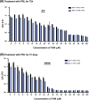
Prolactin treatment of MCF-7 breast cancer cells reduced the cellular viability (proliferative capacity) and improved the responses to the Tamoxifen treatment: MCF-7 cells were treated or left untreated with PRL (250 ng/ml) for 72 h panel (A) or 21 days panel (B). This was followed by treatment with TAM with different concentrations (0–100) mM for 72 h. Cell viability inhibition by various doses of TAM was also measured using an MTT assay at an OD of 570 nm. Results are expressed as mean ± SEM of six replicates of three independent experiments, panels (A) and (B), **p = 0.0089 and ****p < 0.0001(two-way ANOVA), respectively
Fig. 4
Fig. 4
Prolactin receptor expression positively correlates with ESR1 expression in breast cancer patients: The PRLR-ESR1 interaction map shows different degrees of correlation strength by bcGenExMiner, left and middle panels. Right panel: Pearson pairwise correlation graphs show high degree of positively correlated interaction between PRLR and ESR1 genes, r = 0.51 and ****p < 0.0001in all breast cancer patients (4421), panel (A), r = 0.32 and ****p < 0.0001in ER-positive breast cancer patients (3685), panel (B), and r = 0.37 and ****p < 0.0001in ER-negative breast cancer patients (510), panel (C). Source: Database of The Breast Cancer Gene-Expression Miner v4.8 (bcGenExMiner v4.8) (http://bcgenex.ico.unicancer.fr/BC-GEM/GEM-Accueil.php?js=1) [56]
Fig. 5
Fig. 5
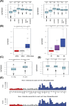
Co-expression of PRLR/ESR1 strongly correlated with well-differed and less aggressive breast cancer subtypes: PRLR-ESR1 gene signature correlation expression level according to breast cancer molecular subtypes, Basal, HER2 + , Luminal A, Luminal B, Normal like, and Unclassified by both HU and PAM50 subtypes classifications showing significant correlation level in Luminal subtypes/ HR + , ****p < 0.0001, panels (A) & (B). PRLR-ESR1 gene signature correlation expression level according to ER status, ER- and ER + breast cancers, panel (C). Panel (D), PRLR-ESR1 gene signature expression level in correlation with histological grades G1, G2, and G3. PRLR-ESR1 genes signature exposed the highest expression profile in breast cancer cell lines that are representative of the luminal (HR + /HER2-) subtype, and the lowest expression intensity was determined in breast cancer cell lines that are representative of basal (A and B) and TNBC subtypes, panel (E). Source: Gene expression-based Outcome for Breast Cancer (GOBO) online database (http://co.bmc.lu.se/gobo/) [54, 55]
Fig. 6
Fig. 6
Co-expression of PRLR/ESR1 associated with favourable prognosis in patients breast cancer patients: PRLR-ESR1 gene signature is significantly associated with prolong OS (p = 2.7^−08), RFS (p = 6.2^−16), and DMFS (p = 5.4^−09), in all breast cancer subtypes, panel (A) and in Luminal A subtype, OS (p = 0.00049), RFS (p = 0.00013), and DMFS (p = 0.0024), panel (B). Source: The Kaplan–Meier Plotter, (https://kmplot.com/analysis/) [53]

Similar articles

References

    1. Fitzmaurice C, et al. Global, regional, and national cancer incidence, mortality, years of life Lost, years lived with disability, and disability-adjusted life-years for 32 cancer groups, 1990 to 2015: a systematic analysis for the global burden of disease study. JAMA Oncol. 2017;3:524–48. 10.1001/jamaoncol.2016.5688. - PMC - PubMed
    1. Ginsburg O, et al. The global burden of women’s cancers: a grand challenge in global health. Lancet. 2017;389:847–60. 10.1016/s0140-6736(16):31392-7. - PMC - PubMed
    1. Anderson RL, et al. A framework for the development of effective anti-metastatic agents. Nat Rev Clin Oncol. 2019;16:185–204. 10.1038/s41571-018-0134-8. - PMC - PubMed
    1. Guiu S, et al. Molecular subclasses of breast cancer: how do we define them? The IMPAKT 2012 Working Group Statement. Ann Oncol. 2012;23:2997–3006. 10.1093/annonc/mds586. - PubMed
    1. Lumachi F, Brunello A, Maruzzo M, Basso U, Basso SM. Treatment of estrogen receptor-positive breast cancer. Curr Med Chem. 2013;20:596–604. 10.2174/092986713804999303. - PubMed

Grants and funding

LinkOut - more resources