. 2024 Dec 2;65(14):32.
doi: 10.1167/iovs.65.14.32.

Consensus Recommendations for Studies of Outflow Facility and Intraocular Pressure Regulation Using Ex Vivo Perfusion Approaches

Ted S Acott  1 Michael P Fautsch  2 Weiming Mao  3   4 C Ross Ethier  5 Alex S Huang  6 Mary J Kelley  1 Mini Aga  1 Sanjoy K Bhattacharya  7 Terete Borras  8 Diane Bovenkamp  9 Uttio Roy Chowdhury  2 Abbot F Clark  10 Mohammed I Dibas  11 Yiqin Du  12 Michael H Elliott  13 Jennifer A Faralli  14 Haiyan Gong  15 Samuel Herberg  16 Murray A Johnstone  17 Paul L Kaufman  18 Kate E Keller  1 Ruth A Kelly  19   20 David Krizaj  21 Markus H Kuehn  22 Hoi Lam Li  15 Raquel Lieberman  23 Shan C Lin  24 Yutao Liu  25 Fiona S McDonnell  26 Colleen M McDowell  18 Gillian J McLellan  18   27 Philip Mzyk  19   20 Kayarat Saidas Nair  28 Darryl R Overby  29 Donna M Peters  14 VijayKrishna Raghunathan  30 Ponugoti Vasantha Rao  19   31 Gavin W Roddy  2 Najam A Sharif  32 Myoung Sup Shim  19   20 Yang Sun  33 Benjamin R Thomson  34 Carol B Toris  35 Colin E Willoughby  36 Hao F Zhang  37 Thomas F Freddo  38 Rudolf Fuchshofer  39 Kamisha R Hill  23 Alireza Karimi  1 Krishnakumar Kizhatil  40 Casey C Kopcyznski  41 Paloma Liton  19 Gaurang Patel  10   42 Michael Peng  3   4 Padmanabhan P Pattabiraman  3 Ganesh Prasanna  30 Ester Reina-Torres  29 E Griffen Samples  43 John R Samples  44 Cynthia L Steel  45 Clemens A Strohmaier  46 Preeti Subramanian  9 Chenna Kesavulu Sugali  3   4 Joseph van Batenburg-Sherwood  29 Cydney Wong  5 Hannah Youngblood  23 Gulab S Zode  47 Elizabeth White  1 W Daniel Stamer  19   20
Affiliations

Consensus Recommendations for Studies of Outflow Facility and Intraocular Pressure Regulation Using Ex Vivo Perfusion Approaches

Ted S Acott et al. Invest Ophthalmol Vis Sci. .

Abstract

Intraocular pressure (IOP) elevation is the primary risk factor and currently the main treatable factor for progression of glaucomatous optic neuropathy. In addition to direct clinical and living animal in vivo studies, ex vivo perfusion of anterior segments and whole eyes is a key technique for studying conventional outflow function as it is responsible for IOP regulation. We present well-tested experimental details, protocols, considerations, advantages, and limitations of several ex vivo model systems for studying IOP regulation. These include: (1) perfused whole globes, (2) stationary anterior segment organ culture, (3) perfused human anterior segment organ culture, (4) perfused animal anterior segment organ culture, (5) perfused human corneal rims, and (6) perfused human anterior segment wedges. These methods, with due consideration paid to their strengths and limitations, comprise a set of very strong tools for extending our understanding of IOP regulation.


Conflict of interest statement

Disclosure: T.S. Acott, None; M.P. Fautsch, None; W. Mao, Arrowhead Pharmaceuticals (C); C.R. Ethier, None; A.S. Huang, None; M.J. Kelley, None; M. Aga, None; S.K. Bhattacharya, None; T. Borras, None; D. Bovenkamp, None; U.R. Chowdhury, None; A.F. Clark, None; M.I. Dibas, None; Y. Du, None; M.H. Elliott, None; J.A. Faralli, None; H. Gong, None; S. Herberg, None; M.A. Johnstone, None; P.L. Kaufman, None; K.E. Keller, None; R.A. Kelly, None; D. Krizaj, None; M.H. Kuehn, None; H.L. Li, None; R. Lieberman, None; S.C. Lin, None; Y. Liu, None; F.S. McDonnell, None; C.M. McDowell, None; G.J. McLellan, None; P. Mzyk, None; K.S. Nair, None; D.R. Overby, None; D.M. Peters, None; V.K. Raghunathan, None; P.V. Rao, None; G.W. Roddy, None; N.A. Sharif, None; M.S. Shim, None; Y. Sun, None; B.R. Thomson, None; C.B. Toris, None; C.E. Willoughby, None; H.F. Zhang, None; T.F. Freddo, None; R. Fuchshofer, None; K.R. Hill, None; A. Karimi, None; K. Kizhatil, None; C.C. Kopcyznski, None; P. Liton, None; G. Patel, None; M. Peng, None; P.P. Pattabiraman, None; G. Prasanna, None; E. Reina-Torres, None; E.G.Samples, None; J.R. Samples, None; C.L. Steel, None; C.A. Strohmaier, None; P. Subramanian, None; C.K. Sugali, None; J. van Batenburg-Sherwood, None; C. Wong, None; H. Youngblood, None; G.S. Zode, None; E. White, None; W.D. Stamer, None

Figures

Figure 1.
Figure 1.
Schematic diagram illustrating the experimental setup for perfusion of intact enucleated eyes. The eye, immersed in a water bath, is connected to both an exchange supply reservoir and an exchange collection reservoir. During fluid exchange, the exchange supply reservoir delivers perfusion medium to the eye, and the exchange collection reservoir collects the exchanged fluid. When the connections between the reservoirs and the eye are closed, the perfusion medium is continuously delivered to the eye by the computerized syringe pump. Simultaneously, the flow rate and eye pressure are monitored for measuring outflow facility. Solid black lines represent fluid connections, dashed black lines represent electrical connections, and red arrows denote two- or three-way stopcocks.
Figure 2.
Figure 2.
Dissection and preparation of human anterior segments for stationary or perfusion culture. Most investigators now carefully remove all the dark material seen covering the outflow pathway in the lower right panel without damaging the outflow pathway.
Figure 3.
Figure 3.
Setup mounting eye on flow cell.
Figure 4.
Figure 4.
Perfusion cell with eye mounted. Top view of a flow cell with human anterior segment mounted. A cross-sectional diagram showing fluid routes and anterior segment placement and clamping. This is the Johnson flow cell design based on a machined, modified Petri dish.
Figure 5.
Figure 5.
Constant flow syringe pump setup. An adjustable precision syringe pump can be used to produce low constant-volume perfusion. The flow rate can be adjusted as desired for the experimental design. Note that all syringe pumps have some misalignment of the lead screws, so that even a “constant flow” perfusion has some unsteadiness in the flow waveform due to the misalignment-induced oscillation of the pusher plate that contacts the syringe plungers. It is prudent to characterize the extent of pressure oscillation for each pump, such as by perfusing a fixed resistance such as a filter and measuring the resultant pressure waveform, which will be proportional to the flow waveform produced by the pump.
Figure 6.
Figure 6.
Perfusion system in the Fautsch Laboratory at Mayo Clinic (Rochester, MN). Shown is a humidified, 37°C, 5% CO2 incubator with PE60 tubing connecting the flow chambers to the syringe pump (blue box) and to a pressure transducer (on the right outside the chamber). It is notable that a tray/pan of water with antibacterial such as Lysol placed in the bottom of each incubator and changed regularly is essential to maintain 100% humidity and to avoid evaporation errors. The monitor indicates pressure transducer readings.
Figure 7.
Figure 7.
Exploded view of a modification of the Johnson and the Erickson-Lamy flow cells. There are four ports and flow channels through the pedestal with tubing nipples on the bottom. Unused ports can be filled with the included small plastic bolts, taking them out of play. This flow cell is machined from PVC, which is relatively resistant to solvent and heat exposure, and glued together or bonded by dissolving the two surfaces to be joined with a solvent such as butyl methacrylate or by acrylic cement. This flow cell is available through Apex Industries, Inc. (Tigard, OR; www/apexind.com; A-008309 Rev B01, Square Eye Cell Assembly–Clear PVC). Recently, for convenience, this cell has been flipped over and placed cornea side down on a small water glass for perfusion instead of cornea side up as is the traditional method. When used right-side up, as in the diagram, a sterile aluminum foil top is used to avoid contamination.
Figure 8.
Figure 8.
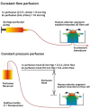
Two approaches to determining outflow facility: constant flow and constant pressure perfusion. The two methods use either constant perfusion rates to measure the resultant pressure with a pressure transducer or constant pressure to measure flow rate, usually gravimetrically. Both methods have advantages.
Figure 9.
Figure 9.
Balances and flow cells for continuous gravimetric assessment of flow in a constant pressure perfusion approach. Two balances weigh fluid in 50-mL conical centrifuge tubes with flow cells inverted on a water glass (lower left). Left is at 2× and right is at 1× pressure. Balances are constantly read at 2-minute intervals and data are inserted into an Excel file.
Figure 10.
Figure 10.
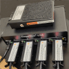
GageMux USB with foot pedal to initiate readings and four USB connections to read the four balances. Data are inserted into an Excel file on a connected laptop.
Figure 11.
Figure 11.
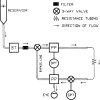
Improved outflow measurement device developed by Ethier et al. The reservoir is mounted on a screw drive to adjust the head. BT indicates a bubble trap, and PP indicates bubble purge ports. DPT is the pressure transducer that reads pressure across the steel tube, and GPT is a pressure transducer that reads equivalent to the IOP at the eye. Arrows show the direction of flow during a measurement.
Figure 12.
Figure 12.
The bovine anterior segment perfusion culture model. A perfusion cell similar to that shown in Figure 4 but scaled larger is shown with a bovine anterior segment attached.,
Figure 13.
Figure 13.
Corneal trephine transplant tissue perfusion culture model. (A, B) The perfusion plate. (C) The perfusion plate and holder. (D) The corneal tissue glued onto the plate. (E) Side view of the corneal tissue after being glued onto the plate. (F, G) Perfusion system setup. (H) Experimental timeline. (Reprinted from Peng M, Margetts TJ, Sugali CK, et al. An ex vivo model of human corneal rim perfusion organ culture. Exp Eye Res. 2022;214:108891.)
Figure 14.
Figure 14.
Glue contamination increased TM stiffness in perfusion cultured human corneal tissue. The stiffness (elastic modulus) of three corneal tissues with no (naïve), mild/non-visible (Glue-B), and severe (Glue-A) glue contamination. (Reprinted from Peng M, Margetts TJ, Sugali CK, et al. An ex vivo model of human corneal rim perfusion organ culture. Exp Eye Res. 2022;214:108891.)
Figure 15.
Figure 15.
(A) The Johnstone model of anterior segment wedge with cannula in Schlemm's canal. The positive pressure in Schlemm's canal is determined by the level of the pressure reservoir (0 or 30 mm Hg) relative to the fluid level in the dish. (B, C) OCT image at 0-mm Hg pressure head (B) and at 30-mm Hg positive pressure head (C). The pressure head can be varied rapidly to achieve dynamic responses. Scale bar: 200 µm. (Reprinted from Johnstone M, Xin C, Acott T, et al. Valve-like outflow system behavior with motion slowing in glaucoma eyes: findings using a minimally invasive glaucoma surgery-MIGS-like platform and optical coherence tomography imaging. Front Med (Lausanne). 2022;9:815866.)
Figure 16.
Figure 16.
Wedge flow for negative Schlemm's canal pressure studies. The pressure head is from the surface of the fluid in the Petri dish to the surface of the fluid in the perfusion reservoir: 0 mm Hg, 8.8 mm Hg (1×), or 17.6 mm Hg (2×). The anterior chamber pressure is higher than the Schlemm's canal pressure as in the normal eye.

References

    1. Quigley HA. Glaucoma. Lancet. 2011; 377: 1367–1377. - PubMed
    1. Quigley HA, Broman AT.. The number of people with glaucoma worldwide in 2010 and 2020. Br J Ophthalmol. 2006; 90: 262–267. - PMC - PubMed
    1. Weinreb RN, Leung CK, Crowston JG, et al. .. Primary open-angle glaucoma. Nat Rev Dis Primers. 2016; 2: 16067. - PubMed
    1. Stamer WD, Acott TS.. Current understanding of conventional outflow dysfunction in glaucoma. Curr Opin Ophthalmol. 2012; 23: 135–143. - PMC - PubMed
    1. Weinreb RN, Aung T, Medeiros FA.. The pathophysiology and treatment of glaucoma: a review. JAMA. 2014; 311: 1901–1911. - PMC - PubMed

Grants and funding