. 2024 Dec 2;65(14):30.
doi: 10.1167/iovs.65.14.30.

Retinal Degeneration Associated With Biallelic RDH12 Variants: Longitudinal Evaluation of Retinal Structure and Visual Function in Pediatric Patients

Affiliations

Retinal Degeneration Associated With Biallelic RDH12 Variants: Longitudinal Evaluation of Retinal Structure and Visual Function in Pediatric Patients

Tomas S Aleman et al. Invest Ophthalmol Vis Sci. .

Abstract

Purpose: The purpose of this study was to determine the natural history of the photoreceptor disease in a large group of pediatric patients with RHD12-associated Leber congenital amaurosis (RDH12-LCA), to estimate the changes expected over the duration of a clinical trial, and to define the relationship between the photoreceptor loss and visual dysfunction.

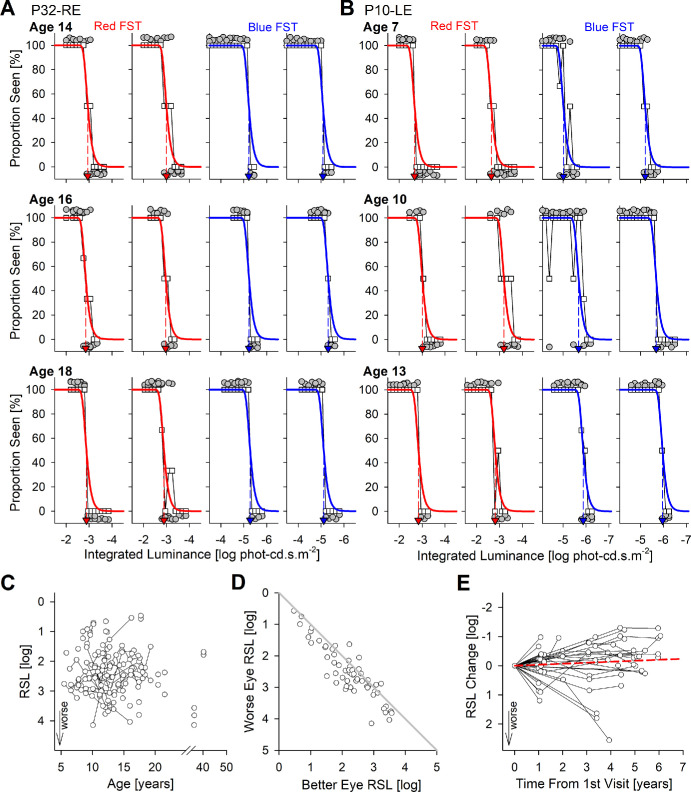
Methods: Forty-six patients representing 36 families were included. The great majority of patients were under the age of 18 years. Patients underwent complete ophthalmic examinations and imaging with various modalities including adaptive optics scanning laser ophthalmoscopy. Visual function was assessed with static and kinetic perimetry, and full-field stimulus test (FST) under dark- and light-adapted conditions.

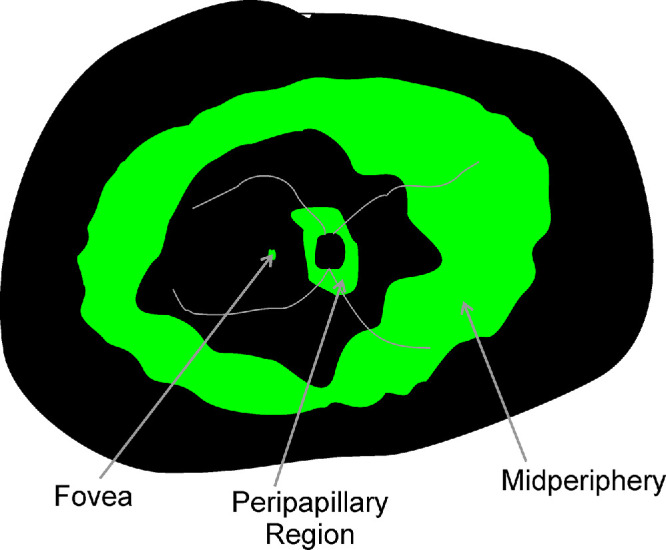
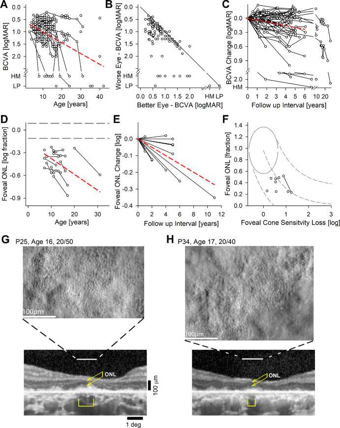
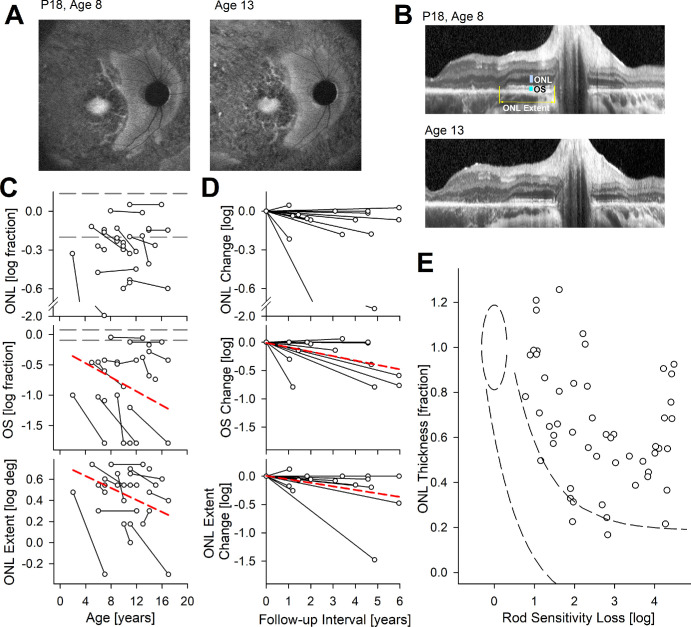
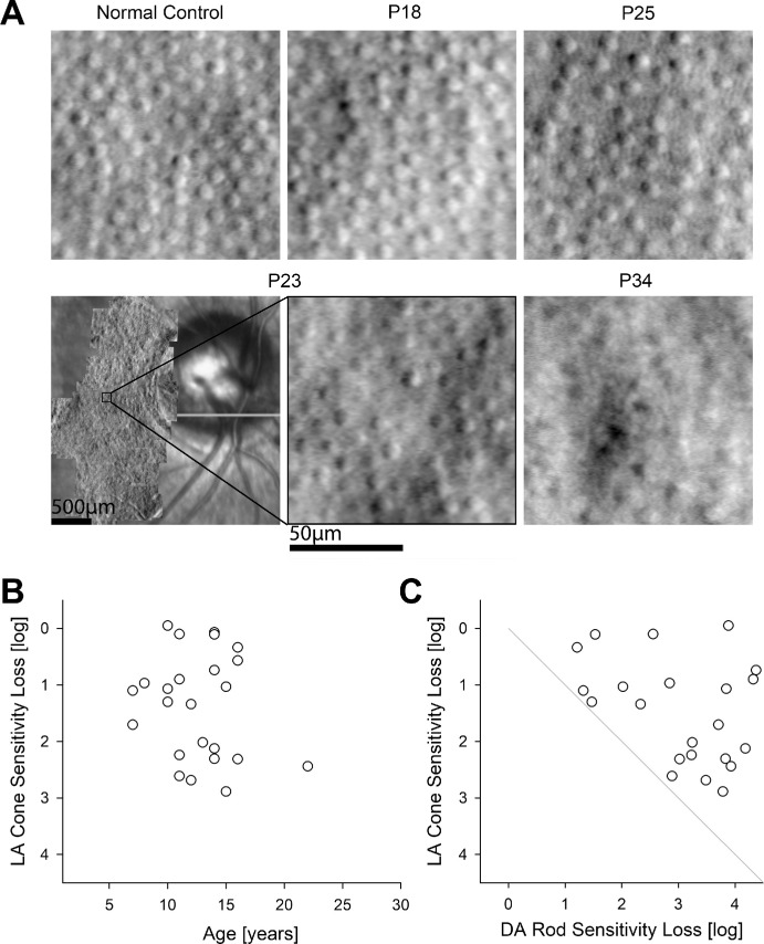
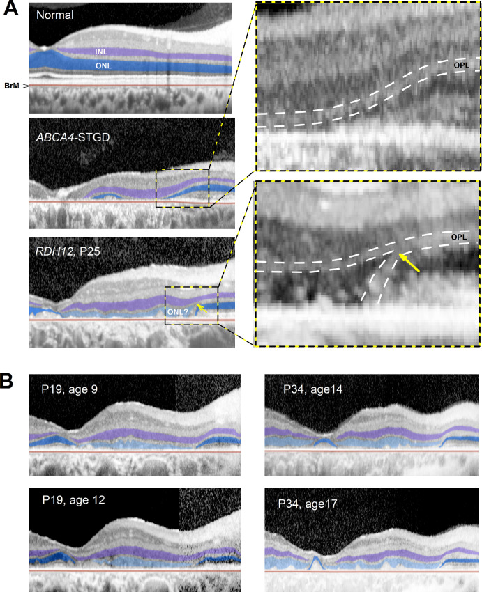
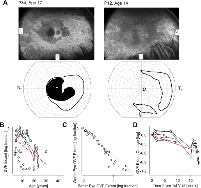
Results: Patients had a severe and early onset retinal degeneration (EORD). Visual acuity losses showed a progression rate of 0.04 logMAR per year. A small foveal island could be retained but showed degeneration over time. Foveal cone sensitivity losses were predictable by the loss of photoreceptors. Peripapillary retina could be retained with no significant progression detectable. Peripapillary rod sensitivity was substantially less than expected from photoreceptor structure pointing to a large improvement potential. FST sensitivities were reliably recordable in pediatric patients and showed a small but significant improvement with age. Locally and globally, loss of rod sensitivity tended to be larger than loss of cone sensitivity.

Conclusions: Foveal cones of RDH12-LCA should be targeted with treatments aimed to slow progression, whereas peripapillary rod photoreceptors should be targeted with treatments aimed to improve night vision. Pediatric FST testing may be complicated by age-related maturation of decision making regarding threshold criteria.


Conflict of interest statement

Disclosure: T.S. Aleman, None; A.J. Roman, None; K.E. Uyhazi, None; Y.Y. Jiang, None; E.C. Bedoukian, None; A. Sumaroka, None; V. Wu, None; M. Swider, None; I. Viarbitskaya, None; R.C. Russell, None; E.O. Shagena, None; A.J. Santos, None; L.W. Serrano, None; K.M. Parchinski, None; R.J. Kim, None; M.L. Weber, None; A.V. Garafalo, None; D.A. Thompson, None; A.M. Maguire, None; J. Bennett, None; D.H. Scoles, None; E.C. O'Neil, None; J.I.W. Morgan, (P) 8226236, 11607125, and US Patent App 63/497,933; A.V. Cideciyan, None

Figures

Figure 1.
Figure 1.
Schematic of retinal regions reported to be relatively preserved (green) in RDH12-LCA: foveal region, parapapillary region, and peripheral region.
Figure 2.
Figure 2.
Best corrected visual acuity (BCVA) and foveal structure and function. (A) BCVA across all subjects, eyes, and visits. Connected symbols represent serial visits. (B) Interocular asymmetry of BCVA. The diagonal gray line represents equal acuity in both eyes. (C) Change of BCVA over time in the subset of eyes with serial visits. (D) Foveal ONL thickness in the subset of eyes with retained visual acuity. (E) Change of foveal ONL thickness over time in the subset of eyes with serial visits. The dashed red lines in panels A, C, D, and E represent mixed-model regression results that produced a statistically significant slope. (F) Relationship between foveal ONL thickness and foveal cone sensitivity loss (CSL). Symbols are the patients with RDH12-LCA. Normal variability is described by ellipses encircling the 95% confidence interval of a bivariate Gaussian distribution. Dashed lines indicate the idealized model of the relationship between structure and function in pure photoreceptor degenerations and the region of uncertainty that results by translating the normal variability along the idealized model. (G, H) Split detection AO images montaged at the foveal region of two patients with retained BCVA. Peak cone densities (based on a sliding 55 µm × 55 µm box) are 28,200, and 23,500 cones.mm−2 for P25 and P34, respectively. Below AO images are foveal OCTs. Horizontal lines demarcate the region corresponding to the AO, the brackets are the regions corresponding to RPE melanization, and the double arrows correspond to the ONL thickness.
Figure 3.
Figure 3.
Peripapillary structure and rod function. (A, B) Preserved peripapillary region on autofluorescence imaging (A) and on OCT (B) in a representative patient P18 at ages 8 and 13 years. Quantified measures of ONL thickness, OS thickness, and ONL extent are shown (B, upper). (C) Peripapillary ONL thickness, OS thickness, and ONL extent as a function of age. ONL and OS thicknesses presented as logarithm of fraction of mean normal values. Connected symbols represent serial visits. Horizontal gray dashed lines represent the normal range. (D) Change in ONL thickness, OS thickness, and ONL extent as a function of time from first visit. Dashed red lines in panels C and D represent mixed-model regression results that produced a statistically significant non-zero slope. (E) Relationship between peripapillary ONL thickness and rod sensitivity loss (RSL). Symbols are from 52 locations with measurable ONL in 14 patients with RDH12-LCA. Normal variability is described by ellipses encircling the 95% confidence interval of a bivariate Gaussian distribution. Dashed lines indicate the idealized model of the relationship between structure and function in pure photoreceptor degenerations and the region of uncertainty that results by translating the normal variability along the idealized model.
Figure 4.
Figure 4.
Peripapillary cones and function. (A) Split detection AO images montaged at the peripapillary region of four patients in comparison with normal. Cone densities were 7770, 7950, 6430, and 4260 cones.mm−2 for P18, P23, P25, and P34, respectively. For P23, peripapillary montage and the chosen region of interest are shown. (B) Peripapillary cone sensitivity loss measured with a white stimulus under light adapted (LA) condition in all available subjects, eyes, and visits. (C) Quantitative comparison of LA cone sensitivity loss with dark-adapted (DA) rod sensitivity loss co-localized in the peripapillary retina. The gray line is the equal rod and cone sensitivity loss.
Figure 5.
Figure 5.
ONL-like hyposcattering layer on OCT in the macula. (A) OCT scans from fovea toward the nasal retina along the horizontal meridian in normal, ABCA4-STGD, and RDH12-LCA eyes. Inner nuclear layer (INL) and outer nuclear layer (ONL) are highlighted with purple and blue, respectively. RDH12-LCA retina has an additional hyposcattering ONL-like layer that is highlighted with light blue. Insets show the magnified regions of transition between macular degeneration and peripapillary sparing. BrM, Bruch membrane; OPL, outer plexiform layer. (B) Longitudinal changes observed over 3 years in 2 patients demonstrating thickening (P19) and thinning (P34) of the ONL-like layer.
Figure 6.
Figure 6.
Peripheral function. (A) Ultra-wide-angle autofluorescence imaging showing mid- and far-peripheral retention of retina (upper row) and retained peripheral vision on kinetic perimetry (lower row) in 2 representative patients P34 and P12. Tr, Ir, and Nr represent temporal, inferior, and nasal retina, and Nf, If, and Tf represent nasal, inferior, and temporal visual fields, respectively. (B) The extent of the kinetic field across all subjects, eyes, and visits. Connected symbols represent serial visits. (C) Interocular asymmetry of kinetic field extent. The diagonal line represents equal extents in both eyes. (D) Logarithm of change of kinetic field extent over time in the subset of eyes with serial visits. The dashed red lines in panels B and D represent mixed-model regression results that produced a statistically significant non-zero slope.
Figure 7.
Figure 7.
Dark-adapted chromatic FST. (A, B) Raw FST data recorded serially at 3 ages in 2 representative pediatric patients P32 (A) and P10 (B). Red and blue colors denote respective stimuli used. Mean proportion seen (white squares) are fit with a modified Weibull function (smooth curves). Threshold is estimated at 50% seen (colored arrow). For each color, laterally paired panels show repeated tests. All yes/no responses are plotted jittered above and below the 100% and 0% lines, respectively (gray filled symbols). (C) Rod sensitivity loss (RSL) estimated from FST across all subjects, eyes, and visits. Greater RSL implies worse vision. Connected symbols represent serial visits. (D) Interocular asymmetry of RSL. The gray line represents equal results in both eyes. (E) Change of RSL over time in the subset of eyes with serial visits. Positive change represents worsening vision. The dashed red line represents mixed-model regression results that produced a statistically significant non-zero slope.

References

    1. den Hollander AI, Roepman R, Koenekoop RK, Cremers FPM.. Leber congenital amaurosis: genes, proteins and disease mechanisms. Prog Retin Eye Res. 2008; 27: 391–419. - PubMed
    1. Kumaran N, Moore AT, Weleber RG, Michaelides M.. Leber congenital amaurosis/early-onset severe retinal dystrophy: clinical features, molecular genetics and therapeutic interventions. Br J Ophthalmol. 2017; 101: 1147–1154. - PMC - PubMed
    1. Huang CH, Yang CM, Yang CH, Hou YC, Chen TC.. Leber's congenital amaurosis: current concepts of genotype-phenotype correlations. Genes (Basel). 2021; 12: 1261. - PMC - PubMed
    1. Russell S, Bennett J, Wellman JA, et al. .. Efficacy and safety of voretigene neparvovec (AAV2-hRPE65v2) in patients with RPE65-mediated inherited retinal dystrophy: a randomised, controlled, open-label, phase 3 trial. Lancet. 2017; 390: 849–860. - PMC - PubMed
    1. Russell SR, Drack AV, Cideciyan AV, et al. .. Intravitreal antisense oligonucleotide sepofarsen in Leber congenital amaurosis type 10: a phase 1b/2 trial. Nat Med. 2022; 28: 1014–1021. - PMC - PubMed

MeSH terms

Substances