Review
. 2024 Dec 24;43(12):115068.
doi: 10.1016/j.celrep.2024.115068. Epub 2024 Dec 16.

Central nervous system vascularization in human embryos and neural organoids

Affiliations
Review

Central nervous system vascularization in human embryos and neural organoids

Sarah M Boutom et al. Cell Rep. .

Abstract

In recent years, neural organoids derived from human pluripotent stem cells (hPSCs) have offered a transformative pre-clinical platform for understanding central nervous system (CNS) development, disease, drug effects, and toxicology. CNS vasculature plays an important role in all these scenarios; however, most published studies describe CNS organoids that lack a functional vasculature or demonstrate rudimentary incorporation of endothelial cells or blood vessel networks. Here, we review the existing knowledge of vascularization during the development of different CNS regions, including the brain, spinal cord, and retina, and compare it to vascularized CNS organoid models. We highlight several areas of contrast where further bioengineering innovation is needed and discuss potential applications of vascularized neural organoids in modeling human CNS development, physiology, and disease.

Keywords: CP: Stem cell research; angiogenesis; blood-brain barrier; central nervous system; endothelium; human pluripotent stem cell; microphysiological system; neural rosette; neuroepithelium; neurogenesis; neurovascular unit; organoid; perineural vascular plexus; periventricular plexus; vasculogenesis.


Conflict of interest statement

Declaration of interests R.S.A. is a co-owner of Neurosetta, LLC, which commercializes micropatterned neural rosette technology.

Figures

Figure 1.
Figure 1.. Anatomy and development of CNS vasculature
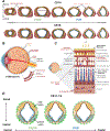
(A) The leftmost diagram shows the major vessels of the human embryo at CS 13 in relation to the forebrain, midbrain (Mb), hindbrain, and spinal cord (neural tube morphology outlined in yellow). Substructures of the nervous system, including the budding cranial nerves (CNI–XII) and cervical spinal nerve nuclei (C1–7) are also shown. The paired dorsal aortas, which run along the rostral-caudal axis of the embryo, give rise to the aortic arches and their derivatives, including the primitive internal carotid artery (ICA) and its finer branches. The primary head vein is also evident at this developmental stage. In the middle diagram, major brain arteries, including the circle of Willis, are shown in a sagittal view of a human embryo at CS 20. Vessels corresponding to the circle of Willis and its branches are shaded in white and labeled in green. The far-right diagram shows the transition of circle of Willis anatomy from CS 20 to CS 21. While most of the circle of Willis is well developed at CS 20, the anterior communicating artery (AComm) forms by CS 21. Vessels corresponding to those shown in the middle diagram are shaded in white. Left and middle diagrams are adapted from Bertulli and Robert and Raybaud. Right diagram is adapted from Takakuwa et al ACA, anterior cerebral artery; ICA, internal carotid artery; Mb, midbrain; MCA, middle cerebral artery; PCA, posterior cerebral artery; PCommA, posterior communicating artery. (B) The PNVP begins to develop at CS 9–10. Blood islands surrounding the developing neural tube form through the fusion of CD34+ hemangioblasts and subsequent differentiation into CD31+ endothelial progenitors. Further fusion results in PNVP formation. The diagram was created with BioRender. (C) Vascularization of the neural tube follows the directionality of neural tube closure, which begins at ~CS 10 at the midbrain/hindbrain boundary and proceeds rostrally and caudally from this closure point. A second putative neural tube closure initiation site is located in the rostral forebrain. PNVP vessels penetrate the neural tube between ~CS 14 and ~CS 23, based on variable literature reports. Vessel penetration proceeds in a ventral-to-dorsal direction and rostrally or caudally from respective neural tube closure sites. The top diagram created with BioRender. The bottom diagram was adapted from Wallingford et al. (D) A transverse section of the neural tube is shown with radial penetration of PNVP vessels toward the luminal surface (right side). A more detailed diagram (left side) shows the progression of penetrating vessels throughout early cortical plate development. The thickness of the neural tissue between the PNVP and the CSF increases over time, and neural progenitors present in the subventricular zone differentiate into neuronal subtypes that occupy deep and superficial cortical layers. Primitive vessels from the PNVP penetrate the glial limiting membrane, and extensions of the subarachnoid space, called the Virchow-Robin compartment, accompany the vessels along their path. Over the course of development, penetrating vessels also branch laterally and form connections with neighboring vessels. The right diagram was adapted from Raybaud. Both diagrams were created with BioRender.
Figure 2.
Figure 2.. Developmental vascular architecture of different CNS regions
(A) Forebrain: between CS 14 and CS 15, the periventricular plexus (PVP; shown in blue near the ventricle), which is initially present in the ventral telencephalon, spreads to the dorsal telencephalon. Vascularization by the PVP also proceeds in a caudal to rostral direction. The perineural vascular plexus (PNVP; shown in green) is present before the establishment of the PVP and forms connections with the PVP. Telencephalic coronal plane slices were adapted from portions of Carnegie embryo sections from the Virtual Human Embryo resource (Endowment for Human Development, https://www.ehd.org/virtual-human-embryo/). (B) Retina: before maturation of the retinal vasculature, two primary blood sources around the retina are the hyaloid artery and its branches (vasa hyaloidea propria), which supply blood to the developing lens and the surface of the inner retina, as well as the choriocapillaris, which supplies the retinal pigment epithelium and photoreceptors. The image was created with BioRender. (C) The retinal vasculature is organized into three layers: the superficial, intermediate, and deep vascular plexuses, located in the GCL and NFL, IPL, and between the INL and OPL, respectively. The image was created with BioRender. (D) Midbrain: the embryonic midbrain is vascularized between ~CS 10/11 and ~CS 14. Midbrain alar and basal plates are shown in yellow and teal, respectively. At this stage, the PNVP (shown in green) is present in all areas of the midbrain along the rostral-caudal axis, with penetrating vessels’ abundance increased in rostral versus caudal regions. The PVP (shown in blue) and its associated penetrating vessels progress in rostral-to-caudal and ventral-to-dorsal orientations as well. The images were created with BioRender.
Figure 3.
Figure 3.. Developmental and adult vascular architecture of different CNS regions, continued
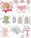
(A) Midbrain, continued: adult vascular structure of a cross-section of the rostral midbrain is shown. Alar or basal plate-derived structures are colored yellow or teal, respectively. The image was adapted from Mihailoff et al. (B) Hindbrain: during development, vessels from the perineural vascular plexus (PNVP) ingress into the neural tissue and grow radially toward the hindbrain ventricular surface. During extension, the vessels turn at near right angles and anastomose, forming a subventricular vascular plexus (SVP). (C) In the cerebellum, blood supply is provided by branches of the basilar and vertebral arteries (BAs and VAs, respectively). The BA branches into two paired arteries, the superior cerebellar artery (SCA) and anterior inferior cerebellar artery (AICA). The vertebral artery branches to form the posterior inferior cerebellar artery (PICA). (D) Schematic of microvasculature of the cerebellar cortex and subcortical white matter, focusing on a single gyrus. The figure was adapted from Akima et al. (E) Spinal cord: between ~CS 7 and ~CS 10, the PNVP has begun to form around the embryonic spinal cord via fusion of blood islands. At ~CS 14, a complete PNVP is seen surrounding the spinal cord, with angiogenic penetrating vessels entering the ventricular zone from the ventral aspect of the cord but avoiding the motor neuron columns. Vessel penetration into the developing spinal cord proceeds in a ventral-to-dorsal orientation, and by ~CS 18, vessels derived from the PNVP are also observed in the MN columns. (F) Choroid plexus (ChP): the ChP villous structure containing vasculature is shown. The choroid epithelium acts as an interface between the CSF and the choroid vasculature (blood-CSF barrier). The fenestrated choroid vasculature lacks tight junctions characteristic of a BBB. Immune cells are shown interacting with both ChP epithelium and endothelium. (G) Schematic of the lateral, third, and fourth ventricles in the adult human brain, along with the main arterial branches of the circle of Willis, which supply the ChPs associated with each ventricle. Dotted arrows from the arteries to the brain are colored according to targeted plexus, and the relative contributions of specific arteries are indicated by dotted line weight. The images were created at least partially with BioRender.
Figure 4.
Figure 4.. Current methods for vascularizing neural organoids
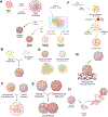
(A) Protocols for generation of generic and region-specific neural organoids. To generate 3D neural organoids, hPSC aggregates are cultured with or without (w/wo) Matrigel encapsulation or in the presence of small-molecule SMAD signaling inhibitors, which induces neural commitment. The organoids are typically cultured in agitated suspension culture, and the resulting structures contain numerous ventricular-like zones, a.k.a., neural rosettes. Incorporating combinations of Wnt activation or inhibition, Shh, FGF, RA, insulin, or chemokine receptor (SDF-1) signaling enables derivation of organoids corresponding to different brain regions (e.g., forebrain, midbrain, hindbrain, and spinal cord). (B) Co-culture of hPSC-derived NPCs and ECs (either hPSC derived or primary) in a 3D format can produce neural organoids with a primitive endothelial network. (C) ECs suspended within a hydrogel can invade an encapsulated neural organoid and self-assemble into a vascular network. (D) Neural organoids derived from hPSCs typically exhibit multiple rosette structures. It is less common to find a neural organoid that contains a single rosette. (E) Vascular-neural assembloids can be created by combining an avascular neural organoid and vascular organoid in a culture medium that supports both tissue types and permits their fusion. Vessels from the vascular organoid invade the neural organoid. (F) Neural organoids with rudimentary vessels can be generated using hPSCs gene edited to enable doxycycline-inducible overexpression of ETV2. When exposed to extrinsic culture conditions which promote neural differentiation and doxycycline, the co-aggregated and unedited hPSCs give rise to neural phenotypes, and the edited hPSCs differentiate into ECs. The result is co-generation of a neural organoid with a primitive network of endothelial vessels. (G) EC vascular beds formed within hydrogels can be used to vascularize co-cultured hPSC-derived neural organoids. (H) Neural organoids derived from hPSCs in vitro can be transplanted into the brain of an animal host to induce vascularization of the organoid, resulting in a perfusable, host-derived vascular network. All diagrams were created with BioRender.

References

    1. Bertulli L, and Robert T (2021). Embryological development of the human cranio-facial arterial system: a pictorial review. Surg. Radiol. Anat. 43, 961–973. 10.1007/s00276-021-02684-y. - DOI - PMC - PubMed
    1. Raybaud C (2010). Normal and Abnormal Embryology and Development of the Intracranial Vascular System. Neurosurg. Clin. N. Am. 21, 399–426. 10.1016/j.nec.2010.03.011. - DOI - PubMed
    1. Hiruma T, Nakajima Y, and Nakamura H (2002). Development of pharyngeal arch arteries in early mouse embryo. J. Anat. 201, 15–29. 10.1046/j.1469-7580.2002.00071.x. - DOI - PMC - PubMed
    1. Takakuwa T, Koike T, Muranaka T, Uwabe C, and Yamada S (2016). Formation of the circle of Willis during human embryonic development. Congenit. Anom. (Kyoto) 56, 233–236. 10.1111/cga.12165. - DOI - PubMed
    1. Wallingford JB, Niswander LA, Shaw GM, and Finnell RH (2013). The continuing challenge of understanding, preventing, and treating neural tube defects. Science 339, 1222002. 10.1126/science.1222002. - DOI - PMC - PubMed

Publication types

MeSH terms

LinkOut - more resources