. 2024 Dec 18;19(12):e0315858.
doi: 10.1371/journal.pone.0315858. eCollection 2024.

Paracentrotus lividus sea urchin gonadal extract mitigates neurotoxicity and inflammatory signaling in a rat model of Parkinson's disease

Affiliations

Paracentrotus lividus sea urchin gonadal extract mitigates neurotoxicity and inflammatory signaling in a rat model of Parkinson's disease

Nehal Shawky Nagy et al. PLoS One. .

Abstract

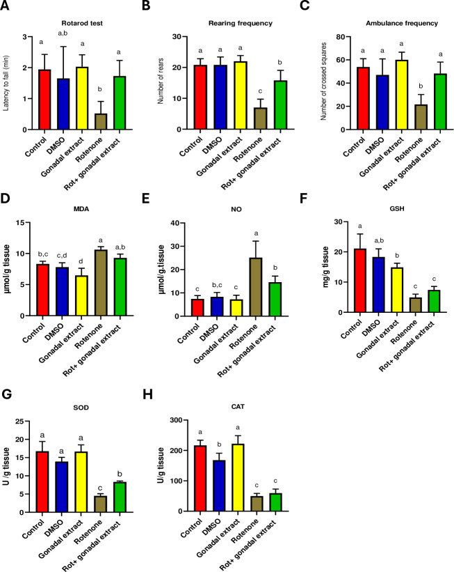
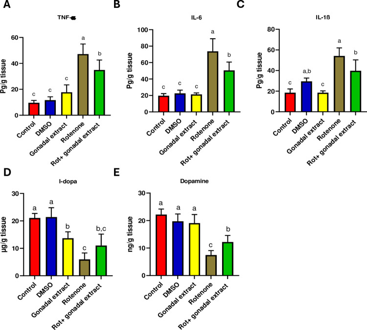
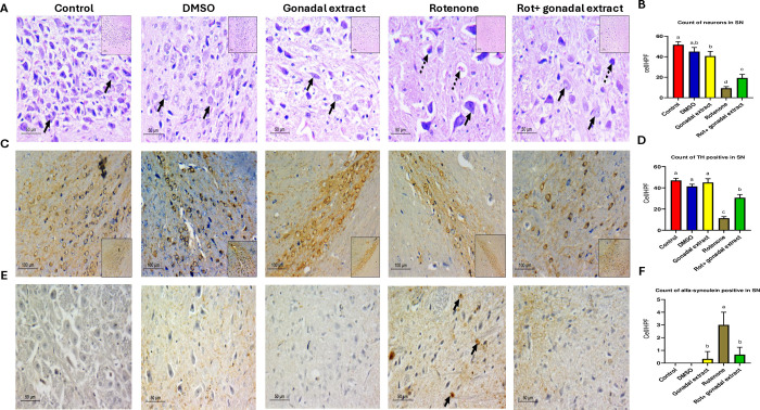
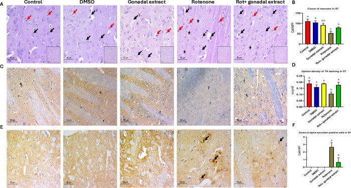
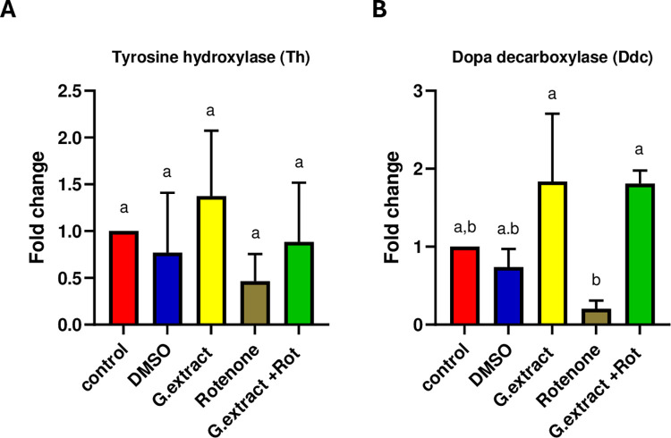
The present study investigates the neuroprotective effects of the sea urchin Paracentrotus lividus gonadal extract on rotenone-induced neurotoxicity in a Parkinson's disease (PD) rat model. Parkinson's disease, characterized by the progressive loss of dopaminergic neurons in the substantia nigra (SN), is exacerbated by oxidative stress and neuroinflammation. The study involved fifty Wistar rats divided into five groups: control, dimethyl sulfoxide (DMSO) control, Paracentrotus lividus gonadal extract-treated, rotenone-treated, and combined rotenone with Paracentrotus lividus gonadal extract-treated. Behavioral assessments included the rotarod and open field tests, while biochemical analyses measured oxidative stress markers (malondialdehyde (MDA), nitric oxide (NO), glutathione (GSH)), antioxidants (superoxide dismutase (SOD), catalase (CAT)), pro-inflammatory cytokines (interleukin-1 beta (IL-1β), interleukin-6 (IL-6), tumor necrosis factor-alpha (TNF-α)), and neurotransmitters (dopamine (DA), levodopa (L-Dopa)). Histological and immunohistochemical analyses evaluated the neuronal integrity and tyrosine hydroxylase (TH) and alpha-synuclein expression. The results showed that Paracentrotus lividus gonadal extract significantly mitigated rotenone-induced motor deficits and improved locomotor activity. Biochemically, the extract reduced oxidative stress and inflammation markers while enhancing antioxidant levels. Histologically, it restored neuronal integrity and reduced alpha-synuclein accumulation. Molecularly, it increased tyrosine hydroxylase and dopa decarboxylase gene expression, essential for dopamine synthesis. These findings suggest that Paracentrotus lividus gonadal extract exerts neuroprotective effects by modulating oxidative stress, neuroinflammation, and dopaminergic neuron integrity, highlighting its potential as a therapeutic agent for Parkinson's disease.


Conflict of interest statement

The authors have declared that no competing interests exist.

Figures

Fig 1
Fig 1
(A-C): Effect of P. lividus gonadal extract on the behavioral alternation induced by rotenone in rats. (A) Rotarod test scores. (B&C) Open field test, measuring rearing frequency & Ambulance frequency. (D-H): Effect of P. lividus gonadal extract on rotenone-induced oxidative impairments in nigrostriatal tissue of rats. Oxidative stress markers (MDA, NO, and GSH) and antioxidant enzymes (SOD and CAT) were determined. All the data were analyzed using one-way ANOVA followed by Tukey Pairwise Comparisons. Values are expressed as mean ± SEM; n = 5 rats for each group. Different superscripts on the columns are significantly different at p≤0.05.
Fig 2
Fig 2. Effect of P. lividus gonadal extract on rotenone-induced neuroinflammation and its effect on levels of L-dopa and dopamine in nigrostriatal tissue of male rats.
Proinflammatory cytokines (TNF-α, IL-6 and IL-1β) levels were measured. All the data were analyzed using one-way ANOVA Tukey Pairwise Comparisons. Values are expressed as mean ± SEM; n = 5 rats for each group. Different superscripts on the columns are significantly different at p≤0.05.
Fig 3
Fig 3
A) Pathologic assessment of H&E-stained sections of substantia nigra in different studied groups: control, DSMO, and gonadal extract groups show a compact cellular substania nigra in low power, high power shows viable neurons with large, rounded nuclei with open chromatin and a nucleolus (arrows). The Rotenone group shows less cellular loose substantia nigra, and high power shows degenerated neurons with dark stained nuclei (Dashed arrows) with few viable ones (arrows). The gonadal extract treated rotenone group shows restoration of neurons in SN, high power shows viable neurons (arrows), and fewer degenerated ones (dashed arrows). (H&E, low power x200, scale bar = 100 microns, high power x400, scale bar = 50 microns). B) Count of neurons in substantia nigra. C) Pathologic assessment of tyrosine hydroxylase-stained sections of substantia nigra in different studied groups highlighting positive neurons in each group. Many dopaminergic neurons are seen in deep brown backgrounds in control, DSMO, and gonadal extract groups. The Rotenone group shows a markedly diminished number of positive staining neurons. The gonadal extract-treated rotenone group shows increased expression (IHC, x200, scale bar 100 microns, inset x100). D) Count of TH-positive neurons in substantia nigra. E) Pathologic assessment of α- synuclein stained sections of substantia nigra in different studied groups highlighting positive neurons in each group. Negative staining of neurons is seen in control, DSMO, and gonadal extract groups. The Rotenone group shows multiple positive α—synuclein neurons (arrows) and the gonadal extract-treated rotenone group shows decreased expression. (IHC, x400, scale bar 50 microns). F) Count of α-synuclein positive in substantia nigra. All the data were analyzed using one-way ANOVA followed by Tukey Pairwise Comparisons. Values are expressed as mean ± SEM; n = 3 rats for each group. Different superscripts on the columns are significantly different at p≤0.05.
Fig 4
Fig 4
A) Pathologic assessment of H&E-stained sections of the striatum in different studied groups: control, DSMO, and gonadal extract groups show a cellular striatum, high power shows viable neurons with large, rounded nuclei with open chromatin and a nucleolus (black arrows), few microglial cells (red arrows) and thin capillaries are seen (v). The rotenone group shows evident disturbed architecture and hypocellularity, high power shows few viable neurons (black arrows), and multiple degenerated neurons with dark stained nuclei (Dashed arrows), the background shows vacuolated neuropil (star) and increased microglial cells (red arrows). The gonadal extract-treated rotenone group shows improvement of architecture of the striatum, high power shows viable neurons (arrows) with residual focal neuropil vacuolation (star), and few microglial cells are seen (red arrows) (H&E, low power x200, scale bar = 100 microns, high power x400, scale bar = 50 microns). B) Count of neurons in the striatum in different groups. C) Pathologic assessment of tyrosine hydroxylase-stained sections of the striatum in different studied groups highlighting positive dopaminergic terminals in each group. A high-density deep brown background is seen in the control, DSMO, and gonadal extract groups. The Rotenone group shows decreased staining density, while the gonadal extract-treated rotenone group shows increased staining. (IHC, x200, scale bar 100 microns, inset x100). D) Optical density of TH staining in the striatum. E) Pathologic assessment of α- -synuclein stained sections of striatum in different studied groups highlighting positive neurons in each group. Negative staining of neurons is seen in control, DSMO, and gonadal extract groups. The Rotenone group shows multiple positive α—synuclein neurons (arrows), while the gonadal extract-treated rotenone group shows decreased expression. (IHC, x400, scale bar 50 microns). F) Count of α—synuclein positive cells in the striatum. All the data were analyzed using one-way ANOVA followed by Tukey Pairwise Comparisons. Values are expressed as mean ± SEM; n = 3 rats for each group. Different superscripts on the columns are significantly different at p≤0.05.
Fig 5
Fig 5. Gene expression of tyrosine hydroxylase (Th) and dopa decarboxylase (Ddc) after treatment with P. lividus gonadal extract and RT-qPCR were performed in triplicates.
All the data were analyzed using one-way ANOVA followed by Tukey Pairwise Comparisons. Values are expressed as mean ± SEM. Different superscripts on the columns are significantly different at p≤0.05.

Similar articles

References

    1. Jenner P. Oxidative stress in Parkinson’s disease. Annals of Neurology. 2003;53(S3):S26–S38. doi: 10.1002/ana.10483 - DOI - PubMed
    1. Manoharan S, Guillemin GJ, Abiramasundari RS, Essa MM, Akbar M, Akbar MD. The Role of Reactive Oxygen Species in the Pathogenesis of Alzheimer’s Disease, Parkinson’s Disease, and Huntington’s Disease: A Mini Review. Oxidative Medicine and Cellular Longevity. 2016;2016:8590578. doi: 10.1155/2016/8590578 - DOI - PMC - PubMed
    1. Wei Z, Li X, Li X, Liu Q, Cheng Y. Oxidative Stress in Parkinson’s Disease: A Systematic Review and Meta-Analysis. Frontiers in Molecular Neuroscience. 2018;11. doi: 10.3389/fnmol.2018.00236 - DOI - PMC - PubMed
    1. Ball N, Teo W-P, Chandra S, Chapman J. Parkinson’s Disease and the Environment. Frontiers in Neurology. 2019;10. doi: 10.3389/fneur.2019.00218 - DOI - PMC - PubMed
    1. Poewe W, Seppi K, Tanner CM, Halliday GM, Brundin P, Volkmann J, et al.. Parkinson disease. Nature Reviews Disease Primers. 2017;3(1):17013. - PubMed

MeSH terms