. 2024 Dec 24;121(52):e2405824121.
doi: 10.1073/pnas.2405824121. Epub 2024 Dec 18.

Complement C3d enables cell-mediated immunity capable of distinguishing spontaneously transformed from nontransformed cells

Affiliations

Complement C3d enables cell-mediated immunity capable of distinguishing spontaneously transformed from nontransformed cells

Jeffrey L Platt et al. Proc Natl Acad Sci U S A. .

Abstract

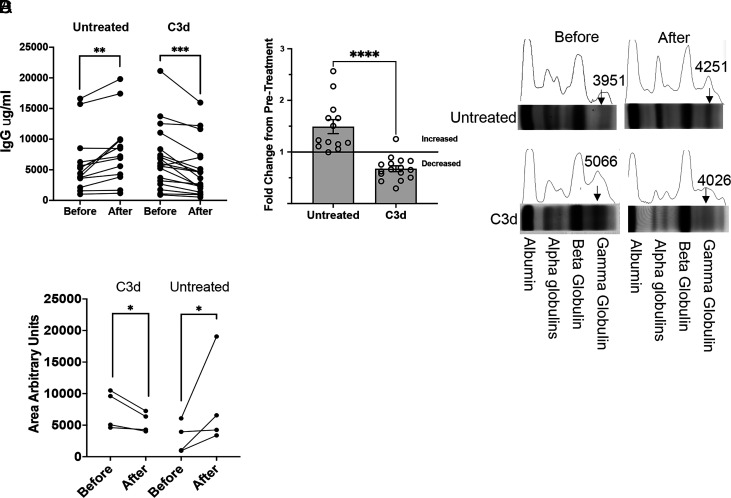
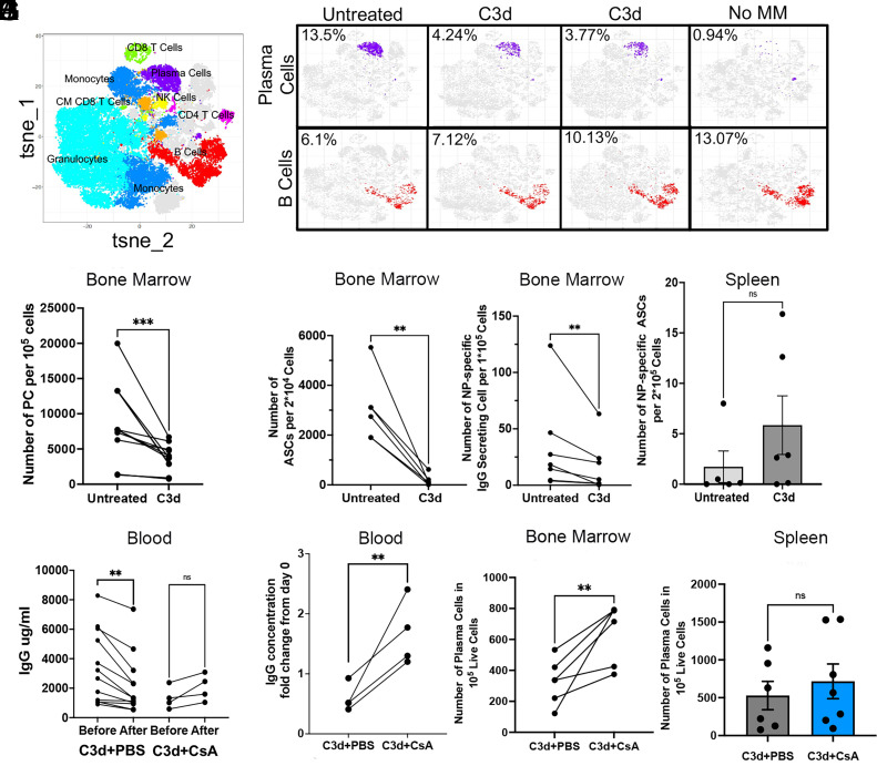
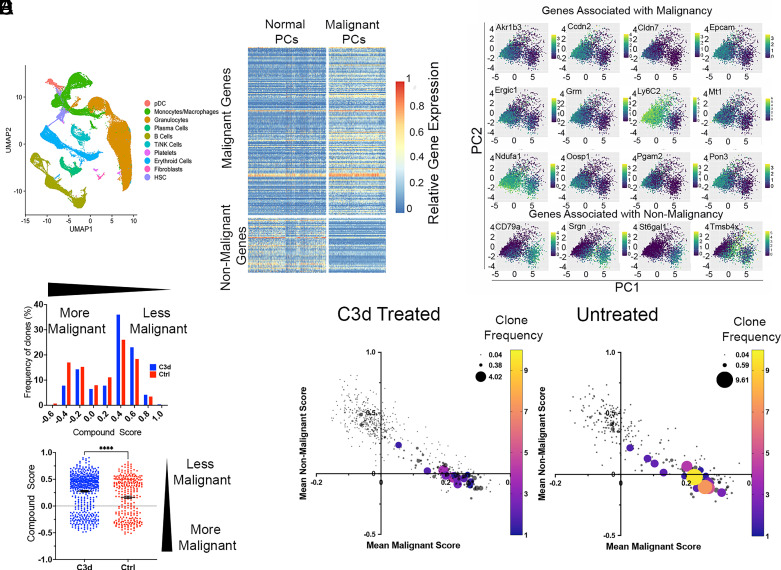
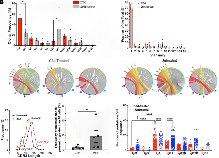
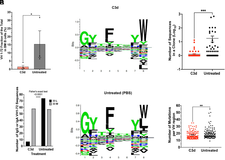
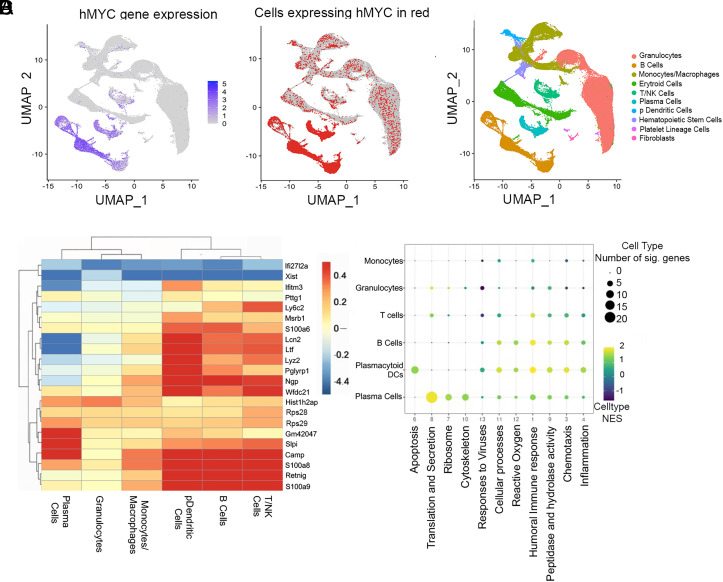
Immune surveillance depends in part on the recognition of peptide variants by T cell antigen receptors. Given that both normal B cells and malignant B cells accumulate mutations we chose a murine model of multiple myeloma to test conditions to induce cell-mediated immunity targeting malignant plasma cell (PC) clones but sparing of normal PCs. Revealing a previously unknown function for intracellular C3d, we found that C3d engaged T cell responses against malignant PC in the bone marrow of mice that had developed multiple myeloma spontaneously. Our results show that C3d internalized by cells augments immune surveillance by several mechanisms. In one, C3d induces a master transcription regulator, E2f1, to increase the expression of long noncoding (lnc) RNAs, to generate peptides for MHC-I presentation, and increase MHC-I expression. In another, C3d increases expression of RNAs encoding ribosomal proteins linked to processing of defective ribosomal products that arise from noncanonical translation and known to promote immunosurveillance. Cancer cells are uniquely susceptible to increased expression and presentation of mutant peptides given the extent of protein misfolding and accumulation of somatic mutations. Accordingly, although C3d can be internalized by any cell, C3d preferentially targets malignant clones by evoking specific T cell-mediated immunity and sparing most nontransformed polyclonal B cells and PC with lower mutation loads. Malignant PC deletion was blocked by cyclosporin or by CD8 depletion confirming that endogenous T cells mediated malignant clone clearance. Besides the potential for therapeutic application our results highlight how intracellular C3d modifies cellular metabolism to augment immune surveillance.

Keywords: Cell-mediated immunity; Complement 3 d; immune-surveillance.


Conflict of interest statement

Competing interests statement:C3d Immunotherapies” Patent submitted on 04/18/2023 “C3d cellular and acellular vaccines for the prevention and treatment of cancer.” PCT NO:US-01/34733-converted and awarded in 2021.

Figures

Fig. 1.
Fig. 1.
C3d injection of Vk*myc mice decreases paraproteinemia. Vk*myc mice older and younger than 1 y with detected M-protein, were injected with 20 microg C3d peptide (N = 16) or PBS (N = 13) intratibia 2 mo before analysis. (A) and (B) Shown is the change in the blood IgG concentration in Vk*myc mice following C3d or control injection. (C) M protein (arrows) was detected by SPEP before and at the end of treatment. M protein, seen as a single narrow band in the gamma region of the gel decreases in mice treated with C3d. Instead, a broad band of polyclonal Ig appears. In control mice the narrow band increases relative to the albumin band. (D) The graph shows the area under the globulin curve comparing analysis of the blood proteins before and after treatment with C3d or with PBS. The units are arbitrary and were determined by selecting the respective areas in image J using the average between lower peaks as the Bottom line. (A) Comparisons were by the double-tailed paired t test. **, P < 0.01, ***, P < 0.001. (B) Comparison was by the two-tailed Mann–Whitney test. ****, P < 0.0001. (D) Comparisons were by the ratio paired T test, P = 0.03.
Fig. 2.
Fig. 2.
C3d induces CMI that in turn depletes malignant cells. Vk*myc mice older and younger than 1 y, with detected M-protein, were injected with 20 μg C3d peptide (N = 6) or PBS (N = 6) intratibia 2 mo before analysis. M protein was detected by SPEP. To accelerate the development of Multiple Myeloma, Vk*myc mice were immunized three times with 4-hydroxy-3-nitrophenyl (NP) conjugated to ovalbumin (NP-OVAL) once every month starting 3 mo before treatment with C3d. (A) Tsne plots of mass cytometry (CYTOF) indicating the various cell clusters. (B) Typical CYTOF tsne plots of plasma cell clusters in purple and B cells (not plasma cells) in red. CYTOF analysis of a representative C57BL/6 mouse of comparable age to the plots of the Vk*myc mice is shown in the far right (Control without MM). (C) Frequency of plasma cells detected by CYTOF of bone marrow. (DF) ELISPOT analysis of IgG antibody secreting cells (D) or of NP-specific IgG ASC (E) in BM, or of NP-specific B cells in the Spleen, SP (F), 2 mo after C3d peptide or with PBS injection intratibia. (GJ) Vk*myc mice treated as explained above were divided in two groups 7 d after C3d injection. One group was treated with Cyclosporin (CsA) (N = 5) administered at a concentration of 3 mg/kg twice weekly for 7 wk until end point, and another group treated with PBS (N = 12) 2 mo after C3d injection. (G and H) Serum IgG concentration before and after treatment. (IJ) Number of plasma cells determined by FACS counting CD19-, B220lo, CD138+ cells in 100,000 live cells in the bone marrow (I) or in the spleen (I). Analyses in (CE) and (GI) were by the double-tailed paired t test, reflecting paired samples. **, P < 0.01;***, P < 0.001. (F and J) Comparison was by the two-tailed Mann–Whitney test, reflecting nonpaired measurements. Significance was considered if P < 0.05.
Fig. 3.
Fig. 3.
C3d decreases malignant plasma cells sparing normal plasma cells. Vk*myc mice were injected with 20 microg C3d peptide (N = 6) or with PBS (N = 5) intratibia 2 mo before analysis. Malignancy and nonmalignancy associated gene expression was analyzed in 3,995 individual plasma cells obtained from the bone marrow. (A) UMAP plots of sc RNA gene expression depicting the different cell populations analyzed. (B) Graph depicts the differential expression (DE) of a set of malignancy associated or nonmalignancy associated genes in the upper and lower percentile 20 for each category and in each group of mice. Each horizontal line represents a gene, each vertical line represents a cell. (C) Graphs show PCA of the expression of selected genes in the upper and lower percentile 20 associated with malignancy or nonmalignancy in all the plasma cells analyzed from both C3d-treated and untreated mice. The genes chosen contributed most of the difference between malignant and nonmalignant cells. (D) Distribution of clones according to a compound score obtained by subtracting the malignant score value from the nonmalignant score value for each cell. Upper graph depicts a histogram with bins separated by 0.2 compound score units. Frequencies (Y-axis) are shown as a percent of the clones (obtained from C3d treated or controls) included in the analysis. Lower graph is a scatter plot of clones according to their compound score. Statistical analysis by the Mann–Whitney test yielded P < 0/0001, (****). (E) Two-dimensional plots depicting malignant (X- axis) and nonmalignant (Y-axis) gene expression scores from treated and untreated mice. The size and color of the circles represent the number of copies of each clonotype defined according to the CDR3 sequence, VH and JH identity.
Fig. 4.
Fig. 4.
C3d treatment decreases the frequency of highly mutated malignant B cell clones and Ig class switched B cells in the BM but increases the frequency of normal maturing B cells. Vk*myc mice were injected with 20 microg C3d peptide (N = 6) or with PBS intratibia 2 mo before analysis (N = 5). Single cell V(D)J sequences were obtained using the 10X genomics kits followed by NGS. IgH+L sequences were further analyzed using the ARGalaxy immune repertoire pipeline as well as the NCBI Ig Blast and IMGT suites. (A) Frequency distribution of Ig isotypes in Vk-MYC* mice treated or not (Unt) with C3d. Comparison of the frequencies of IgM-producing clones in the BM by the Mann–Whitney test of mice treated or not with C3d indicated P = 0.0157. IgG1 frequencies also differed according to the treatment (P = 0.0476 Mann–Whitney test). (B) IgVH family frequencies in B cells in the bone marrow of Vk-MYC* mice treated or not (Unt) with C3d. Differences in the frequencies according to treatment did not reach significance by multiple Mann–Whitney tests. (C and D) Typical Circos analysis (ARGalaxy immune repertoire pipeline) of the V-J recombination in H chains obtained from bone marrow of mice C3d treated (C) or not (D) with C3d. The ribbons represent the top most frequent VH/JH gene combinations and they are chosen by the program automatically. The width is proportional to the overall frequency of VH/JH combinations. Around the circle the colored bars reflect each JH or VH. Figure Shows there are fewer colored ribbons in C3d-treated mice (C) as compared to untreated (D) mice that have wider and/or more frequent VH-JH ribbons, indicative of clonal skewing. (E) CDR3 AA length distribution curves obtained from bone marrow B cells of mice treated or not with C3d. Untreated CDR3 lengths are skewed and show a peak of 8 AA length. Comparison of the curves by the Wilcoxon matched ranks test indicates an exact P value of 0.0092. The area under the curves (AUC) is also indicated. (F) The graph shows the frequency of Ig clones (larger than 10 cells) with VH mutation frequencies above 2%. Each dot represents the average frequency of mutated clonotypes in all the V(D)J sequences obtained from one mouse marrow. Data analysis by the Mann–Whitney test indicated a P = 0.0317. (G). Number of mutations per VH region in heavy chain sequences obtained from B cells isolated from the bone marrow of Vk-MYC* mice treated or not (Unt) with C3d. Each dot represents mutation frequencies in VH sequences in one cell. Each clonotype is counted only once. Frequencies were analyzed by ANOVA followed by the Kruskal–Wallis multiple comparisons test. Comparisons that yielded P < 0.0001 are noted by ****.
Fig. 5.
Fig. 5.
C3d induces clonal-specific deletion of antigen-specific MM cells. Vk*myc mice were immunized 3 times with 4-hydrox-3-nitrophenyl(acetyl) (NP) coupled to Ovalbumin with 2 mo intervals, starting at 8 to 10 wk. Mice were injected with 20 microg C3d peptide (N = 6) or with PBS (N = 5) intratibia 2 mo before analysis. Analysis of BM cells was by 10X genomics. (A) Graph depicts the frequency of VH1-72 usage in each mouse. VH1-72 is the main VH region recruited by NP antigens. Comparison was by the Mann–Whitney test, P = 0.0286. (B) The number of IgG or IgA VH1-72 sequences in clones obtained from mice treated or not with C3d was compared by Fisher’s Exact test, P < 0.0001. (C and D) Seq2Logo depicting the CDR1 aminoacid motifs in B cell clones with VH1-72 from mice treated (C) or not (D) with C3d. Stacks represent AA with the size of the letters proportional to their prevalence. Shown is the Kullback–Leibler Logo. AA below “0” depict depleted AA. Apparent is the depletion of L at position 8 (corresponding to AA33 in the VH sequence) in mice treated with C3d. The W33L mutation causes a 10X increase in affinity to the NP hapten. Seq2Logo is available at http://www.cbs.dtu.dk/biotools/Seq2Logo. (E) Graph depicts the size of each clone shown as the Log10 of the number of sequences in each clone (dot), (C3d N = 115; PBS N = 110). Clones were defined by 80% similarity in CDR3 AA, same VH and JH. Results were compared by the Mann–Whitney test, P = 0.0006. (F) Number of mutations per VH1-72 regions in heavy chain sequences obtained from B cells isolated from the bone marrow of Vk-MYC* mice treated or not with C3d. Each dot represents the VH mutation frequency/cell. Each clonotype is counted only once. Only VH regions with mutation frequencies above 10% are shown. Results were analyzed by ANOVA followed by the Kruskal–Wallis multiple comparisons test.
Fig. 6.
Fig. 6.
C3d increases expression of genes encoding ribosomal proteins and Long Noncoding RNAs. Vk*myc mice were injected with 20 microg C3d peptide (N = 6) or with PBS (N = 5) intratibia 2 mo before single cell RNA analysis of bone marrow cells. (A and B) UMAP plots of sc RNA gene expression depicting the different cell populations analyzed indicating hMYC expression to identify cells of the B cell lineage that activated hMYC. (C) Genes differentially expressed in C3d-treated relative to untreated mice in all the bone marrow cell populations indicated. Scale on the right refers to the Log2 fold changes. (D) GSEA pathway analysis of differentially expressed genes in various bone marrow cell populations. Major pathways are identified below and the cell types on the Left. The normalized enrichment score is shown in a colored scale and the number of genes differentially expressed in each pathway are represented in proportion to the size of the circle.

Update of

References

    1. Nielsen S. C. A., et al. B cell clonal expansion and convergent antibody responses to SARS-CoV-2. Res. Sq. 10, 27220 (2020) 10.21203/rs.3.rs-27220/v1. - DOI - PMC - PubMed
    1. Khodadoust M. S., et al. , Antigen presentation profiling reveals recognition of lymphoma immunoglobulin neoantigens. Nature 543, 723–727 (2017), 10.1038/nature21433. - DOI - PMC - PubMed
    1. Kleczko E. K., et al. , Immune checkpoint activity regulates polycystic kidney disease progression. JCI Insight 8, e161318 - PMC - PubMed
    1. Pio R., Corrales L., Lambris J. D., The role of complement in tumor growth. Adv. Exp. Med. Biol. 772, 229–262 (2014), 10.1007/978-1-4614-5915-6_11. - DOI - PMC - PubMed
    1. Markiewski M. M., Lambris J. D., Is complement good or bad for cancer patients? A new perspective on an old dilemma Trends Immunol. 30, 286–292 (2009), 10.1016/j.it.2009.04.002. - DOI - PMC - PubMed

MeSH terms

Substances

LinkOut - more resources