. 2024 Dec 24;121(52):e2411640121.
doi: 10.1073/pnas.2411640121. Epub 2024 Dec 18.

Corticosteroids reduce pathological angiogenesis yet compromise reparative vascular remodeling in a model of retinopathy

Affiliations

Corticosteroids reduce pathological angiogenesis yet compromise reparative vascular remodeling in a model of retinopathy

Masayuki Hata et al. Proc Natl Acad Sci U S A. .

Abstract

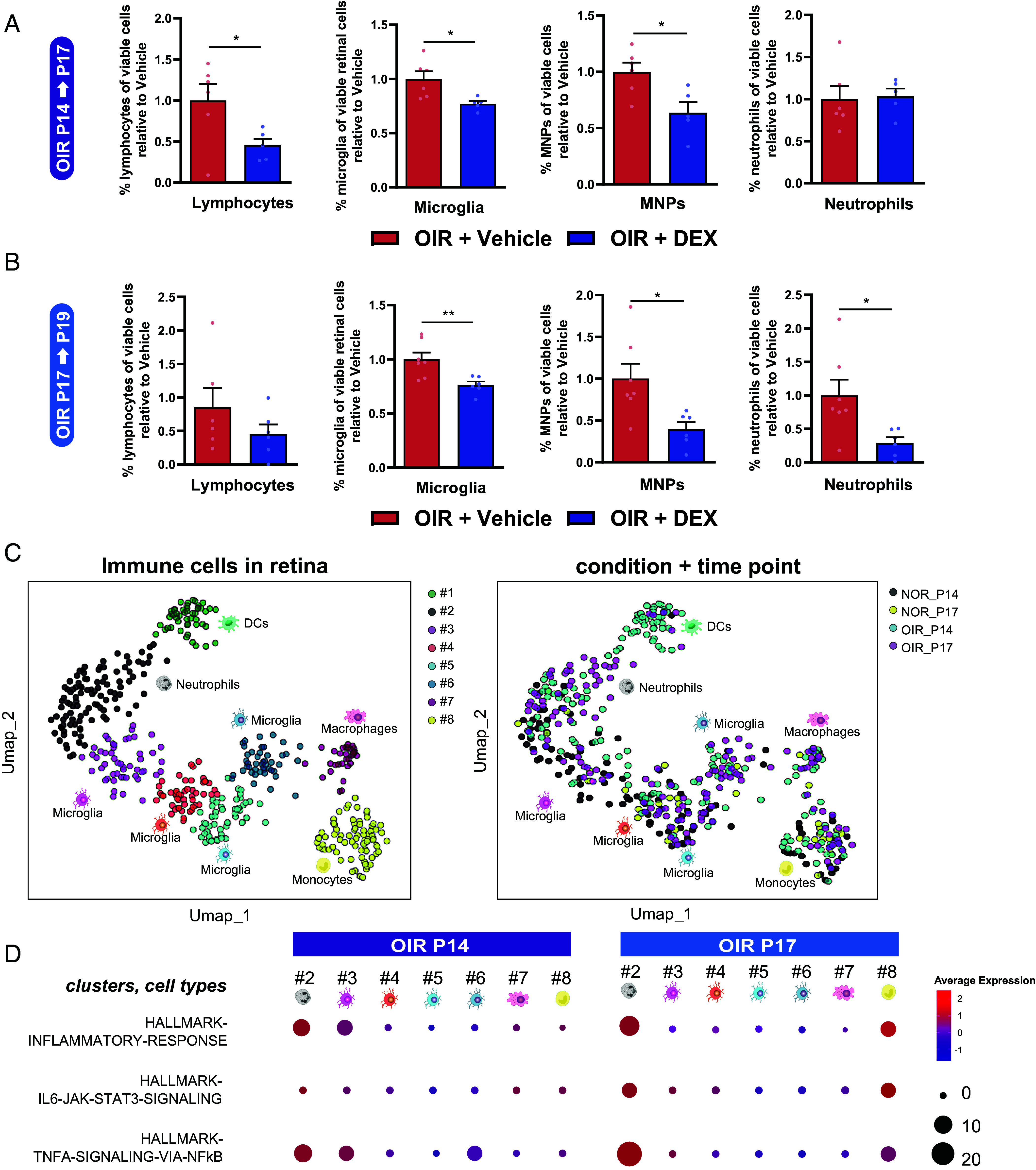
Tissue inflammation is often broadly associated with cellular damage, yet sterile inflammation also plays critical roles in beneficial tissue remodeling. In the central nervous system, this is observed through a predominantly innate immune response in retinal vascular diseases such as age-related macular degeneration, diabetic retinopathy, and retinopathy of prematurity. Here, we set out to elucidate the dynamics of the immune response during progression and regression of pathological neovascularization in retinopathy. In a mouse model of oxygen-induced retinopathy, we report that dexamethasone, a broad-spectrum corticosteroid, suppresses initial formation of pathological preretinal neovascularization in early stages of disease, yet blunts reparative inflammation by impairing distinct myeloid cell populations, and hence reduces beneficial vascular remodeling in later stages of disease. Using genetic depletion of distinct components of the innate immune response, we demonstrate that CX3C chemokine receptor 1-expressing microglia contribute to angiogenesis. Conversely, myeloid cells expressing lysozyme 2 are recruited to sites of damaged blood vessels and pathological neovascularization where they partake in a reparative process that ultimately restores circulatory homeostasis to the retina. Hence, the Janus-faced properties of anti-inflammatory drugs should be considered, particularly in stages associated with persistent neovascularization.

Keywords: angiogenesis; dexamethasone; inflammation; retina; retinopathy.


Conflict of interest statement

Competing interests statement:The authors declare no competing interest.

Figures

Fig. 1.
Fig. 1.
Intravitreal dexamethasone treatment suppresses pathological neovascularization in early stages but prevents reparative vascular remodeling in later stages of disease. (A) Schematic representation of the mouse model of OIR and the distinct phases of the pathological vascularization (vaso-obliteration from P7 to P12, neovascularization from P12 to P17, and neovascular regression from P17). (B) Dot plots of the top 15 enriched Gene ontologies (GO) terms related to the biological processes for bulk RNA-seq from OIR and normoxic mouse retinas at P14, P17, and P30 (N = 2 to 3 mice per condition). The size of the dots represents the number of genes in the significant DE gene list associated with the GO term, and the color of the dots represents the P-adjusted values. Inflammation-related GO terms are highlighted. (CF) mRNA expression of inflammation-related genes (C) Tnf, (D) Il6, (E) Ccl2, and (F) Il1b throughout the progression of OIR. Data are presented as fold change compared with P12 normoxic retinas (N = 3 to 10 depending on the group). Statistics were calculated comparing OIR versus normoxia for each given time point. (G) Schematic representation of intravitreal administration of dexamethasone (0.3 μg) to P14 pups during OIR. (H) mRNA expression of Tnf, Il6, Ccl2, and Il1b (N = 4 per condition) in P17 retinas of OIR treated with or without dexamethasone (OIR + DEX, OIR + Vehicle, respectively) relative to normoxia. (I) IBA1+ phagocytes colocalized with isolectin-B4+ ECs were found in P17 flatmounts of OIR treated with or without dexamethasone (DEX and Vehicle, respectively) in neovascular tuft areas and outside tuft areas (N = 16 to 24 depending on the group). (J and K) Neovascular area (J) and avascular area (K) as assessed at P19 flatmounts of OIR treated with or without dexamethasone (N = 13 to 15 depending on the group). (L) Schematic representation of intravitreal administration of dexamethasone (0.3 μg) to P17 pups during OIR. (M) mRNA expression of Tnf, Il6, Ccl2, and Il1b (N = 4 per condition) in P19 retinas of OIR treated with or without dexamethasone (OIR + DEX, OIR + Vehicle, respectively) relative to normoxia. (N) IBA1+ phagocytes colocalized with isolectin-B4+ ECs were found in P19 flatmounts of OIR treated with or without dexamethasone (DEX and Vehicle, respectively) in neovascular tuft areas and outside tuft areas (N = 12 to 24 depending on the group). (O and P) Neovascular area (O) and avascular area (P) as assessed at P19 flatmounts of OIR treated with or without dexamethasone (N = 14 to 17 depending on the group). Student’s unpaired t test (CF, IK and NP) and one-way ANOVA with Tukey’s multiple-comparison test (H and M) were used; *P < 0.05, **P < 0.01, ***P < 0.001, and ****P < 0.0001; error bars represent mean ± SEM.
Fig. 2.
Fig. 2.
Distinct populations of innate immune cells are elevated during neovascularization and vascular remodeling in ischemic retinopathy. (A and B) Flow cytometric analyses of OIR retinas treated with and without dexamethasone (OIR+DEX and OIR+Vehicle, respectively). Percentages of viable lymphocytes (CD45+/CD11b-/CD3ɛ+), microglia (CD45+/CD11b+/CX3CR1+), MNP (MNPs; CD45+/CD11b+/Ly6Cint/high/Ly6Glow), or neutrophils (CD45+/CD11b+/Ly6Cint/Ly6Ghigh) assessed at P17 (A) and P19 (B) by FACS (N = 5 to 7 depending on the group). (C) Uniform manifold approximation and projection (UMAP) plot for single-cell RNAseq of retinal immune cells from normoxic and OIR retinas at P14 and P17 (from two normoxic and three OIR sets of data). UMAP split according to immune cell populations (Left panel) and condition+timepoint (Right panel). Each dot represents one cell. (D) Dot plot representing results of Gene Set Variation Analysis (GSVA) for the inflammation-associated gene sets from Hallmark in each of the seven populations identified in (C) at P14 and P17 of OIR. Student’s unpaired t test (A and B) were used; *P < 0.05 and **P < 0.01; error bars represent mean ± SEM.
Fig. 3.
Fig. 3.
LysM+ monocytes mediate vascular remodeling in later stages of OIR while CX3CR1-expressing cells contribute to angiogenesis. (A) UMAP visualization of the expression of two major myeloid marker genes Lyz2 (Left panel) and Cx3cr1 (Right panel) in retinal immune cells during OIR. (B) Time course of Cx3cr1CreER/+ and Cx3cr1 CreER/+:R26iDTR/+ OIR mice. For both Cx3cr1 CreER/+ and Cx3cr1 CreER/+:R26 iDTR/+ mice, tamoxifen (TAM) was administered daily between P3 and P5, and diphtheria toxin intravitreally (ivt) at either P13 or P16. (C) Representative FACS plots of CX3CR1+ microglia of retinas from Cx3cr1 CreER/+ and Cx3cr1 CreER/+:R26 iDTR/+ mice following intravitreal diphtheria toxin injection. (D) Neovascular area and avascular area as assessed at P17 flatmounts of OIR treated with or without dexamethasone at P13 (N = 6 or 7 depending on the group). (E) Neovascular area and avascular area as assessed at P19 flatmounts of OIR treated with or without dexamethasone at P16 (N = 7 or 11 depending on the group). (F) Time course of LysMCre/+ and LysMCre/+:R26iDTR/+ OIR mice. For both LysMCre/+ and LysMCre/+:R26iDTR/+ mice, TAM was administered daily between P3 and P5, and diphtheria toxin ivt at either P13 or P16. (G) Representative FACS plots of MNPs of retinas from LysMCre/+ and LysMCre/+:R26iDTR/+ mice following intravitreal diphtheria toxin injection. (H) Neovascular area and avascular area as assessed at P17 flatmounts of OIR treated with or without dexamethasone at P13 (N = 6 or 8 depending on the group). (I) Neovascular area and avascular area as assessed at P19 flatmounts of OIR treated with or without dexamethasone at P16 (N = 4 or 8 depending on the group). Student’s unpaired t test (D, E, H and I) was used; *P < 0.05, **P < 0.01, and ***P < 0.001; error bars represent mean ± SEM.

References

    1. Yu C., Roubeix C., Sennlaub F., Saban D. R., Microglia versus monocytes: Distinct roles in degenerative diseases of the retina. Trends Neurosci. 43, 433–449 (2020). - PMC - PubMed
    1. Rashid K., Akhtar-Schaefer I., Langmann T., Microglia in retinal degeneration. Front. Immunol. 10, 1975 (2019). - PMC - PubMed
    1. Davies M. H., Stempel A. J., Powers M. R., MCP-1 deficiency delays regression of pathologic retinal neovascularization in a model of ischemic retinopathy. Invest. Ophthalmol. Vis. Sci. 49, 4195–4202 (2008). - PMC - PubMed
    1. Dejda A., et al. , Neuropilin-1 mediates myeloid cell chemoattraction and influences retinal neuroimmune crosstalk. J. Clin. Invest. 124, 4807–4822 (2014). - PMC - PubMed
    1. Marchetti V., et al. , Differential macrophage polarization promotes tissue remodeling and repair in a model of ischemic retinopathy. Sci Rep 1, 76 (2011). - PMC - PubMed

MeSH terms