. 2024 Dec 24;121(52):e2415048121.
doi: 10.1073/pnas.2415048121. Epub 2024 Dec 18.

A minimal complex of KHNYN and zinc-finger antiviral protein binds and degrades single-stranded RNA

Affiliations

A minimal complex of KHNYN and zinc-finger antiviral protein binds and degrades single-stranded RNA

Zoe C Yeoh et al. Proc Natl Acad Sci U S A. .

Abstract

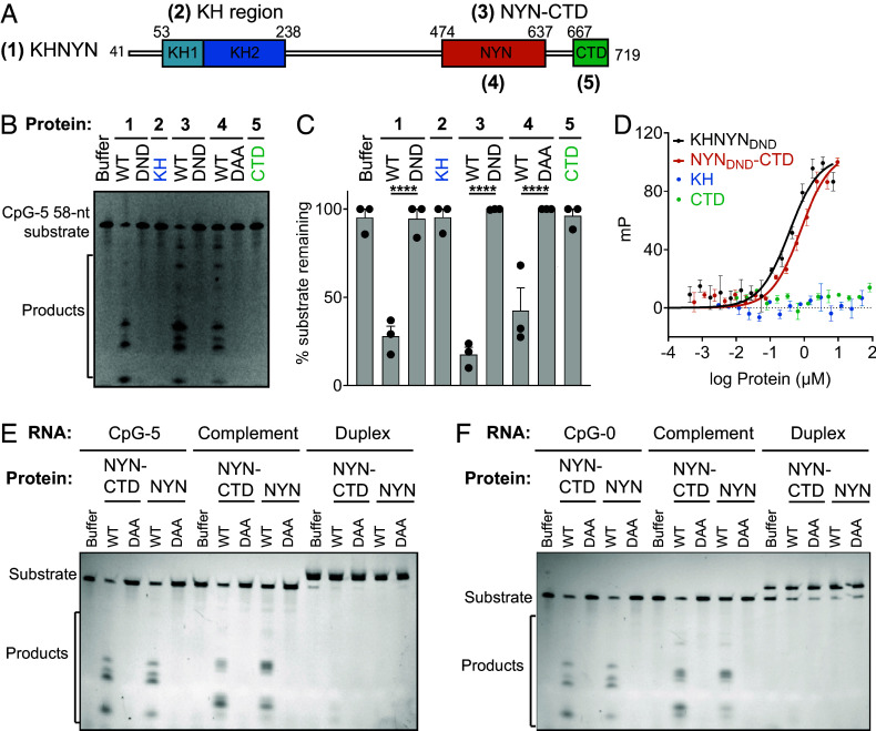
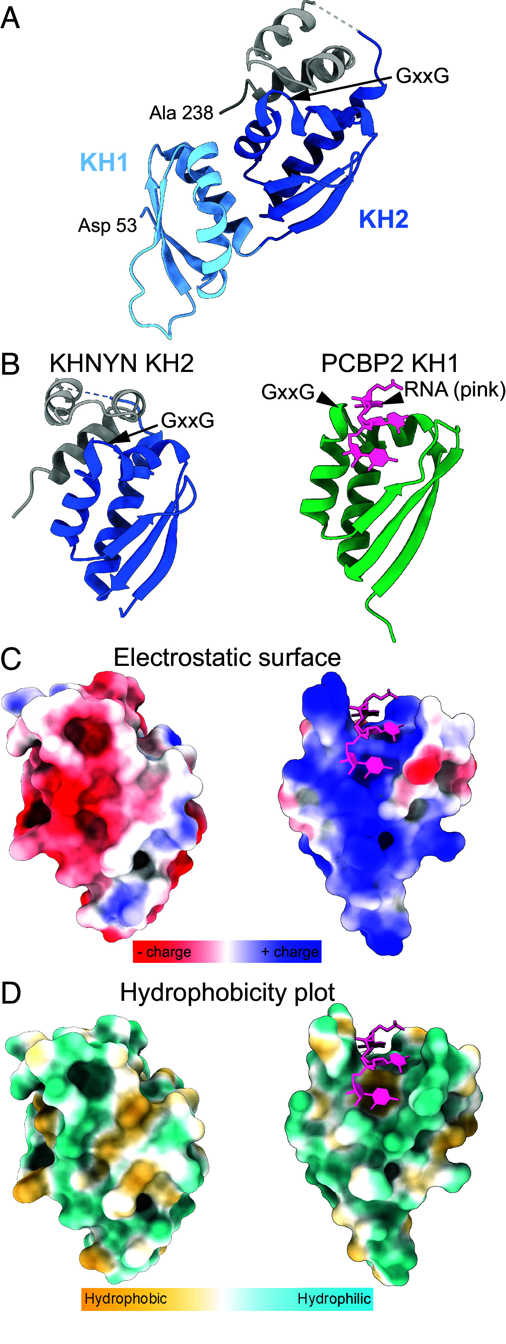
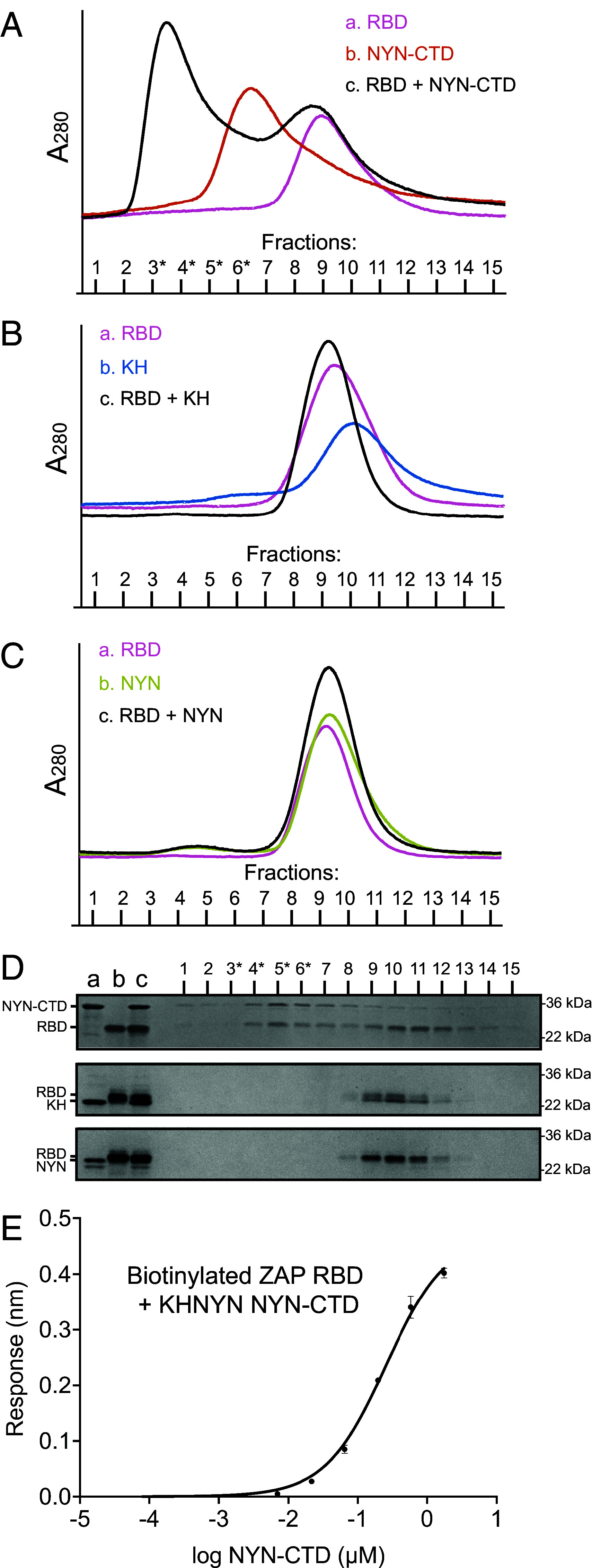
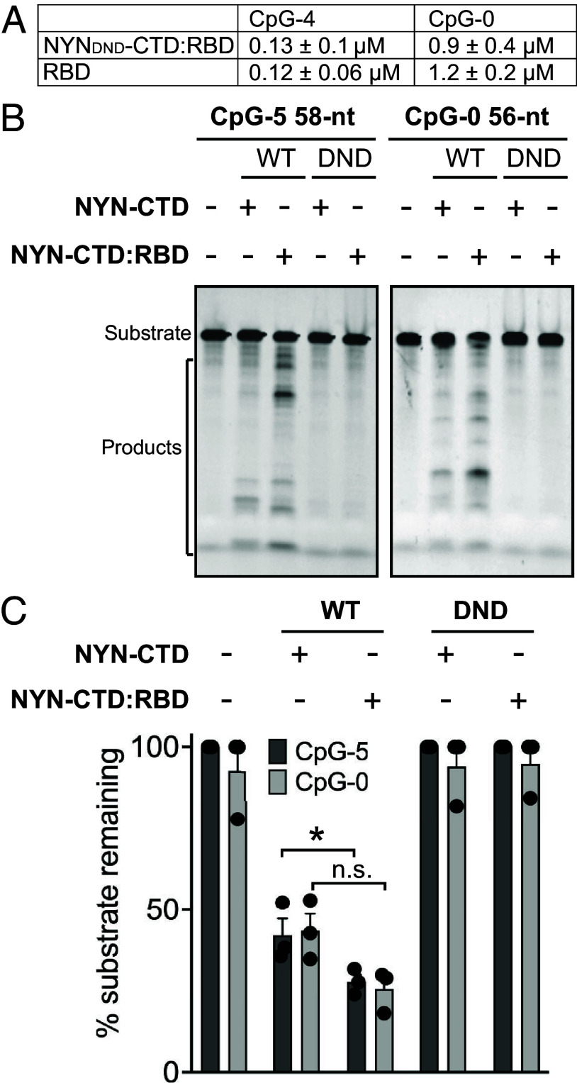
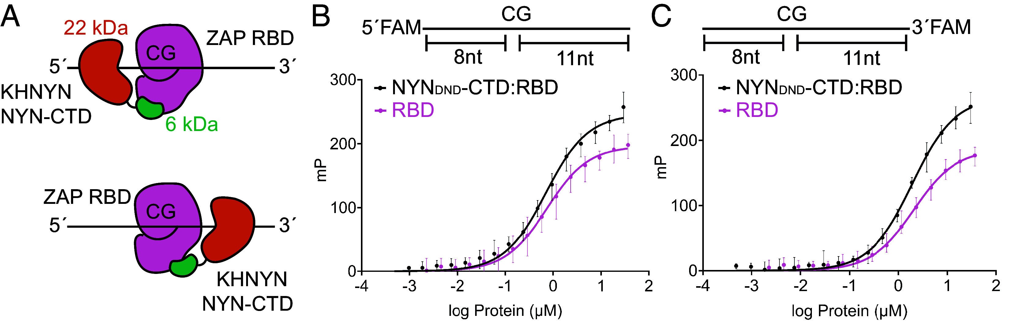
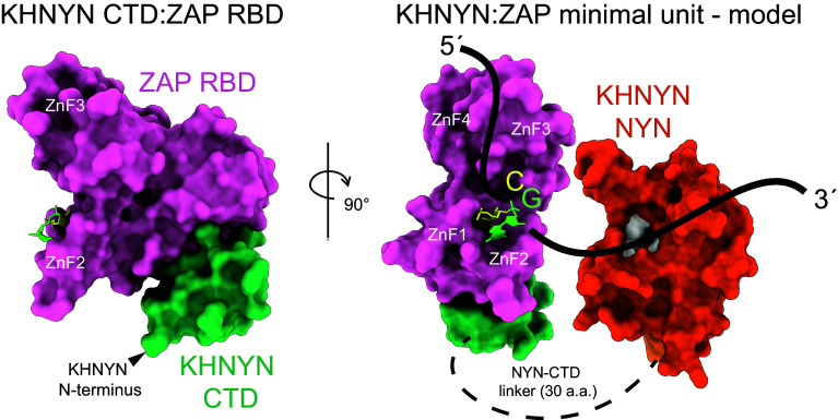
Detecting viral infection is a key role of the innate immune system. The genomes of some RNA viruses have a high CpG dinucleotide content relative to most vertebrate cell RNAs, making CpGs a molecular marker of infection. The human zinc-finger antiviral protein (ZAP) recognizes CpG, mediates clearance of the foreign CpG-rich RNA, and causes attenuation of CpG-rich RNA viruses. While ZAP binds RNA, it lacks enzymatic activity that might be responsible for RNA degradation and thus requires interacting cofactors for its function. One of these cofactors, KHNYN, has a predicted nuclease domain. Using biochemical approaches, we found that the KHNYN NYN domain is a single-stranded RNA ribonuclease that does not have sequence specificity and digests RNA with or without CpG dinucleotides equivalently in vitro. We show that unlike most KH domains, the KHNYN KH domain does not bind RNA. Indeed, a crystal structure of the KH region revealed a double-KH domain with a negatively charged surface that accounts for the lack of RNA binding. Rather, the KHNYN C-terminal domain (CTD) interacts with the ZAP RNA-binding domain (RBD) to provide target RNA specificity. We define a minimal complex composed of the ZAP RBD and the KHNYN NYN-CTD and use a fluorescence polarization assay to propose a model for how this complex interacts with a CpG dinucleotide-containing RNA. In the context of the cell, this module would represent the minimum ZAP and KHNYN domains required for CpG-recognition and ribonuclease activity essential for attenuation of viruses with clusters of CpG dinucleotides.

Keywords: RNA; anti-viral restriction; nuclease; protein crystal structure.


Conflict of interest statement

Competing interests statement:The authors declare no competing interest.

Figures

Fig. 1.
Fig. 1.
Recombinant KHNYN binds RNA and is a ribonuclease. (A) Full-length KHNYN (1) schematic highlighting protein fragments used in this study: KH region (2, light and dark blue), NYN-CTD (3, orange and green), NYN (4, orange), and CTD (5, green). (B) Ribonuclease assay with a 5´-fluorescein-labeled 58-nt RNA containing five CpG dinucleotides (SI Appendix, Table S2, oligo 1) and purified KHNYN or KHNYN fragments using wild type (WT) and two NYN domain mutants (“DAA” D484/D565A/D566A or “DND” D484/D565N/D566). (C) Quantification of the ribonuclease assays shown in panel B. The fraction of uncleaved substrate was calculated as the ratio of uncut to cut RNA in each lane. Error bars represent the SE of three independent experiments. P-values were calculated with two-way ANOVA, ****P < 0.0001, ***P < 0.0005. (D) Binding of KHNYNDND (black), NYNDND-CTD (orange), KH (blue), or CTD (green) to 5’-fluorescein-labeled 21-nt RNA (10 nM) containing four CpG dinucleotides (SI Appendix, Table S2, oligo 5). Error bars are the SE of 3 technical replicates. The Kd values of 0.36 ± 0.02 μM for KHNYNDND and Kd = 1.5 ± 0.7 μM for NYNDND-CTD were calculated from an average signal from three experimental replicates with a one-site binding model. (E) In vitro ribonuclease assay with wild-type (WT) and nuclease dead (DAA) NYN-CTD and NYN domains using the 5´-fluorescein labeled CpG-5 RNA in (B), its complement (oligo 2), or the annealed RNA duplex. (F) In vitro ribonuclease assay as in (E) but with a 5´-fluorescein-labeled 56-nt RNA containing no CpG dinucleotides (oligo 3), its complement (oligo 4), or the annealed RNA duplex.
Fig. 2.
Fig. 2.
KHNYN has two KH domains that differ from RNA-binding KH domains. (A) KH region of KHNYN with KH1 in light blue, KH2 in dark blue, and extended three-helix region in gray. (BD) Comparison of KHNYN KH2 domain (Left) and the RNA complex of a PCBP2 KH domain [Right, PDB: 2PY9 (28)], viewed in identical orientations following superposition (RMSD = 2.2 Å for 64 Cα atoms). (B) Cartoon rendering with the RNA shown in ribbon model representation (pink). (C) Electrostatic potential mapped on the protein surfaces. The KHNYN KH2 surface is strongly negative, whereas the PCBP2 KH domain surface is strongly positive. Red, negative charges; blue, positive charges; white, neutral. (D) Hydrophobicity mapped on the protein surfaces as a yellow-white-cyan color map. An RNA base binds in a PCBP2 hydrophobic pocket that does not exist in the KHNYN KH2 domain. Yellow, hydrophobic; cyan, hydrophilic.
Fig. 3.
Fig. 3.
KHNYN CTD binds ZAP RBD. (AC) Size exclusion chromatography (SEC) analysis of purified ZAP RBD mixed with KHNYN domains: RBD + NYN-CTD (A), RBD + KH (B), and RBD + NYN (C). The elution profiles of ZAP RBD (pink) and a KHNYN domain (NYN-CTD, orange; KH, blue; NYN, chartreuse) are shown for each protein alone and when combined (black), with fractions indicated on the x-axis. In panel A, fractions where the elution peak has shifted are indicated with asterisks (*). (D) Coomassie-stained SDS-PAGE gel of fractions collected from the elution of the ZAP RBD mixtures with proteins in A-C. Fractions from a peak shift are indicated with asterisks (*). (E) BLI measurement of ZAP RBD binding to KHNYN NYN-CTD. Biotinylated ZAP RBD was immobilized onto streptavidin biosensors. The Kd value of 0.31 ± 0.2 μM was calculated from three experimental replicates. Error bars are SE of three technical replicates.
Fig. 4.
Fig. 4.
ZAP RBD drives KHNYN NYN-CTD RNA binding and nuclease activity. (A) Comparison of the binding of ZAP RBD or a NYNDND-CTD:RBD complex to CpG-replete or CpG-free RNA oligomers by fluorescence polarization. The oligomers (10 nM) were a 5´-fluorescein-labeled 21-nt RNA containing either four CpG dinucleotides (SI Appendix, Table S2, oligo 5) or no CpG dinucleotides (SI Appendix, Table S2, oligo 7). Binding constants were calculated from three experimental replicates. Error bars are variance of three technical replicates. (B) In vitro ribonuclease assay with wild-type (WT) and catalytically inactive (DND) KHNYN NYN-CTD and KHNYN NYN-CTD:ZAP RBD using either a 5´-fluorescein-labeled RNA containing five CpG dinucleotides (SI Appendix, Table S2, oligo 1) or a 5´-fluorescein-labeled RNA containing no CpG dinucleotides (SI Appendix, Table S2, oligo 3). A representative image is shown. (C) Quantification of the ribonuclease assays in (B). The fraction of uncleaved substrate was calculated as the ratio of uncut to cut RNA in each lane. Error bars represent the SE of three experimental replicates. P-values were calculated with two-way ANOVA, *P < 0.05, n.s. is not significant.
Fig. 5.
Fig. 5.
KHNYN NYN-CTD:ZAP RBD complex engages RNA 3´ to ZAP bound to a CpG-containing RNA. (A). Schematic of two modes for NYN-CTD to engage an RNA containing one CpG bound to ZAP RBD. KHNYN NYN-CTD, orange-green; ZAP RBD, pink. (B) Fluorescence polarization (FP) binding curves for KHNYN NYNDND-CTD:ZAP RBD complex (black line) or ZAP RBD (pink line) with a 5´-fluorescein-labeled (6-fluorescein, FAM) 21-nt RNA containing one CpG dinucleotide (SI Appendix, Table S2, oligo 8). The Kd value was 0.74 ± 0.19 μM for ZAP RBD and 0.69 ± 0.11 μM for KHNYN NYNDND-CTD:ZAP RBD). (C) FP binding curves for a KHNYN NYNDND-CTD:ZAP RBD complex (black line) or ZAP RBD (pink line) with a 3´-fluorescein-labeled 21-nt RNA containing one CpG dinucleotide (oligo 9). The Kd value was 2.0 ± 0.12 μM for ZAP RBD and 1.9 ± 0.13 μM for KHNYN NYNDND-CTD:ZAP RBD. Affinities of the proteins for each RNA oligomer are similar but NYN-CTD binding to ZAP RBD has a greater impact on tumbling of 3´-FAM than 5´-FAM, indicating NYN-CTD engagement of the RNA 3´ of the CpG position. Error bars represent the range of four experimental replicates, each with three technical replicates.
Fig. 6.
Fig. 6.
Model of ZAP:KHNYN core complex with RNA. ZAP RBD (pink) in complex with CTD (green) and CpG dinucleotide (yellow-neon green) (PDB ID: 6UEJ, 9BGL) and a predicted model of KHNYN NYN domain (orange) with active site residues Asp484, Asp565, and Asp566 (white), and line depicting a single-stranded RNA containing one CpG (black) indicating NYN binding on the 3´ side of CpG.

Similar articles

Cited by

References

    1. Greenbaum B. D., Levine A. J., Bhanot G., Rabadan R., Patterns of evolution and host gene mimicry in influenza and other RNA viruses. PLOS Pathog. 4, e1000079 (2008). - PMC - PubMed
    1. Takata M. A., et al. , CG dinucleotide suppression enables antiviral defence targeting non-self RNA. Nature 550, 124–127 (2017). - PMC - PubMed
    1. Gao G., Guo X., Goff S. P., Inhibition of retroviral RNA production by ZAP, a CCCH-type zinc finger protein. Science 297, 1703–1706 (2002). - PubMed
    1. Guo X., Carroll J.-W.N., MacDonald M. R., Goff S. P., Gao G., The zinc finger antiviral protein directly binds to specific viral mRNAs through the CCCH zinc finger motifs. J. Virol. 78, 12781–12787 (2004). - PMC - PubMed
    1. Bick M. J., et al. , Expression of the zinc-finger antiviral protein inhibits alphavirus replication. J. Virol. 77, 11555–11562 (2003). - PMC - PubMed

LinkOut - more resources