. 2024 Dec 20;10(51):eadn7540.
doi: 10.1126/sciadv.adn7540. Epub 2024 Dec 18.

dCasMINI-mediated therapy rescues photoreceptors degeneration in a mouse model of retinitis pigmentosa

Affiliations

dCasMINI-mediated therapy rescues photoreceptors degeneration in a mouse model of retinitis pigmentosa

Qing Wang et al. Sci Adv. .

Abstract

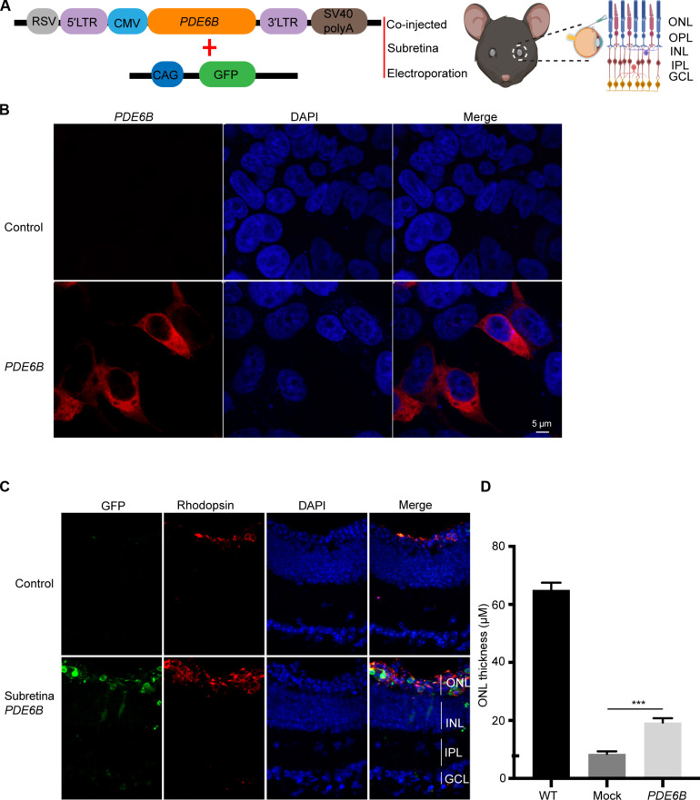
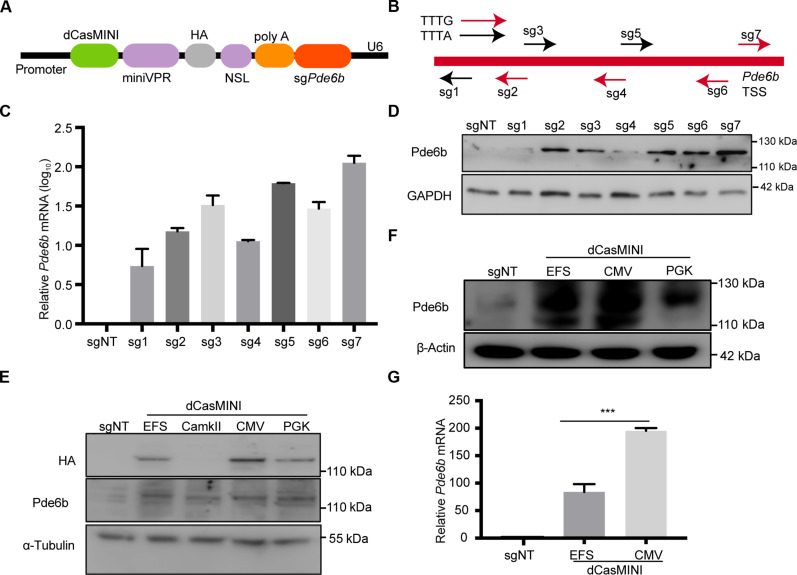
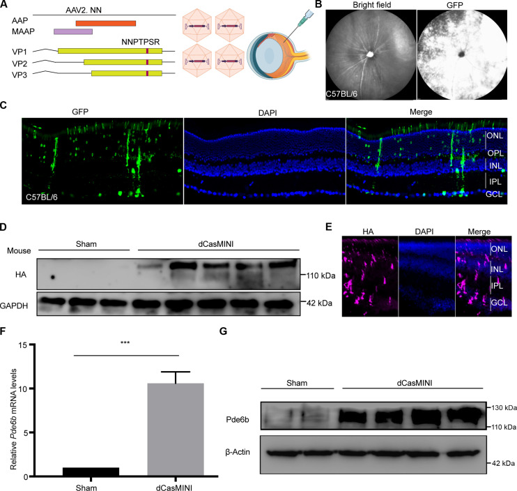
Retinitis pigmentosa (RP) is characterized by degeneration of rod and cone photoreceptors that progresses to irreversible blindness. Now, there are no mutation-agnostic approaches to treat RP. Here, we utilized a single adeno-associated virus (AAV)-based CRISPR activation system to activate phosphodiesterase 6B (Pde6b) to mitigate the severe degeneration in Pde6anmf363 mice. We demonstrate that transcriptional activation of Pde6b can rescue the loss of Pde6a, with preservation of retinal structure, restoration of electroretinography responses, and improvement of visual function as assessed by optokinetic response and looming-induced escape behaviors. These findings demonstrate the therapeutic potential of a dCasMINI-mediated activation strategy that provides a mutation-independent treatment for retinal degeneration. This study offers a promising therapeutic approach for RP and potentially other forms of genetic diseases.


Figures

Fig. 1.
Fig. 1.. Pde6b can compensate Pde6a dysfunction in Pde6anmf363 mice.
(A) Schematic diagram of experimental design showing PDE6B plasmid co-injected with CAG-GFP followed by electroporation. (B) Representative images of immunofluorescence staining with antibodies against PDE6B in 293 cells 48 hours after transfection. (C) Representative retinal slices electroporated PDE6B and GFP for 7 weeks immunostained for 4′,6-diamidino-2-phenylindole (DAPI) and rhodopsin. GFP signal marks the electroporated patch and that the area that did not receive electroporated plasmids serves as an internal control. Right: Four-week statistics for ONL thickness of control and PDE6B-electroporated region. (D) Seven-week statistics for ONL thickness of control and PDE6B-electroporated region. IPL, inner plexiform layer; GCL, ganglion cell layer; LTR, long terminal repeat; RSV, Rous sarcoma virus; INL, inner nuclear layer; OPL, outer plexiform layer. ***P < 0.001.
Fig. 2.
Fig. 2.. Screening of sgRNAs targeted for of Pde6b in vitro.
(A) Schematic of vector constructs expressing dCasMINI and sgRNA targeting Pde6b. (B) Diagram illustrating the sgRNA distributions and PAMs for mouse Pde6b. TSS, transcriptional start site. For PAM, arrows represent the directionality of the designed sgRNAs. Black, TTTA; red, TTTG. (C) qPCR analysis of Pde6b (N = 4) mRNA levels in N2a cells transfected with different sgRNAs. (D) Representative immunoblots of N2a cell-transfected lysates with different sgRNAs and detected with anti-Pde6b antibody. (E) Representative immunoblots of 293 cell-transfected lysates with sgRNA7 with EFS, CamKII, CMV, and PGK promoter-driven dCasMINI detected with anti-Pde6b antibody. (F) Representative immunoblots of N2a cell-transfected lysates with sgRNA7 with EFS, CMV, and PGK promoter-driven dCasMINI and detected with anti-PDE6B antibody. (G) qPCR analysis of Pde6b (N = 4) mRNA levels in N2a cells transfected with sgRNA7 with EFS and CMV promoter driven by dCasMINI. Data are means ± SEM. ***P < 0.001.
Fig. 3.
Fig. 3.. dCasMINI-mediated Pde6b expression in vivo.
(A) Schematic of AAV2.NN backbone and AAV2.NN vectors used for intravitreal injection. (B) Bright-field and SLO image of mouse retina showing transduction and spread of vectors 3 weeks after AAV2.NN-CAG-GFP injection. (C) Representative retinal cross-sections immunolabeled for GFP after AAV2.NN-CAG-GFP injection for 4 weeks. (D and E) WB (D) and IF (E) showing expression of dCasMINI 1 month after intravitreal injection of EFS-dCasMINI into Pde6anmf363 mice. (D) Representative immunoblots of total lysates and retina cross-sections probed with the antibody against HA. (E) Representative retinal cross sections of 8-week-old mice from each group immunolabeled for HA. (F) Quantitative PCR analysis of Pde6b (N = 4) mRNA levels in sham retina and dCasMINI-treated retina. (G) Representative immunoblots of total sham retina and dCasMINI-treated retina lysates and detected with anti-Pde6b antibody. Data are means ± SEM. ***P < 0.001.
Fig. 4.
Fig. 4.. Rescue of retinal structure by dCasMINI-activated Pde6b.
(A and B) Representative OCT images of the sham retina, AAV2.NN-injected retina, and AAV2.NN-dCasMINI–injected retina in Pde6anmf363 mice. The green circle showed the OCT scan area surrounding the optical nerve head. GCC indicated in the top red line, and ONL indicated in the bottom red line. (C) Statistics for ONL thickness. *P < 0.05. (D) DAPI staining of a longitudinal cryosection through the mouse retina. Staining of nuclear DNA demonstrates the retinal layers: PC, layer of outer and inner segments of photoreceptor cells. (E) Statistics for ONL thickness. (F) Representative image of sham and AAV2.NN-dCasMINI–injected retina staining with recoverin and PNA. Data are presented as means ± SEM; ***P < 0.001, two-tailed unpaired t test. OS, outer segment.
Fig. 5.
Fig. 5.. Pde6b activation improves ERG in Pde6a nmf363 mice.
(A and B) Experimental schematic of in vivo plasmid intravitreal injection at P7. ERG was obtained at 4 weeks after injection. (B) Left eye was sham group, and right eye was injected AAV2.NN. (C) Representative the scotopic 0.01, 0.1, and1 cd·s·m−2 dim-light rod-specific ERG traces sham eye and AAV2.NN–injected eye in Pde6anmf363 mouse. (D) Quantification of maximum b-wave amplitude for (C). (E) Left: Constructs schematic of in vivo AAV-dCasMINI-sgPDE6B injection. Right: Left eye is sham group and right eye injected AAV2.NN-dCasMINI. (F) Representative the scotopic 0.01, 0.1, and 1 cd·s·m−2 dim-light rod-specific ERG traces C57BL/6 eye, AAV2.NN-dCasMINI injected eye, and sham eye in Pde6anmf363 mouse. (G and H) Quantification of maximum a-wave amplitude (G) and b-wave amplitude (H) for (F). Data are presented as means ± SEM; **P < 0.001, two-tailed unpaired t test. WPI, weeks post-immunization.
Fig. 6.
Fig. 6.. dCasMINI-mediated gene activation preserves visual behavior.
(A) Experimental schematic of in vivo plasmid intravitreal injection at P7 and visual behaviors was assessed at 4 weeks after injection. (B) Experiment setup for OKR. (C) Number of responders and nonresponders is shown on the y axis with respective percentages. Statistical analysis was performed using Fisher’s exact test. *P < 0.05. (D) Experiment setup for looming induced escape behaviors. (E) Number of responders and nonresponders to looming is shown on the y axis with respective percentages. Statistical analysis was performed using Fisher’s exact test. (F) Statistics for response latency of AAV2.NN- (N = 5) and dCasMINI-treated mice (N = 11). Data are presented as means + SEM; ***P < 0.001, two-tailed unpaired t test.

References

    1. Hartong D. T., Berson E. L., Dryja T. P., Retinitis pigmentosa. Lancet 368, 1795–1809 (2006). - PubMed
    1. Kennan A., Aherne A., Humphries P., Light in retinitis pigmentosa. Trends Genet. 21, 103–110 (2005). - PubMed
    1. Kamde S. P., Anjankar A., Retinitis pigmentosa: Pathogenesis, diagnostic findings, and treatment. Cureus 15, e48006 (2023). - PMC - PubMed
    1. Hanany M., Rivolta C., Sharon D., Worldwide carrier frequency and genetic prevalence of autosomal recessive inherited retinal diseases. Proc. Natl. Acad. Sci. U.S.A. 117, 2710–2716 (2020). - PMC - PubMed
    1. Streilein J. W., Ocular immune privilege: therapeutic opportunities from an experiment of nature. Nat. Rev. Immunol. 3, 879–889 (2003). - PubMed

Publication types

MeSH terms

Substances