. 2025 Jan 8;147(1):305-317.
doi: 10.1021/jacs.4c10326. Epub 2024 Dec 18.

Indirect Formation of Peptide Bonds as a Prelude to Ribosomal Transpeptidation

Affiliations

Indirect Formation of Peptide Bonds as a Prelude to Ribosomal Transpeptidation

Harvey J A Dale et al. J Am Chem Soc. .

Abstract

The catalytic competency of the ribosome in extant protein biosynthesis is thought to arise primarily from two sources: an ability to precisely juxtapose the termini of two key substrates─3'-aminoacyl and N-acyl-aminoacyl tRNAs─and an ability to ease direct transpeptidation by their desolvation and encapsulation. In the absence of ribosomal, or enzymatic, protection, however, these activated alkyl esters undergo efficient hydrolysis, while significant entropic barriers serve to hamper their intermolecular cross-aminolysis in bulk water. Given that the spontaneous emergence of a catalyst of comparable size and sophistication to the ribosome in a prebiotic RNA world would appear implausible, it is thus natural to ask how appreciable peptide formation could have occurred with such substrates in bulk water without the aid of advanced ribozymatic catalysis. Using a combination of fluorine-tagged aminoacyl adenylate esters, in situ monitoring by 19F{1H} NMR spectroscopy, analytical deconvolution of kinetics, pH-rate profile analysis, and temperature-dependence studies, we here explore the mechanistic landscape of indirect amidation, via transesterification and O-to-N rearrangement, as a highly efficient, alternative manifold for transpeptidation that may have served as a prelude to ribosomal peptide synthesis. Our results suggest a potentially overlooked role for those amino acids implicated by the cyanosulfidic reaction network with hydroxyl side chains (Ser and Thr), and they also help to resolve some outstanding ambiguities in the broader literature regarding studies of similar systems (e.g., aminolyzes with Tris buffer). The evolutionary implications of this mode of peptide synthesis and the involvement of a very specific subset of amino acids are discussed.


Conflict of interest statement

The authors declare no competing financial interest.

Figures

Figure 1
Figure 1
Underlying chemistry of ribosomal peptide synthesis.
Figure 2
Figure 2
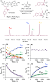
Chemical scheme, example 19F{1H}IG NMR spectrum (376 MHz), and key temporal evolution profiles from the monitoring of a typical aminolysis of MepA-l-PheF (Em) with l-serinamide LS in aqueous solution (this example: H2O, 20 °C, pH = 7.3, I = 2.0 M). (A) Temporal evolution of experimental mole fractions of Em,2′, Em,3′, PAm, and Paa. (B) Temporal evolution of experimentally observed 2′/3′ speciation of Em, with f{Em,3′} = [Em,3]/[Em]T = fractional population of the 3′-isomer of Em over all charge states. (C) Temporal evolution of the mole fraction of Em,T with fit to the kinetic model: Em,TPAm (kAmψ), Em,TPaa (kHydψ). (D) Temporal evolution of the mole fractions of PAm and Paa with fit to the kinetic model above. [Em]T = [Em,2] + [Em,3] = total concentration of Em in all charge states. kTψ = total pseudo-first-order rate constant for the decay of Em,T. kAmψ = f{Am}.kTψ = kAm.[LS]T = pseudo-first-order rate constant for the aminolysis of Em,T. kAm′ = total second-order rate constant for the aminolysis of Em,T. kHydψ = f{Hyd}. kTψ = pseudo-first-order rate constant for the hydrolysis of Em,T. [Em,j′] = [Em,j′(0)] + [Em,j′(+)], where [Em,j′(i)] = concentration of j′-regioisomer with a charge of i on the α-amino group. X{i} = mole fraction of species i.
Figure 3
Figure 3
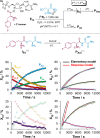
Kinetic scheme, temporal evolution profiles, and comparative fits to two distinct kinetic models for key species in a typical aminolysis of MepA-l-PheF (Em) with l-serinamide under a low pH regime (this example: D2O, 30 °C, pH*(30 °C) = 5.7, I = 2.0 M). Elementary model = Em,TPAm (kAmψ), Em,TPaa (kHydψ). Stepwise model: = Em,TIEs (kAmψ), IEsPAm (krψ), Em,TPaa (kHydψ).
Figure 4
Figure 4
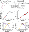
Kinetic analysis of the aminolysis of Em with l-serinamide LS, and concurrent hydrolysis, over the range pH* = 5.7–9.3 (20 °C, D2O, I = 2.0 M, KCl; [Em]T,0 = 4.0 mM). (A) pH*–kAm profile for [LS]T = 600 mM (blue) and 1200 mM (red), with fit to experimental data. (B) pH*–kHydψ profile, with same color coding as (A), and fit to experimental data. (C) Empirical selectivity for aminolysis, kAmψ/kTψ, and total pseudo-first-order rate of decay kTψ as a function of pH*(20 °C), with fits to experimental data, for [LS]T = 600 mM. (D) as for (C) but with [LS]T = 1200 mM. Fitted parameters (kAm,+, kAm,0, kOH,+, kOH,0; pKa*(Em+)) and their standard errors (Table S1, Supporting Information) were determined by global, unweighted nonlinear fitting of the pH*–kAm and pH*–kHydψ profiles (OriginPro 2024b). Relative uncertainties in kHydψ and kAm were conservatively estimated to be ±10% (see Supporting Information). Uncertainties in kTψ and kAmψ/kTψ (not fitted directly) were calculated by propagation. See Supporting Information for further discussion.
Figure 5
Figure 5
Simplified structural depictions of Em+ and Em0 and the N-formylated derivative Emf. Note that 2′–3′ isomerization (not shown) appears to be limitingly rapid for both Em and Emf, relative to hydrolysis and aminolysis (pH* > 5.5), and so, all rate constants and isotope effects in Tables 1 and 2 are weighted averages. See Supporting Information for details.
Figure 6
Figure 6
Temperature dependence (10–30 °C) of the hydrolysis and aminolysis of Em with l-serinamide LS, as a function of pH* (D2O, I = 2.0 M, KCl, [Em]T,0 ≈ 4 mM). (A) Temperature dependence of the pH*–kAm profile (aminolysis), with (unweighted) fits to experimental data. (B) Temperature dependence of the pH*–kHydψ profile (hydrolysis), with (unweighted) fits to experimental data. (C) Temperature and pH* dependence of (i) the empirical selectivity for aminolysis kAmψ/kTψ and (ii) the total pseudo-first-order rate of decay kTψ, with fits to experimental data, for [LS]T = 1200 mM. (D) Linearized Eyring plots of the second-order rate constants kAm,+, kAm,0, kOH,+, and kOH,0. (E) 13C{1H} NMR titrations (δ13CO) of l-serinamide (100 mM; D2O, I = 2.0 M, KCl) at different temperatures ([dpKaH/dT] = −0.025 °C–1; cf. glycinamide). pH*(10 °C) = 6.38–9.42 (pKa*(LS+) = 8.17). pH*(20 °C) = 6.01–9.14 (pKa*(LS+) = 7.94). pH*(30 °C) = 5.69–8.95 (pKa*(LS+) = 7.69). [LS]T = 600–2000 mM. Fitted parameters (kAm,+, kAm,0, kOH,+, kOH,0; pKa*(Em+)) and their standard errors (Table 1, Supporting Information) were determined by global, unweighted nonlinear fitting of pH*–kAm and pH*–kHydψ (OriginPro 2024b). See the Figure 4 caption and Supporting Information for details of error analysis.
Figure 7
Figure 7
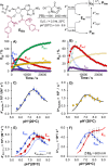
Example 19F{1H}IG NMR monitoring data and kinetic analysis for the aminolysis, and concurrent hydrolysis, of MepA-(l-PheF)2 (EBis) with l-serinamide in D2O (20 °C, pH* = 5.6–8.6, I = 2.0 M; [LS]T = 600–2000 mM; [EBis]T,0 ≈ 4 mM). (A) Temporal evolution profiles, with kinetic fits shown, for all key species under a low pH* regime (this example: pH* = 5.6, [LS]T = 2000 mM). (B) Expanded view of (A), with a focus on the intermediate kinetics of Em,T and IEs. (C) pH*–kAm,Bis profile with (unweighted) fit to experimental data (kAm,Bis = kAm,Bisψ/[LS]T). (D) pH*–kHyd,Bisψ profile with (unweighted) fit to experimental data. (E) Co-plot of the pH*–kAm (Em) and pH*–kAm,Bis (EBis) profiles; dotted line reflects the pH*–kAm,Bis profile without any contribution from the aminolysis of the doubly protonated state, EBis++ (i.e., kAm,Bis,++ = 0). (F) Co-plot of the pH*–kTψ (Em) and pH*–kT,Bisψ (EBis) profiles ([LS]T = 600 mM). Kinetic model for pH* > 6.5 = EBisEm,T + PAm (kAm,Bisψ), EBisEm,T + Paa (kHyd,Bisψ), Em,TPAm (kAmψ), Em,TPaa (kHydψ). Kinetic model for pH* < 6.5: = EBisEm,T + IEs (kAm,Bisψ), EBisEm,T + Paa (kHyd,Bisψ), Em,TIEs (kAmψ), Em,TPaa (kHydψ), IEsPAm (krψ). All steps are (pseudo; ψ) first-order. Fitted parameters (kAm,Bis,++, kAm,Bis,+, kAm,Bis,0, kOD,Bis,++, kOD,Bis,+) and their standard errors in Table 4. pKa*(EBis++) and pKa*(EBis+) constrained during fitting; values taken from 19F{1H} NMR chemical shift analysis. See Supporting Information for details.
Figure 8
Figure 8
Structural depictions of EBis (simplified) and IEs in their various charge states. Note that [EBis+] = [EBis,2′++] + [EBis,3′++]. For simplicity, we assume that IEs+ and I′Es+ have identical microscopic acidities.

Similar articles

References

    1. Ban N.; Nissen P.; Hansen J.; Moore P. B.; Steitz T. A. The Complete Atomic Structure of the Large Ribosomal Subunit at 2.4 Å Resolution. Science 2000, 289, 905–920. 10.1126/science.289.5481.905. - DOI - PubMed
    2. Nissen P.; Hansen J.; Ban N.; Moore P. B.; Steitz T. A. The Structural Basis of Ribosome Activity in Peptide Bond Synthesis. Science 2000, 289, 920–930. 10.1126/science.289.5481.920. - DOI - PubMed
    3. Schlünzen F.; Zarivach R.; Harms J.; Bashan A.; Tocilj A.; Albrecht R.; Yonath A.; Franceschi F. Structural basis for the interaction of antibiotics with the peptidyl transferase centre in eubacteria. Nature 2001, 413, 814–821. 10.1038/35101544. - DOI - PubMed
    4. Harms J.; Schluenzen F.; Zarivach R.; Bashan A.; Gat S.; Agmon I.; Bartels H.; Franceschi F.; Yonath A. High Resolution Structure of the Large Ribosomal Subunit from a Mesophilic Eubacterium. Cell 2001, 107, 679–688. 10.1016/S0092-8674(01)00546-3. - DOI - PubMed
    5. Hansen J. L.; Schmeing T. M.; Moore P. B.; Steitz T. A. Structural insights into peptide bond formation. Proc. Natl. Acad. Sci. U.S.A. 2002, 99, 11670–11675. 10.1073/pnas.172404099. - DOI - PMC - PubMed
    6. Beringer M.; Bruell C.; Xiong L.; Pfister P.; Bieling P.; Katunin V. I.; Mankin A. S.; Böttger E. C.; Rodnina M. V. Essential Mechanisms in the Catalysis of Peptide Bond Formation on the Ribosome. J. Biol. Chem. 2005, 280, 36065–36072. 10.1074/jbc.M507961200. - DOI - PubMed
    7. Trobro S.; Åqvist J. Mechanism of peptide bond synthesis on the ribosome. Proc. Natl. Acad. Sci. U.S.A. 2005, 102, 12395–12400. 10.1073/pnas.0504043102. - DOI - PMC - PubMed
    8. Bowman J. C.; Petrov A. S.; Frenkel-Pinter M.; Penev P. I.; Williams L. D. Root of the Tree: The Significance, Evolution, and Origins of the Ribosome. Chem. Rev. 2020, 120, 4848–4878. 10.1021/acs.chemrev.9b00742. - DOI - PubMed
    1. Polacek N.; Mankin A. S. The Ribosomal Peptidyl Transferase Center: Structure, Function, Evolution, Inhibition. Crit. Rev. Biochem. Mol. Biol. 2005, 40, 285–311. 10.1080/10409230500326334. - DOI - PubMed
    2. Beringer M.; Rodnina M. V. The Ribosomal Peptidyl Transferase. Mol. Cell 2007, 26, 311–321. 10.1016/j.molcel.2007.03.015. - DOI - PubMed
    1. Huang K. S.; Weinger J. S.; Butler E. B.; Strobel S. A. Regiospecificity of the Peptidyl tRNA Ester within the Ribosomal P Site. J. Am. Chem. Soc. 2006, 128, 3108–3109. 10.1021/ja0554099. - DOI - PubMed
    1. Bose T.; Fridkin G.; Davidovich C.; Krupkin M.; Dinger N.; Falkovich A. H.; Peleg Y.; Agmon I.; Bashan A.; Yonath A. Origin of life: protoribosome forms peptide bonds and links RNA and protein dominated worlds. Nucleic Acids Res. 2022, 50, 1815–1828. 10.1093/nar/gkac052. - DOI - PMC - PubMed
    1. Noller H. F. Ribosomal RNA and Translation. Annu. Rev. Biochem. 1991, 60, 191–227. 10.1146/annurev.bi.60.070191.001203. - DOI - PubMed
    2. Green R.; Noller H. F. Ribosomes and Translation. Annu. Rev. Biochem. 1997, 66, 679–716. 10.1146/annurev.biochem.66.1.679. - DOI - PubMed
    3. Davidovich C.; Belousoff M.; Wekselman I.; Shapira T.; Krupkin M.; Zimmerman E.; Bashan A.; Yonath A. The Proto-Ribosome: An Ancient Nano-machine for Peptide Bond Formation. Isr. J. Chem. 2010, 50, 29–35. 10.1002/ijch.201000012. - DOI - PMC - PubMed

LinkOut - more resources