. 2025 Mar 3;85(5):973-986.
doi: 10.1158/0008-5472.CAN-24-0798.

Antibody-Drug Conjugates Targeting the EGFR Ligand Epiregulin Elicit Robust Antitumor Activity in Colorectal Cancer

Affiliations

Antibody-Drug Conjugates Targeting the EGFR Ligand Epiregulin Elicit Robust Antitumor Activity in Colorectal Cancer

Joan Jacob et al. Cancer Res. .

Abstract

As colorectal cancer remains a leading cause of cancer-related death, identifying therapeutic targets and approaches is essential to improve patient outcomes. The EGFR ligand epiregulin (EREG) is highly expressed in RAS wild-type (WT) and mutant colorectal cancer, with minimal expression in normal tissues, making it an attractive target for antibody-drug conjugate (ADC) development. In this study, we produced and purified an EREG mAb, H231, which had high specificity and affinity for human and mouse EREG. H231 also internalized to lysosomes, which is important for ADC payload release. ImmunoPET and ex vivo biodistribution studies showed significant tumor uptake of zirconium-89-labeled H231, with minimal uptake in normal tissues. H231 was conjugated to either cleavable dipeptide or tripeptide chemical linkers attached to the DNA-alkylating payload duocarmycin DM, and the cytotoxicity of EREG ADCs was assessed in a panel of colorectal cancer cell lines. EREG ADCs incorporating tripeptide linkers demonstrated the highest potency in EREG-expressing colorectal cancer cells irrespective of RAS mutations. Preclinical safety and efficacy studies showed that EREG ADCs were well tolerated, neutralized EGFR pathway activity, caused significant tumor growth inhibition or regression, and increased survival in colorectal cancer cell line and patient-derived xenograft models. These data suggest that EREG is a promising target for the development of ADCs for treating colorectal cancer and other cancer types that express high levels of EREG. Although the efficacy of clinically approved anti-EGFR mAbs is largely limited by RAS mutational status, EREG ADCs may show promise for both RAS mutant and WT patients, thus improving existing treatment options. Significance: EREG-targeting antibody-drug conjugates demonstrate acceptable safety and robust therapeutic efficacy in RAS mutant and wild-type colorectal cancer, suggesting their potential as an alternative to EGFR-targeted therapy to benefit a broader patient population.


Conflict of interest statement

Conflict of Interest: Y.A. and K.T. are named inventors on all or some of the patent applications (WO2018218004A1, US11629122B2, EP3630189A4 and WO2023122587A3) relating to the linker technologies described in this article. K.T. is a co-founder of and holds equity in CrossBridge Bio. All other authors declare no potential conflicts of interest.

Figures

Figure 1.
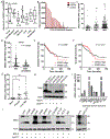
Figure 1.. EREG is highly expressed in colorectal tumors and cancer cell lines.
A, EREG RNA-Seq expression data from patient tumors of the colorectal adenocarcinoma (COADREAD) TCGA cohort. Values are Log2 RNA-Seq by expectation–maximization (Log2 RSEM). B, EREG RNA-Seq RSEM values for matched tumor and adjacent normal from the COADREAD cohort (N=32). Distribution of C, MSI-H (N=23), MSI-L (N=30), and MSS (N=129) subtypes and D, KRAS WT (N=109) and MUT (N=56) status based on EREG expression. Values are read per kilobase of transcript per million (RPKM). Kaplan-Meier analyses of E, disease-free survival (EREG-High, N=86; EREG-Low, N=245) and F, overall survival (EREG-High, N=92; EREG-Low, N=279) in patients from the COADREAD cohort. P values were obtained by the log-rank test. G, EREG RNA-seq values from cohort of 18 matched normal colonic epithelium, primary adenocarcinoma, and synchronous liver metastases (NCBI GSE50760). H, Western blot endogenous EREG protein expression in patient-derived xenograft (PDX) models. *, indicates mutant NRAS. I, EREG RNA-seq values for select CRC cell lines used in this study from Cancer Cell Line Encyclopedia (CCLE). J, Western blot of endogenous EREG protein expression across CRC cell lines. Quantitative data presented as mean ± SEM and statistical analysis performed using one-way analysis of variance (ANOVA) and Tukey’s multiple comparison test or paired Student’s t test for two groups. ** P< 0.01, *** P< 0.001.
Figure 2.
Figure 2.. Characterization of H231 mAb binding, neutralization, and internalization.
A, Cell-based binding assays show H321 mAb binds recombinant hEREG- and mEREG-overexpressing 293T cells and not vector cells. H231 and not cmAb binds endogenous EREG in B, DLD-1 and C, LoVo cells. Data presented as mean ± SD. D, Western blot of DLD-1 cells with high EREG expression stably transduced with CRISPR-Cas9 vector containing sgRNA to generate EREG knockout (KO) line. E, H231 and cmAb conjugated to phycoerythrin (PE) were utilized for the flow cytometric analysis to quantify surface pro-EREG ligands/cell using QuantBRITE PE kit. F, Confocal images shows H231 binds to EREG and co-localizes with lysosome marker, LAMP1, after 90 mins at 37 °C in EREG-expressing LoVo, HCT116, and DLD-1 cells and not DLD-1 EREG KO cells. Yellow indicates colocalization. G, Overexpressed (O.E.) EREG co-localizes with lysosomes in EGFR-negative SW620 CRC cells after H231 treatment. H, Western blot of LoVo and LIM1215 cells treated in the presence of 15 μg/ml cmAb or H231 for 5 min with or without 300 ng/ml EREG.
Figure 3.
Figure 3.. ImmunoPET imaging and biodistribution of H231 in EREG-high colorectal tumors.
A, Cell-based binding assay shows equivalent binding for unconjugated H231 mAb and lysine-conjugated H231-DFO on DLD-1 cells. B, Representative PET/CT images acquired 5 days post-injection of 89Zr-radiolabeled EREG-targeting H231 mAb or non-targeting cmAb in DLD-1 tumor xenografts. Solid and dashed arrows indicate the lung and liver, respectively. C, Quantification of PET images tumor/muscle ratio (%ID/cc) of 89Zr-H231 (N=4) and 89Zr-cmAb (N=3). D, Biodistribution data (ID/g%) measured ex vivo in tumor and normal tissues (89Zr-H231, N=5; 89Zr-cmAb (N=4). Statistical analysis using Student’s t-test. Quantitative data presented as mean ± SD. *P< 0.05, **P< 0.01, *** P< 0.001.
Figure 4.
Figure 4.. Generation of EREG-targeting antibody-drug conjugates.
A, Schematic depiction of microbial transglutaminase (MTGase)-mediated conjugation of a branched linker to Q295 of the Fc region of H231 mAb followed by strain promoted alkyne-azide cycloaddition of duocarmycin DM (DuoDM) payload attached to either a cleavable dipeptide valine-citrulline (VC) or tripeptide glutamic acid-glycine-citrulline (EGC) linker to generate H321 ADCs (DAR = 4). B, Deconvoluted ESI-mass spectra. First panel, H231 mAb; second panel, H231-branched linker conjugate, third panel, H231-VC-cDuoDM ADC; fourth panel, H231-EGC-cDuoDM ADC; fifth panel, H231-EGC-qDMDM gluc ADC. All homogeneous ADCs have a DAR of 4. *Fragment ions detected in ESI-MS analysis.
Figure 5.
Figure 5.. Therapeutic potency and selectivity of EREG ADCs in CRC cell lines.
A, Binding of H231 ADCs and non-targeting control ADC (cADC, DAR=4) with same linker-payload. B, Efficacy of H231 ADCs modified with different linkers, H231, cmAb, and cADC in DLD-1 cells. C, Dose-dependent cytotoxicity of H231 EGC-qDuoDM gluc ADC in LoVo cells. H231, cmAb, and cADC have minimal effects. Efficacy of H231 VC-cDuoDM, H231 EGC-cDuoDM, and H231 EGC-qDuoDM gluc in a panel of CRC cell lines with different levels of EREG expression presented as D, IC50 values and E, Area under the curve (AUC). #, indicates IC50 values cannot be calculated at tested concentrations due to minimal effects. Quantitative data presented as mean ± SD.
Figure 6.
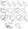
Figure 6.. EREG ADC safety assessment and antitumor efficacy in CRC cell line and patient-derived xenograft models.
Immunocompetent C57BL/6 mice were treated with 3 weekly 5 mg/kg doses of H231 mAb, cADC, or H231 EGC-DuoDM gluc ADC as indicated, or PBS vehicle. Blood was drawn and mice were euthanized 2 weeks after 3rd dose. A, Bodyweight measurements of C57BL/6 mice (n=3, vehicle; n=4 for all other groups). B, White blood cell (WBC) counts. C, Alanine aminotransferase (ALT) and D, aspartate aminotransferase (AST) liver enzyme analysis and E, kidney creatinine in serum. F, Antitumor efficacy of EREG ADCs (5 mg/kg) in LoVo nu/nu xenografts (n=9, vehicle; n=5 for all other groups). G, Kaplan-Meier survival plot and log-rank test for LoVo xenograft study in F. Vehicle and cADC showed similar survival. H, Antitumor efficacy of EREG ADCs (5mg/kg) as indicated in DLD-1 nu/nu xenografts (n=6, vehicle and H231 EGC-qDuoDM; n=5, cADC and H231 EGC-qDuoDM gluc). I, Kaplan–Meier survival plot and log-rank test for DLD-1 xenograft study in H. (J-M) Antitumor efficacy in patient-derived tumor xenograft (PDX) models. All treatments dosed at 5mg/kg. J, XST-GI-010 individual animals (n=13, vehicle; n=5, cADC and H231 EGC-cDuoDM; n=7, H231 EGC-qDuoDM) and K, average tumor volume at day 23. L, CRC-001 individual animals (n=5, vehicle; n=6, H231 EGC-qDuoDM) and M, average tumor volume at Day 27. N, Antitumor efficacy of higher dose H231 EGC-qDuoDM gluc compared to cetuximab and control treatment arms in DLD-1 nu/nu xenografts as indicated (n=5/group). All groups were treated with two weekly doses of 10 mg/kg. O, Kaplan–Meier survival plot and log-rank test for DLD-1 xenograft study in N. Statistical analysis performed using Student’s unpaired two-tailed t test for two groups or one-way analysis of variance (ANOVA) and Dunnett’s multiple comparison test. Quantitative data presented as mean ± SD. *P< 0.05, **P< 0.01, ***P< 0.001.

Update of

References

    1. Siegel RL, Wagle NS, Cercek A, Smith RA, Jemal A. Colorectal cancer statistics, 2023. CA Cancer J Clin 2023;73:233–54 - PubMed
    1. Xie YH, Chen YX, Fang JY. Comprehensive review of targeted therapy for colorectal cancer. Signal Transduct Target Ther 2020;5:22. - PMC - PubMed
    1. Fakih MG, Salvatore L, Esaki T, Modest DP, Lopez-Bravo DP, Taieb J, et al. Sotorasib plus Panitumumab in Refractory Colorectal Cancer with Mutated KRAS G12C. N Engl J Med 2023;389:2125–39 - PubMed
    1. Riese DJ 2nd, Cullum RL. Epiregulin: roles in normal physiology and cancer. Semin Cell Dev Biol 2014;28:49–56 - PMC - PubMed
    1. Sunaga N, Kaira K, Imai H, Shimizu K, Nakano T, Shames DS, et al. Oncogenic KRAS-induced epiregulin overexpression contributes to aggressive phenotype and is a promising therapeutic target in non-small-cell lung cancer. Oncogene 2013;32:4034–42 - PMC - PubMed