Case Reports
. 2024 Dec 18;17(12):e262470.
doi: 10.1136/bcr-2024-262470.

Lower limb necrotising fasciitis descending from malignant colonic perforation: a rare pattern

Affiliations
Case Reports

Lower limb necrotising fasciitis descending from malignant colonic perforation: a rare pattern

Elliot Robbie et al. BMJ Case Rep. .

Abstract

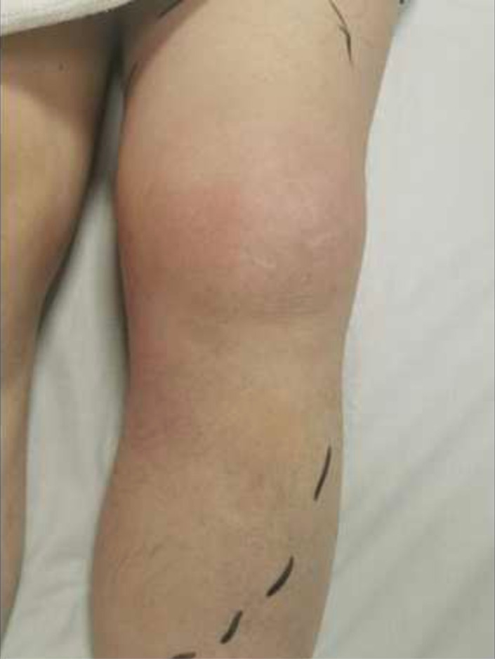
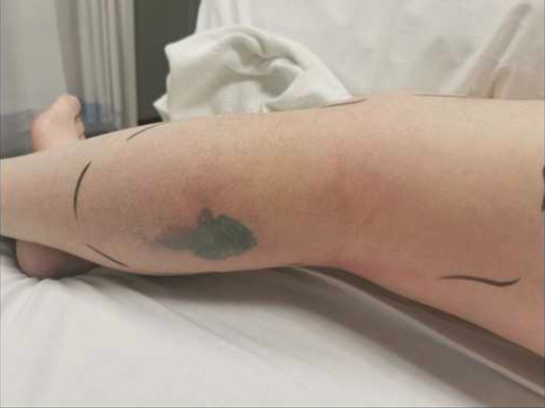
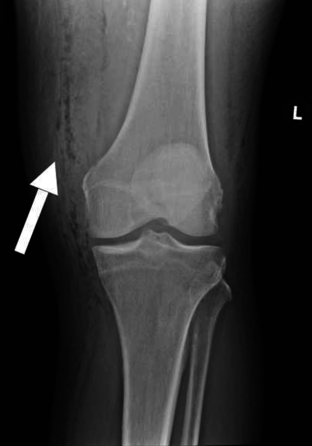
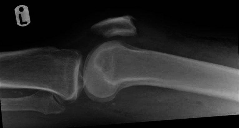
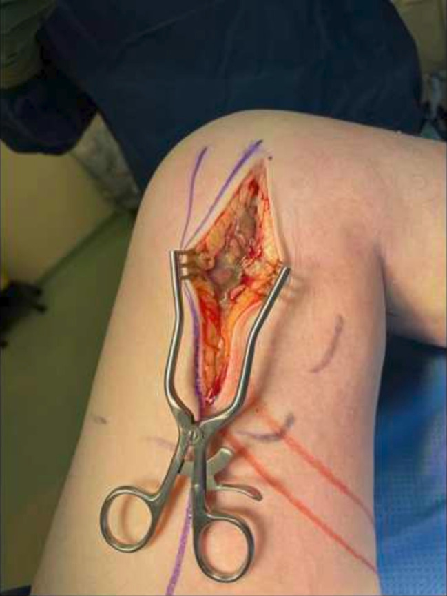
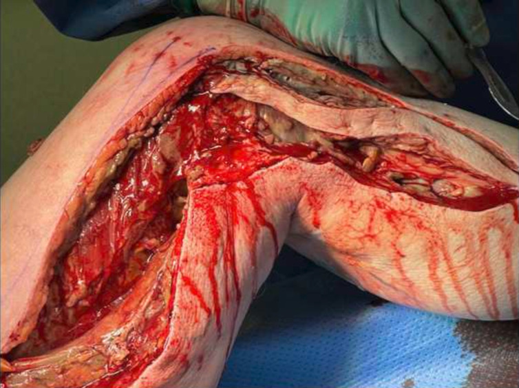
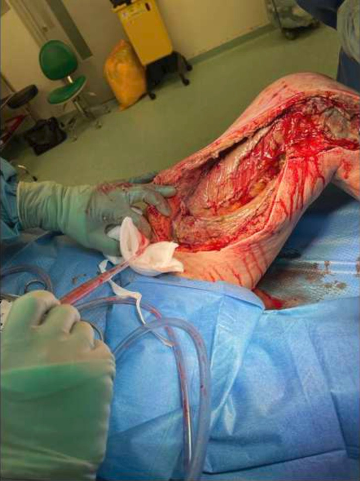
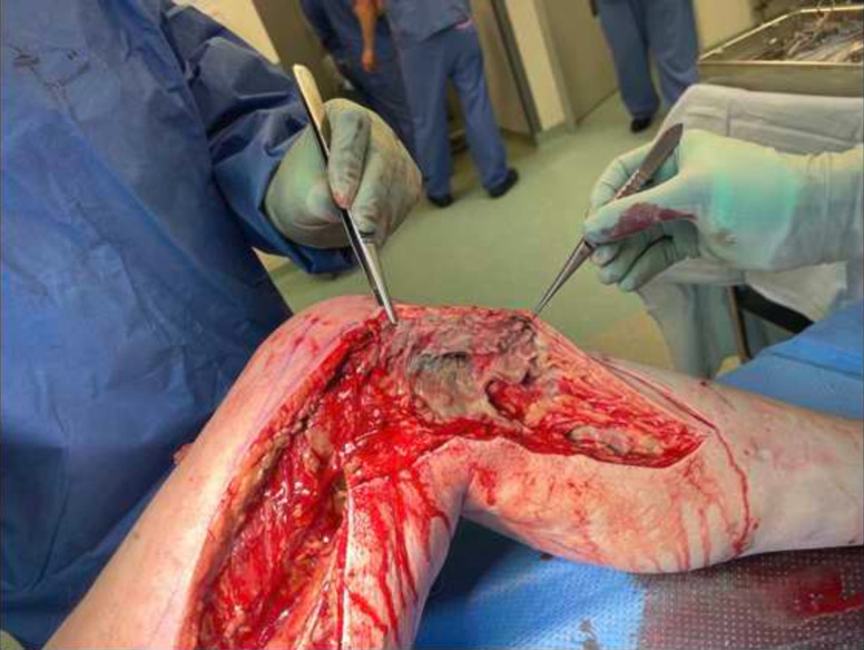
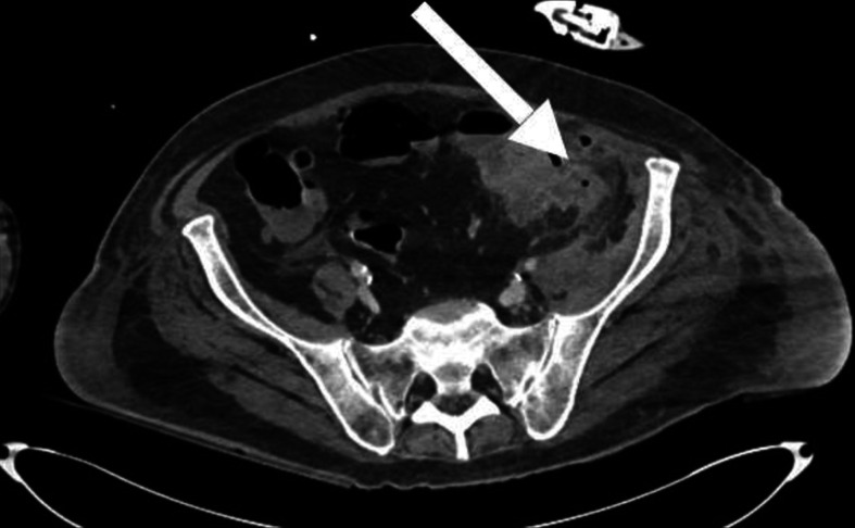
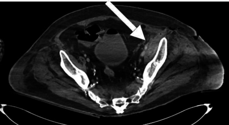
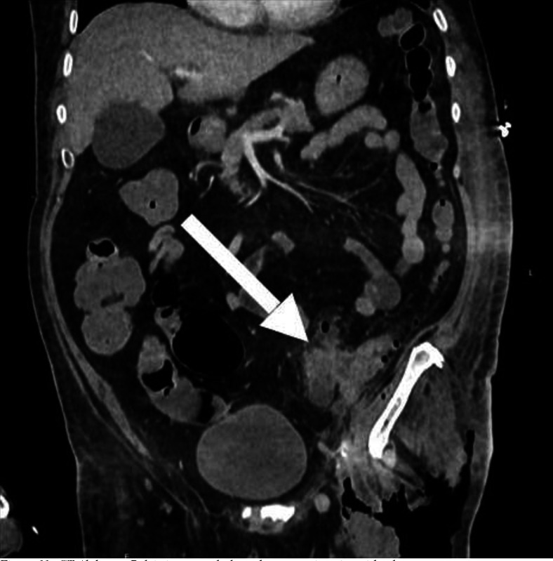
Necrotising fasciitis (NF) is a rare surgical emergency characterised by soft tissue necrosis and systemic compromise. Typically it originates following adjacent skin and soft tissue insult; however, our unusual case required a high index of clinical suspicion to avert potential mortality. A man in his 60s with diabetes mellitus presented with 2 weeks of knee pain, swelling and necrotic skin on the posterior calf. X-ray demonstrated subcutaneous emphysema. Initial debridement confirmed extensive necrotising fasciitis of the whole lower limb with tracking through femoral canal into the abdomen. CT confirmed sigmoid colon perforation. Both life-saving Hartmann's and hip disarticulation procedures were performed with good outcomes. Histology confirmed locally invasive sigmoid colon adenocarcinoma. Our case highlights lower limb necrotising fasciitis as a rare complication secondary to sigmoid perforation associated with malignancy. In cases where the infection nidus cannot be identified, an abdominal source should be considered.

Keywords: Bone and joint infections; Colon cancer; Gas/Free Gas; Orthopaedic and trauma surgery.


Conflict of interest statement

Competing interests: None declared.

Figures

Figure 1
Figure 1. Left knee erythema and swelling.
Figure 2
Figure 2. Area of necrosis on the left calf.
Figure 3
Figure 3. Anterior left knee X-ray showing gas within the fascial planes over the medial aspect.
Figure 4
Figure 4. Lateral left knee X-ray showing gas within the proximal fascial planes.
Figure 5
Figure 5. Initial incision revealing necrotic tissue.
Figure 6
Figure 6. Extension of incisions revealing extensive necrotising fasciitis.
Figure 7
Figure 7. Proximal spread of necrotising fasciitis seen through femoral canal.
Figure 8
Figure 8. Initial debridement of left proximal thigh.
Figure 9
Figure 9. CT abdominal pelvis in transverse plane demonstrating sigmoid colon mass.
Figure 10
Figure 10. CT abdominal pelvis in transverse plane demonstrating sigmoid colon mass.
Figure 11
Figure 11. CT abdominal pelvis in coronal plane demonstrating sigmoid colon mass.

Similar articles

References

    1. Taviloglu K, Yanar H. Necrotizing fasciitis: strategies for diagnosis and management. World J Emerg Surg. 2007;2:19. doi: 10.1186/1749-7922-2-19. - DOI - PMC - PubMed
    1. Puvanendran R, Huey JCM, Pasupathy S. Necrotizing fasciitis. Can Fam Physician. 2009;55:981–7. - PMC - PubMed
    1. Voros D, Pissiotis C, Georgantas D, et al. Role of early and extensive surgery in the treatment of severe necrotizing soft tissue infection. Br J Surg. 1993;80:1190–1. doi: 10.1002/bjs.1800800943. - DOI - PubMed
    1. Frazee BW, Fee C, Lynn J, et al. Community-acquired necrotizing soft tissue infections: a review of 122 cases presenting to a single emergency department over 12 years. J Emerg Med. 2008;34:139–46. doi: 10.1016/j.jemermed.2007.03.041. - DOI - PubMed
    1. Neeki MM, Dong F, Au C, et al. Evaluating the Laboratory Risk Indicator to Differentiate Cellulitis from Necrotizing Fasciitis in the Emergency Department. West J Emerg Med. 2017;18:684–9. doi: 10.5811/westjem.2017.3.33607. - DOI - PMC - PubMed

Publication types

LinkOut - more resources