. 2024 Dec 18;12(12):e009444.
doi: 10.1136/jitc-2024-009444.

LKB1 dictates sensitivity to immunotherapy through Skp2-mediated ubiquitination of PD-L1 protein in non-small cell lung cancer

Affiliations

LKB1 dictates sensitivity to immunotherapy through Skp2-mediated ubiquitination of PD-L1 protein in non-small cell lung cancer

Liting Lv et al. J Immunother Cancer. .

Abstract

Background: Loss-of-function mutations of liver kinase B (LKB1, also termed as STK11 (serine/threonine kinase 11)) are frequently detected in patients with non-small cell lung cancer (NSCLC). The LKB1 mutant NSCLC was refractory to almost all the antitumor treatments, including programmed cell death protein 1 (PD-1)/programmed death-ligand 1 (PD-L1) blockade therapy. Unfortunately, mechanisms underlying resistance to immunotherapy are not fully understood. In this study, we deciphered how LKB1 regulated sensitivity to anti-PD-1/PD-L1 immunotherapy.

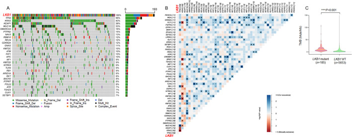
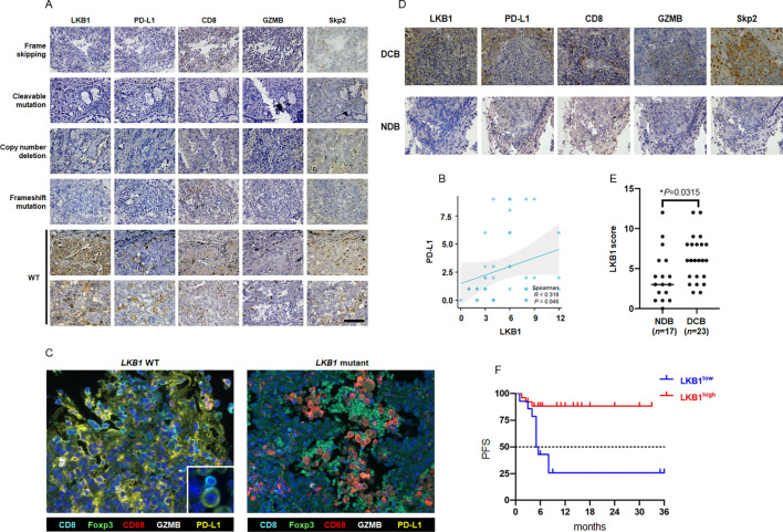
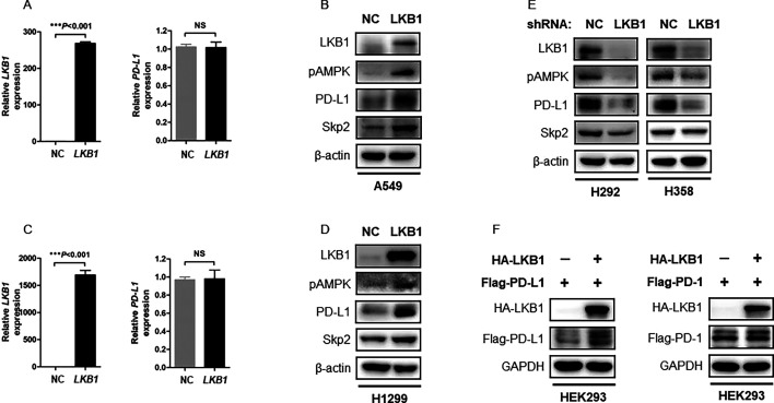
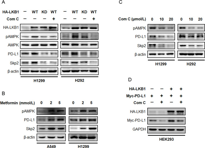
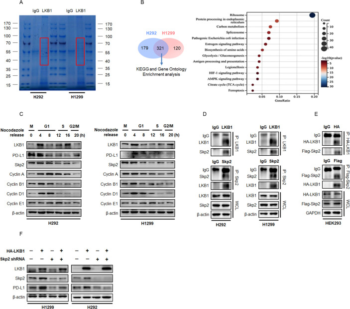
Methods: We investigated the mutational landscape of LKB1 mutant NSCLC in next generation sequencing (NGS) data sets. Expression of LKB1, PD-L1 and S-phase kinase-associated protein 2 (Skp2) in NSCLC samples were assessed by immunohistochemistry (IHC). The tumor microenvironment (TME) profiling of LKB1 wild type (WT) and mutant NSCLC was performed using fluorescent multiplex IHC. Mass spectrometry and enrichment analysis were used to identify LKB1 interacting proteins. Mechanistic pathways were explored by immunoblotting, ubiquitination assay, cycloheximide chase assay and immunoprecipitation assay.

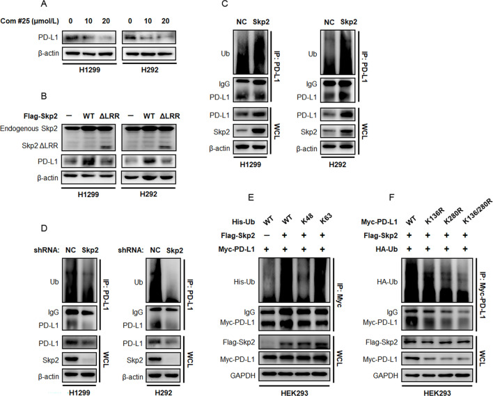
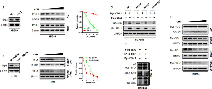
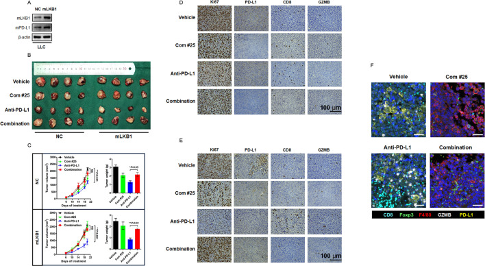
Results: By using NGS data sets and histological approaches, we demonstrated that LKB1 status was positively associated with PD-L1 protein expression and conferred a T cell-enriched "hot" TME in NSCLC. Patients with good responses to anti-PD-1/PD-L1 immunotherapy possessed a high level of LKB1 and PD-L1. Skp2 emerged as the molecular hub connecting LKB1 and PD-L1, by which Skp2 catalyzed K63-linked polyubiquitination on K136 and K280 residues to stabilize PD-L1 protein. Inhibition of Skp2 expression by short hairpin RNA or its E3 ligase activity by compound #25 abrogated intact expression of PD-L1 in vitro and generated a T cell-excluded "cold" TME in vivo. Thus, the LKB1-Skp2-PD-L1 regulatory loop was crucial for retaining PD-L1 protein expression and manipulation of this pathway would be a feasible approach for TME remodeling.

Conclusion: LKB1 and Skp2 are required for intact PD-L1 protein expression and TME remodeling in NSCLC. Inhibition of Skp2 resulted in a conversion from "hot" TME to "cold" TME and abrogated therapeutic outcomes of immunotherapy. Screening LKB1 and Skp2 status would be helpful to select recipients who may benefit from anti-PD-1/PD-L1 immunotherapy.

Keywords: Immune Checkpoint Inhibitor; Immunotherapy; Lung Cancer; Next generation sequencing - NGS; Tumor infiltrating lymphocyte - TIL.


Conflict of interest statement

Competing interests: WL is an employee of Liaoning Kanghui Biotechnology. WL analyzed the next generation sequencing data and did not have access to interpret the bioinformatic and experimental results. The remaining authors did not have any financial association with Liaoning Kanghui Biotechnology or other biomedical companies.

Figures

Figure 1
Figure 1. The mutational landscape of LKB1-mutant NSCLC. (A) A patient cohort consisting of 193 cases of NSCLC harboring LKB1 mutations was analyzed. Tumor samples were arranged from left to right. Alterations of LKB1 co-occurring genes were annotated for each sample according to the color panel below the image. The somatic mutation frequencies for each candidate gene were plotted on the right panel. (B) Co-mutation gene pattern analysis of the LKB1 mutant cohort. The gene pairs in blue color indicated the probability of co-mutation, and the red color indicated the probability of mutually exclusive. (C) The distribution of TMB in LKB1 mutant and LKB1 WT cohorts. LKB1, liver kinase B; NSCLC, non-small cell lung cancer; TMB, tumor mutation burden; WT, wild type.
Figure 2
Figure 2. Effect of LKB1 on TME and sensitivity to anti-PD-1/PD-L1 immunotherapy in patients with NSCLC. (A) A total number of 40 cases of surgical resected NSCLC were analyzed for LKB1, PD-L1, CD8, GZMB and Skp2 expression by IHC staining. Representative IHC images of LKB1 mutant tumor (the top four panels) and LKB1 WT (the bottom two panels) tumor were shown. The distinct alterations in LKB1 were listed in the left side of images. (B) The linear regression analysis of IHC H-score to determine the correlation between LKB1 and PD-L1. (C) Analysis of TME components in LKB1-WT and LKB1-mutant NSCLC by fluorescent mIHC. Each fluorescent channel indicated a specific biomarker for immune cells or tumor cells. Cell nucleus was visualized by DAPI staining. (D) Representative IHC images of LKB1, PD-L1, CD8, GZMB and Skp2 protein expression in patients with NSCLC receiving anti-PD-1/PD-L1 immunotherapy with different therapeutic outcomes. (E) The distribution of LKB1 IHC-H score in patients with NSCLC with favorable response (DCB) and unfavorable response (NDB) to anti-PD-1/PD-L1 immunotherapy. ((F) Progression-free survival analysis of patients with NSCLC receiving anti-PD-1/PD-L1 immunotherapy with different LKB1 status. DCB, durable clinical benefit; GZMB, granzyme B; IHC, immunohistochemistry; LKB1, liver kinase B; mIHC, multiplex immunohistochemistry; NDB, no durable benefit; NSCLC, non-small cell lung cancer; PD-1, programmed cell death protein-1; PD-L1, programmed death-ligand 1; PFS, progression-free survival; Skp2, S-phase kinase-associated protein 2; TME, tumor microenvironment; WT, wild type.
Figure 3
Figure 3. LKB1 selectively regulates PD-L1 protein expression. (A) qPCR assay of the LKB1-null A549 cells transfected with NC or LKB1 plasmids. Expression of LKB1 and PD-L1 mRNA was determined by qPCR analysis (NS: not significant). (B) Immunoblotting of A549 cell lysates transfected with NC or LKB1 plasmids. β-actin was used as equal loading control. (C) Measurement of PD-L1 mRNA expression in H1299 cells transfected with NC or LKB1 plasmids (NS: not significant). (D) Immunoblotting of H1299 cell lysates transfected with NC or LKB1 plasmids. (E) Immunoblotting of cellular extracts from H292 and H358 stable cells infected with NC shRNA or LKB1 shRNA lentivirus. (F) HEK293 cells were transfected with HA-LKB1, together with Flag-PD-L1 or Flag-PD-1 plasmids. 48 hours after transfection, cells were lyzed for immunoblotting analysis of Flag-tagged PD-L1 or PD-1 protein expression. GAPDH was used as equal loading control. LKB1, liver kinase B; mRNA, messenger RNA; NC, negative control; PD-1, programmed cell death protein-1; PD-L1, programmed death-ligand 1; qPCR, quantitative real-time PCR; shRNA, short hairpin RNA; Skp2, S-phase kinase-associated protein 2.
Figure 4
Figure 4. LKB1/AMPK pathway determines PD-L1 protein expression. (A) H1299 and H292 cells were transfected with HA-LKB1 WT or dominant negative HA-LKB1 KD mutant, and treated with 20 µmol/L compound C for 24 hours. Immunoblotting assay was performed to detect the expression indicated proteins after treatment. (B) A549 and H1299 cells were treated with metformin, an AMPK agonist, for 48 hours. Expression of pAMPK, PD-L1 and Skp2 was determined by immunoblotting. (C) H1299 and H292 cells were treated with increasing concentrations of compound C and evaluated for pAMPK, PD-L1 and Skp2 expression by Western blot. (D) HEK293 cells were transfected with HA-LKB1 and Myc-PD-L1, treated with or without compound C to block LKB1/AMPK pathway. Protein level of ectopically expressed Myc-PD-L1 was assessed by immunoblotting. AMPK, AMP-activated protein kinase; KD, kinase dead; LKB1, liver kinase B; PD-L1, programmed death-ligand 1; Skp2, S-phase kinase-associated protein 2; WT, wild type.
Figure 5
Figure 5. LKB1 regulates PD-L1 expression through Skp2 in NSCLC. (A) Representative image of Coomassie blue stained SDS-PAGE gel. Proteins that interacted with LKB1 were pulled down by anti-LKB1 antibody, separated by electrophoresis and visualized by Coomassie blue staining. Proteins potentially interacted with LKB1 were highlighted in red box and the gels were excised and proceed for MS analysis. (B) Venn diagram showing the overlap of LKB1 interacting proteins between H292 (n=500) and H1299 (n=441) cells. Enrichment analysis on overlap proteins using clusterProfiler in GO terms. Ranking of the interacting proteins among these enriched signatures according to their frequency. (C) Immunoblotting of whole cell lysates (WCL) derived from H292 and H1299 cells synchronized in M phase by nocodazole followed by releasing back into cell cycle. (D) Reciprocal immunoprecipitation of endogenous LKB1 and Skp2 in H292 and H1299 cells. β-actin was used as equal loading control. (E) Reciprocal immunoprecipitation of ectopically expressed HA-tagged LKB1 and Flag-tagged Skp2 in HEK293 cells. GAPDH was used as equal loading control. (F) H1299 and H292 cells were stably expressed HA-LKB1 with or without shRNA targeting Skp2. Expression of LKB1, Skp2 and PD-L1 was determined by immunoblotting. GO, Gene Ontology; IgG, immunoglobulin G; LKB1, liver kinase B; MS, mass spectrometry; NSCLC, non-small cell lung cancer; PD-L1, programmed death-ligand 1; shRNA, short hairpin RNA; Skp2, S-phase kinase-associated protein 2.
Figure 6
Figure 6. Skp2 promotes ubiquitination of PD-L1 protein. (A) H1299 and H292 cells were treated with increasing concentrations of compound #25 for 48 hours and evaluated for endogenous PD-L1 protein expression. (B) H1299 and H292 cells were transfected with Flag-tagged Skp2 WT or its E3 ligase deficient ΔLRR mutant. The effect of Skp2 on endogenous PD-L1 protein expression was assessed by immunoblotting. (C) Endogenous PD-L1 protein in H1299 and H292 cells was pulled down by an anti-PD-L1 antibody and PD-L1 ubiquitination status was determined with an anti-Ub antibody. (D) Effect of shRNA-mediated Skp2 inhibition on PD-L1 protein ubiquitination in H1299 and H292 cells. (E) HEK293 cells were transfected with Myc-PD-L1, Flag-Skp2, together with His-Ub WT, His-Ub K48-only or His-Ub K63-only mutants. PD-L1 protein was precipitated by an anti-Myc tag antibody and its ubiquitination status was probed with an antibody targeting His-tag protein. (F) HEK293 cells were transfected with HA-Ub, Flag-Skp2, together with Myc-PD-L1 WT, Myc-PD-L1 K136R mutant, Myc-PD-L1 K280R mutant, or K136/280R double mutant. Recombinant Myc-PD-L1 protein was precipitated by an anti-Myc tag antibody and its ubiquitination state was probed with an antibody targeting HA tag protein. IgG, immunoglobulin G; NC, negative control; PD-L1, programmed death-ligand 1; shRNA, short hairpin RNA; Skp2, S-phase kinase-associated protein 2; Ub, ubiquitin; WCL, whole cell lysates; WT, wild type.
Figure 7
Figure 7. Effect of Skp2 on PD-L1 protein stability. (A) Protein synthesis was stalled by CHX treatment. The half-life of PD-L1 protein in H1299 cells with overexpression of Skp2 or NC was measured by CHX chase assay. Representative PD-L1 protein intensity curve following CHX treatment was shown. (B) The endogenous Skp2 in H1299 cells were knockdown by shRNA. The effect of Skp2 inhibition on PD-L1 protein stability was evaluated by CHX chase assay. Representative PD-L1 protein intensity curve following CHX treatment was shown. (C) Myc-PD-L1 WT or its KR mutants, together with or without Flag-Skp2, were expressed in HEK293 cells. Expression of Myc-PD-L1 WT or recombinant Myc-PD-L1 KR protein was evaluated by immunoblotting. (D) Myc-PD-L1 WT or its KR mutants was expressed in HEK293 cells. PD-L1 protein stability was determined by CHX chase assay. (E) Myc-PD-L1, HA-β-TrCP plasmids, with or without Flag-Skp2, were expressed into HEK293 cells. The PD-L1 binding to HA-β-TrCP or Flag-Skp2 was determined by immunoprecipitation assay. β-TrCP, β-transducin repeats-containing protein; CHX, cycloheximide; IgG, immunoglobulin G; NC, negative control; PD-L1, programmed death-ligand 1; shRNA, short hairpin RNA; Skp2, S-phase kinase-associated protein 2; WT, wild type.
Figure 8
Figure 8. Inhibition of Skp2 antagonized PD-L1 blockade therapy and generated a “cold” TME. (A) Mouse LKB1 was stably expressed in LLC cells and its expression was confirmed by Western blot analysis. (B) Representative image of the gross observation of isolated subcutaneous tumor in mice treated with Vehicle, compound #25 (40 mg/kg daily), anti-mouse PD-L1 antibody (200 µg per week), or their combination. (C) The syngeneic tumor volumes were calculated and recorded at indicated time points. NS, not significant (combination vs vehicle). **p<0.01 (anti-PD-L1 vs vehicle). At the end of experiment, tumor nodules were carefully isolated and weighted. *p<0.05 and **p<0.01 (anti-PD-L1 vs combination). (D) Representative IHC staining of Ki67, PD-L1, CD8 and GZMB for NC syngeneic tumor after indicated treatment. (E) Representative IHC staining of Ki67, PD-L1, CD8 and GZMB for mLKB1 syngeneic tumor after indicated treatment. (F) mIHC staining of TME component after indicated treatment in vivo. Each fluorescent channel indicated a specific biomarker for immune cells or tumor cells. Cell nucleus was visualized by DAPI staining. GZMB, granzyme B; IHC, immunohistochemistry; LKB1, liver kinase B; LLC, Lewis lung cancer; mIHC, multiplex immunohistochemistry; NC, negative control; PD-L1, programmed death-ligand 1; Skp2, S-phase kinase-associated protein 2; TME, tumor microenvironment.

References

    1. Iwai Y, Ishida M, Tanaka Y, et al. Involvement of PD-L1 on tumor cells in the escape from host immune system and tumor immunotherapy by PD-L1 blockade. Proc Natl Acad Sci U S A. 2002;99:12293–7. doi: 10.1073/pnas.192461099. - DOI - PMC - PubMed
    1. Errico A. Immunotherapy: PD-1-PD-L1 axis: efficient checkpoint blockade against cancer. Nat Rev Clin Oncol. 2015;12:63. doi: 10.1038/nrclinonc.2014.221. - DOI - PubMed
    1. Clift R, Souratha J, Garrovillo SA, et al. Remodeling the Tumor Microenvironment Sensitizes Breast Tumors to Anti-Programmed Death-Ligand 1 Immunotherapy. Cancer Res. 2019;79:4149–59. doi: 10.1158/0008-5472.CAN-18-3060. - DOI - PubMed
    1. Patel SP, Kurzrock R. PD-L1 Expression as a Predictive Biomarker in Cancer Immunotherapy. Mol Cancer Ther. 2015;14:847–56. doi: 10.1158/1535-7163.MCT-14-0983. - DOI - PubMed
    1. Lin H, Wei S, Hurt EM, et al. Host expression of PD-L1 determines efficacy of PD-L1 pathway blockade-mediated tumor regression. J Clin Invest. 2018;128:805–15.:96113. doi: 10.1172/JCI96113. - DOI - PMC - PubMed

MeSH terms