. 2024 Dec 18;12(12):e009523.
doi: 10.1136/jitc-2024-009523.

Blocking IL1RAP on cancer-associated fibroblasts in pancreatic ductal adenocarcinoma suppresses IL-1-induced neutrophil recruitment

Affiliations

Blocking IL1RAP on cancer-associated fibroblasts in pancreatic ductal adenocarcinoma suppresses IL-1-induced neutrophil recruitment

Nils Hansen et al. J Immunother Cancer. .

Abstract

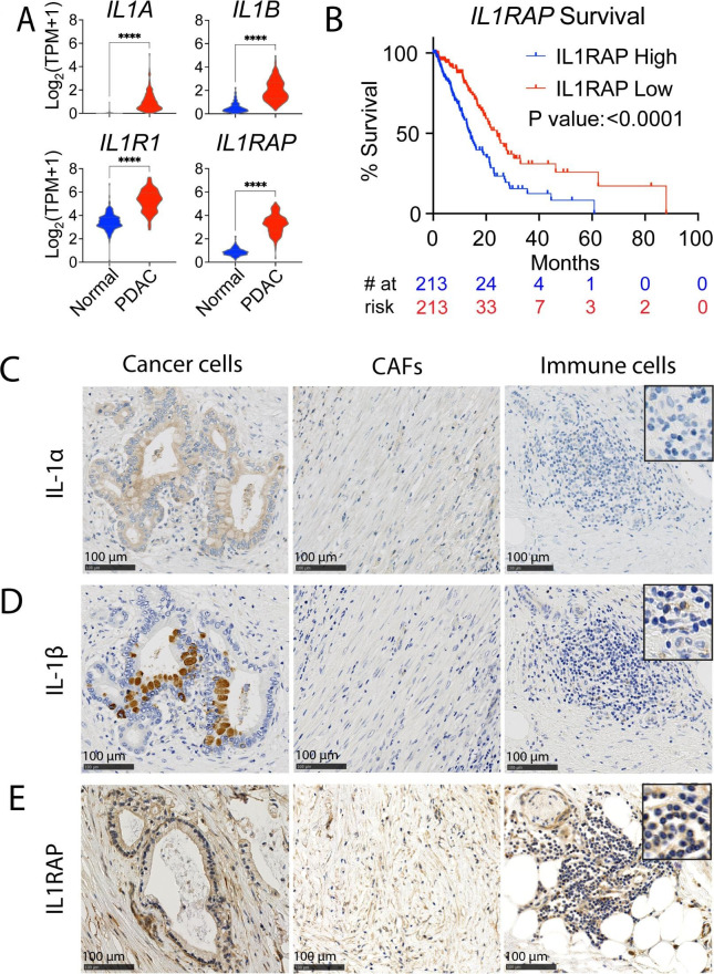
Background: Pancreatic ductal adenocarcinoma (PDAC) represents a major clinical challenge due to its tumor microenvironment, which exhibits immune-suppressive properties that facilitate cancer progression, metastasis, and therapy resistance. Interleukin 1 (IL-1) signaling has been implicated as a driver in this process. Mechanistically, both IL-1α and IL-1β bind to the IL-1 receptor type 1, forming a complex with IL-1-receptor accessory protein (IL1RAP), which triggers downstream signaling pathways. The IL1RAP blocking antibody nadunolimab is currently in clinical development, but the precise consequences of inhibiting IL-1 signaling in PDAC remains elusive.

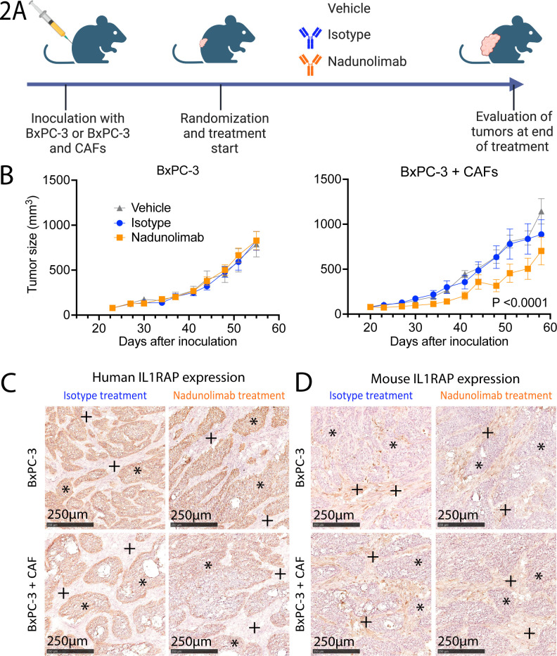
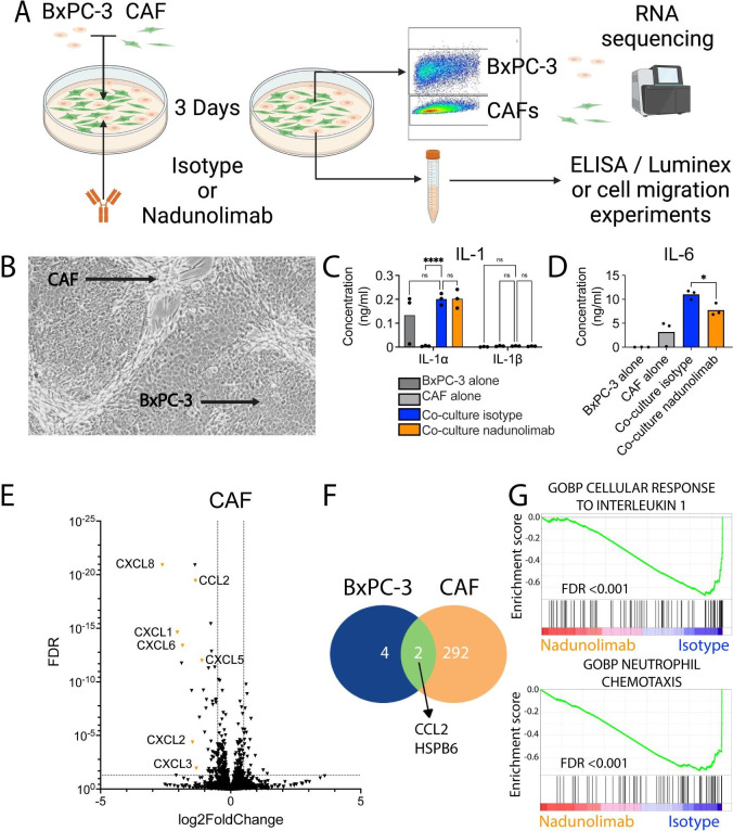
Methods: To evaluate the biological relevance of blocking IL1RAP using nadunolimab in a PDAC animal model, human PDAC cells and cancer-associated fibroblasts (CAFs) were co-transplanted into mice. To study the underlying mechanisms of IL1RAP blockade ex vivo, co-cultured PDAC cells and CAFs were treated with nadunolimab prior to RNA sequencing. Migration assays were performed to assess how nadunolimab affects interactions between CAFs and myeloid immune cells. Finally, to establish a clinical correlation between IL1RAP expression and nadunolimab treatment effects, we analyzed tumor biopsies from a clinical phase I/II study in which nadunolimab was administered to patients.

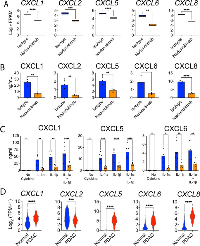
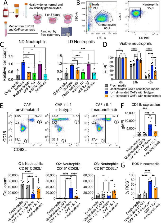
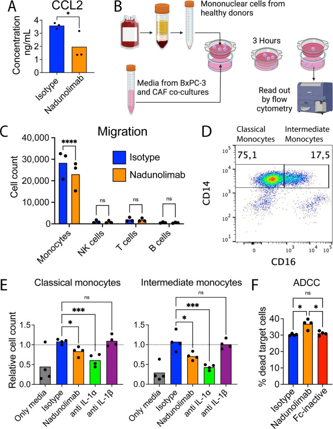
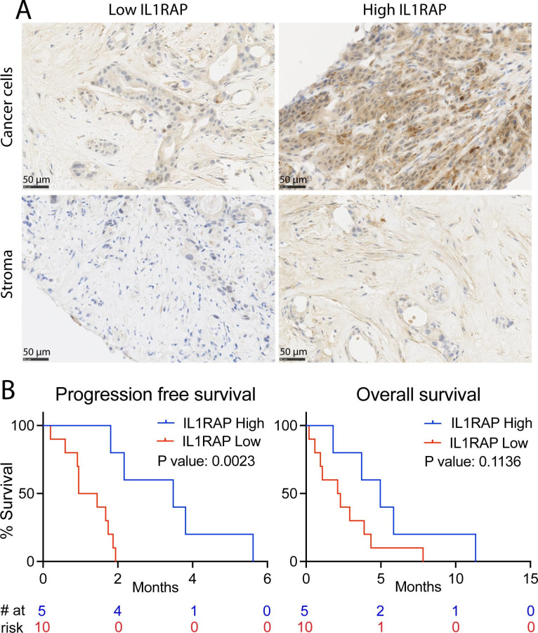
Results: In the xenograft mouse model, nadunolimab exhibited antitumor effects only when human CAFs were co-transplanted with PDAC cells. IL-1 stimulation induced CAFs to secrete chemokines that recruited neutrophils and monocytes. The secretion of this chemokine and the migration of myeloid cells were inhibited by nadunolimab. Media conditioned by IL-1-stimulated CAFs sustained a neutrophil population with a tissue invasion phenotype, an effect that was reversed by nadunolimab. In a cohort of metastatic late-stage PDAC patients receiving nadunolimab as monotherapy, high IL1RAP expression in tumors was associated with extended progression-free survival.

Conclusions: Our study demonstrates that targeting IL1RAP on CAFs inhibits IL-1-induced chemokine secretion and recruitment of neutrophils and monocytes, thereby counteracting the immunosuppressive microenvironment in PDAC. These findings highlight the therapeutic potential of targeting IL1RAP in PDAC.

Keywords: Cytokine; Monoclonal antibody; Monocyte; Neutrophil.


Conflict of interest statement

Competing interests: NH, PS, CG, SM, KvW, CRM, DL, and MJ are current employees of, and/or hold stocks or stock options in Cantargia AB, Lund, Sweden. The use of nadunolimab for cancer treatment is patented within WO 2015/13602.

Figures

Figure 1
Figure 1. IL1RAP is broadly expressed in pancreatic tumors. (A) Gene expression data from GTEx and TCGA public datasets, comparing gene expression in normal pancreatic tissue (N=167) with pancreatic ductal adenocarcinoma (PDAC) tumors (N=150). (B) Survival analysis of PDAC patients grouped by median IL1RAP expression in the Know Your Tumor data set. (C–E) IHC stains for IL-1α, IL-1β, and IL1RAP in a PDAC tumor, showing expression on cancer cells (left images), cancer-associated fibroblasts (CAFs) (middle images), and immune cells (right images). The images shown are representative stains out of five tumors tested. A 2.5× magnification of selected cells is shown in the upper right corner. Significance calculated by Mann-Whitney test in (A) and by Log-rank (Mantel-Cox) test in (B). Significance levels are indicated as ****p<0.0001. IHC, Immunohistochemistry.
Figure 2
Figure 2. Nadunolimab elicits a CAF-dependent antitumor effect. (A) Experimental outline of in vivo experiments with BxPC-3 cells transplanted subcutaneously into mice, alone or mixed with CAFs before injection. The recipient mice were subsequently divided into groups (N=9–10) and treated with a murine IgG2a variant of the human-specific IL1RAP antibody nadunolimab, or matching isotype control (10 mg/kg, twice per week). Tumor volumes were monitored, and tumor tissue harvested at experimental endpoint (created with BioRender.com). (B) Tumor size of mice receiving BxPC-3 cells alone (left panel) or mixed with CAFs (right panel). Mean tumor volume and SE of the mean are plotted. Significance between isotype and nadunolimab-treated groups over the treatment period is indicated and calculated by two-way ANOVA test of the three treatment arms with Tukey correction. (C–D) IHC stain of tumors at the experimental endpoint, using human specific (C) and mouse specific (D) IL1RAP antibodies. *BxPC-3-regions, +CAF-regions. ANOVA, analysis of variance; CAF, cancer-associated fibroblast; IHC, immunohistochemistry.
Figure 3
Figure 3. Nadunolimab treatment primarily affects cancer-associated fibroblasts (CAFs). (A) The pancreatic ductal adenocarcinoma (PDAC) cell line BxPC-3 and CAFs were cultured separately or in a mixed co-culture, with 20 µg/mL nadunolimab or isotype control antibody for 3 days. The BxPC-3 cells and CAFs were sorted by flow cytometry based on EpCAM expression, and followed by RNA sequencing. Cell culture media was harvested for further experiments (created with BioRender.com). (B) Image of a co-culture with CAF and BxPC-3 cells indicated. (C) Concentration of IL-1 in media from single and co-cultures with nadunolimab or isotype control. (D) Concentration of IL-6 in media. (E) Volcano plot of genes dysregulated in CAFs from co-cultures treated with nadunolimab. (F) Number of genes significantly dysregulated (FDR<0.01) following nadunolimab treatment. (G) GSEA enrichment plots showing gene sets negatively enriched in CAFs following nadunolimab treatment. Significance in (C) and (D) was calculated using one-way ANOVA with Šidák correction and the indicated comparisons and bar shows mean value with N=3. To identify significantly dysregulated gens in (E) and (F), we used a Wald test with Benjamini-Hochberg multiple test correction. Significance levels are indicated as *p<0.05, ****p<0.0001. ANOVA, analysis of variance; FDR, false discovery rate; GOBP, gene ontology biological processes; GSEA, gene set enrichment analysis; ns, not significant.
Figure 4
Figure 4. IL-1-induced CAF secretion of CXCR1/2 ligands is suppressed by IL1RAP blockage. (A) RNA expression of genes encoding chemokines in CAF and BxPC-3 co-cultures treated with nadunolimab or isotype control (average of N=4 with boxes showing minimum and maximum values; data from the RNA sequencing experiment are presented in figure 3). (B) Concentration of corresponding chemokines in the media from CAF and BxPC-3 co-cultures treated with nadunolimab or isotype control. The full data set is shown in online supplemental figure S4A. (C) CXCL1, CXCL5 and CXCL6 levels in media after 24 hours culture of CAFs, stimulated with 0.1 ng/mL IL-1α and IL-1β in combination with nadunolimab or isotype control (20 µg/mL). (D) Expression of CXCL chemokines in normal pancreas (N=167) and PDAC tumors (N=150) in the GTEx and TCGA data sets. Significance is calculated by one-way ANOVA test with Šidák correction in (A–C) and by Mann-Whitney test in (D). Mean values in A to C are shown with N=3-6. Significance levels are indicated as *p<0.05; **p<0.01; ***p<0.001; ****p<0.0001. ANOVA, analysis of variance; CAF, cancer-associated fibroblast. FPKM, fragments per million; PDAC, pancreatic ductal adenocarcinoma; ns, not significant. TPM, transcripts per million.
Figure 5
Figure 5. Blockage of IL1RAP on cancer-associated fibroblasts (CAFs) reduces neutrophil chemotaxis, survival, and activation. (A) Outline of the migration assay using normal density (ND) and low density (LD) neutrophils from healthy blood. After separation based on density, erythrocytes were lysed, and granulocytes were collected. Cells were stained with fluorescently labeled antibodies and allowed to migrate over a cell culture insert toward conditioned media, followed by cell counting by flow cytometry (created with BioRender.com). (B) Representative pseudo-color plots from the neutrophil migration assay. Neutrophils were gated as CD15+CD49D cells from the granulocyte population in FSC and SSC. (C) Neutrophil chemotaxis using nadunolimab, IL-1 blocking antibodies (20 µg/mL), and anakinra (500 ng/mL) during the media conditioning. Each experiment was normalized to media from co-cultures without addition of antibodies (N=4). (D) Viability of healthy donor ND neutrophils after culture in CAF-conditioned media diluted 1:4 (N=4 or 7). (E) Flow cytometry of CD16 and CD62L expression on neutrophils after 24 hours culture. Upper panels show pseudo-color plots of the neutrophil populations. Lower panels show quantification of neutrophil numbers in the respective gates from the top panels (N=7). (F) CD11b expression on neutrophils after 24 hours culture with CAF conditioned media (N=7). (G) Percentage of neutrophils with ROS production after 24 hours culture with CAF conditioned media (N=3). Mean values are shown, and significance was calculated using one-way analysis of variance (ANOVA) with Dunnet correction except in (D) where two-way ANOVA with Dunnet correction was used, with the indicated pair-wise comparisons. *p<0.05, **p<0.01, ***p<0.001, ****p<0.0001. CAF, cancer-associated fibroblast; FSC-A, forward scatter area; gMFI, geometric mean fluorescence intensity; ns, not significant. PI, propidium iodide; ROS, reactive oxygen species; SSC-A, side scatter area.
Figure 6
Figure 6. Nadunolimab reduces monocyte chemotaxis and induces antibody-dependent cellular cytotoxicity (ADCC). (A) Concentration of CCL2 in supernatants of BxPC-3 and CAF co-cultures. See online supplemental figure S7 for the full analysis. (B) Outline of a transwell migration assay with PBMCs, stained with fluorescently labeled antibodies and allowed to migrate toward co-culture conditioned media with nadunolimab or isotype antibody added to the co-culture. Migrated cells were assessed and counted by flow cytometry (created with BioRender.com). (C) Counts of migrated monocytes (CD14+), NK cells (CD56+), T cells (CD3+), and B cells (CD19+). (D) Representative plot of CD14 and CD16 expression on monocytes with the gating strategy used to assess classical and intermediate monocyte populations in the migration assay. (E) Monocyte migration with co-culture conditioned media and blockage of IL1RAP or IL-1 in the BxPC-3 and CAF co-cultures. Mean values are shown with N=3 or 4. Experiments were normalized to media from co-cultures without addition of antibodies. (F) Percentage of dead target cells after an ADCC assay with BxPC-3 target cells, monocytes (CD14 enriched PBMCs), and antibodies at 20 µg/mL. Significance calculated by one-way ANOVA with Šidák correction in (A) and (C), Dunnet correction in (E) and (F), and indicated as *p<0.05; ***p<0.001; ****p<0.0001. ANOVA, analysis of variance; CAF, cancer-associated fibroblast; ns, not significant. NK, natural killer; PBMCs, peripheral blood mononuclear cells.
Figure 7
Figure 7. High IL1RAP expression is associated with longer survival in nadunolimab-treated patients. (A) Representative histology slides where cancer cells (upper panels) and stroma cells (lower panels). Tumor and stroma cells were scored as having low IL1RAP expression (Tumor cell H-score ≤100, and stroma staining intensity assessment ≤1, left panels) or high IL1RAP expression (Tumor cell H-score >100 and stroma staining intensity assessment >1, right panels). (B) Progression-free and overall survival of PDAC patients receiving nadunolimab as monotherapy, with survival analysis based on low or high combined IL1RAP expression in the tumor. The combined score was defined as high when both the tumor cells and stromal cells scored high according to the above criteria (N=5). All other samples were categorized as having a low combined score (N=10). Significance in the survival analysis was tested by log-rank (Mantel-Cox) test.

References

    1. Bengtsson A, Andersson R, Ansari D. The actual 5-year survivors of pancreatic ductal adenocarcinoma based on real-world data. Sci Rep. 2020;10:16425. doi: 10.1038/s41598-020-73525-y. - DOI - PMC - PubMed
    1. Muller M, Haghnejad V, Schaefer M, et al. The Immune Landscape of Human Pancreatic Ductal Carcinoma: Key Players, Clinical Implications, and Challenges. Cancers (Basel) 2022;14:995. doi: 10.3390/cancers14040995. - DOI - PMC - PubMed
    1. Morgan A, Griffin M, Kameni L, et al. Medical Biology of Cancer-Associated Fibroblasts in Pancreatic Cancer. Biology (Basel) 2023;12:1044. doi: 10.3390/biology12081044. - DOI - PMC - PubMed
    1. Truong L-H, Pauklin S. Pancreatic Cancer Microenvironment and Cellular Composition: Current Understandings and Therapeutic Approaches. Cancers (Basel) 2021;13:5028. doi: 10.3390/cancers13195028. - DOI - PMC - PubMed
    1. Biffi G, Oni TE, Spielman B, et al. IL1-Induced JAK/STAT Signaling Is Antagonized by TGFβ to Shape CAF Heterogeneity in Pancreatic Ductal Adenocarcinoma. Cancer Discov. 2019;9:282–301. doi: 10.1158/2159-8290.CD-18-0710. - DOI - PMC - PubMed

MeSH terms

Substances

LinkOut - more resources