

Right ventricular outflow tract doppler flow abnormalities suggestive of pulmonary embolism - case series and review
- PMID: 39695024
- PMCID: PMC11655731
- DOI: 10.1186/s13089-024-00377-2
Right ventricular outflow tract doppler flow abnormalities suggestive of pulmonary embolism - case series and review
Abstract
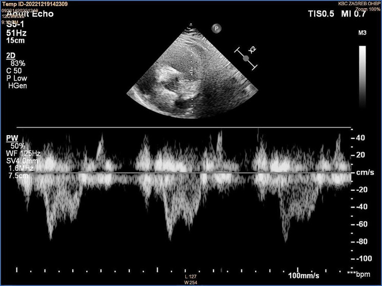
Background: Pulmonary embolism (PE) is one of the most challenging diagnoses in emergency medicine, mainly because symptoms range from asymptomatic disease to sudden death. The role of echocardiography in the workup of suspected PE has been supportive and used primarily to assess the right ventricular (RV) size and function, which is important for risk stratification. Several echocardiographic parameters described in the literature lack the desired accuracy. Recently, a potential value of less well-recognized RV outflow tract (RVOT) Doppler variables has been reported. The early systolic notching (ESN) pattern was observed in 92% of patients with high and intermediate risk PE, making it a promising sign in selected PE patients.
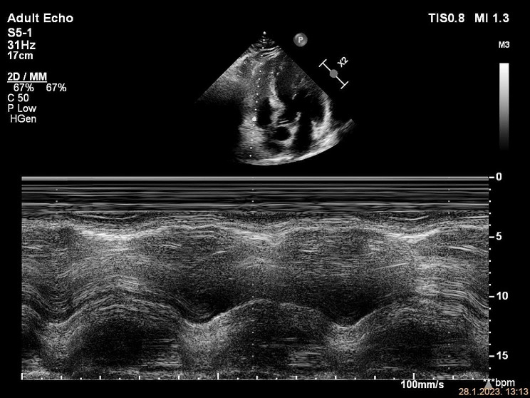
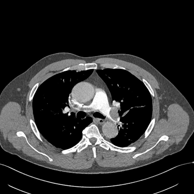
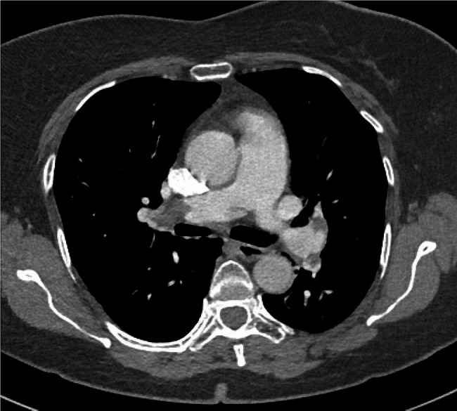
Case presentation: In this case series, we demonstrate a typical ESN pattern on RVOT Doppler evaluation in three patients with intermediate-risk PE presenting to our emergency department (ED). None of the patients had been previously diagnosed with pulmonary hypertension or other chronic pulmonary and cardiac disease. The pre-test probability was low. Massive proximal emboli were found on CT angiograms, involving pulmonary truncus or main pulmonary arteries. Previously, the ESN pattern was identified on a focused echocardiogram, which was the only echocardiographic indicator of increased pulmonary vascular resistance.
Conclusions: RVOT Doppler flow pattern of ESN has potential clinical utility for the detection of PE in ED patients. ESN could identify patients at higher risk, which are otherwise stratified as low risk according to the latest guidelines. Moreover, this case series illustrates that even in the absence of other echocardiographic findings of RV strain, the presence of ESN should alert to the possibility of acute PE. Further prospective studies are needed to assess its diagnostic value in a selected subgroup of patients, similar to the cases presented, that would have no other obvious reason for the altered RVOT Doppler curve.
Keywords: Early systolic notching; Emergency medicine; Focused echocardiography; Pulmonary embolism; Pulsed wave doppler; Right ventricular outflow tract.
© 2024. The Author(s).
Conflict of interest statement
Declarations. Ethics approval and consent to participate: Informed written consent for case reports was obtained for all patients. Consent for publication: Informed written consent for publication of each case report was obtained for all patients. Competing interests: The authors declare that they have no known competing financial interests or personal relationships that could have appeared to influence the work reported in this paper.
Figures


Similar articles
-
A Doppler Echocardiographic Pulmonary Flow Marker of Massive or Submassive Acute Pulmonary Embolus.J Am Soc Echocardiogr. 2019 Jul;32(7):799-806. doi: 10.1016/j.echo.2019.03.004. Epub 2019 May 2. J Am Soc Echocardiogr. 2019. PMID: 31056367
-
Diagnostic Accuracy of Early Systolic Notching in Pulmonary Embolism.J Ultrasound Med. 2022 Mar;41(3):637-644. doi: 10.1002/jum.15744. Epub 2021 May 14. J Ultrasound Med. 2022. PMID: 33987920
-
Analysis of the 60/60 Sign and Other Right Ventricular Parameters by 2D Transthoracic Echocardiography as Adjuncts to Diagnosis of Acute Pulmonary Embolism.Cureus. 2021 Mar 10;13(3):e13800. doi: 10.7759/cureus.13800. Cureus. 2021. PMID: 33842172 Free PMC article.
-
Assessment of right ventricular systolic function by tissue Doppler echocardiography.Dan Med J. 2012 Mar;59(3):B4409. Dan Med J. 2012. PMID: 22381093 Review.
-
What echocardiographic findings differentiate acute pulmonary embolism and chronic pulmonary hypertension?Am J Emerg Med. 2023 Oct;72:72-84. doi: 10.1016/j.ajem.2023.07.011. Epub 2023 Jul 10. Am J Emerg Med. 2023. PMID: 37499553 Review.
References
-
- Raskob GE, Angchaisuksiri P, Blanco AN et al (2014) Thrombosis: a major contributor to Global Disease Burden. Arterioscler Thromb Vasc Biol 34:2363–2371. 10.1161/ATVBAHA.114.304488 - PubMed
-
- de Miguel-Díez J, Jiménez-García R, Jiménez D et al (2014) Trends in hospital admissions for pulmonary embolism in Spain from 2002 to 2011. Eur Respir J 44:942–950. 10.1183/09031936.00194213 - PubMed
-
- Dentali F, Ageno W, Pomero F et al (2016) Time trends and case fatality rate of in-hospital treated pulmonary embolism during 11 years of observation in Northwestern Italy. Thromb Haemost 115:399–405. 10.1160/th15-02-0172 - PubMed
-
- Keller K, Hobohm L, Ebner M et al (2020) Trends in thrombolytic treatment and outcomes of acute pulmonary embolism in Germany. Eur Heart J 41:522–529. 10.1093/eurheartj/ehz236 - PubMed
LinkOut - more resources
Full Text Sources