. 2024 Dec 18;14(1):492.
doi: 10.1038/s41398-024-03190-6.

The clinically relevant MEK inhibitor mirdametinib combined with D-cycloserine and prediction error disrupts fear memory in PTSD models

Affiliations

The clinically relevant MEK inhibitor mirdametinib combined with D-cycloserine and prediction error disrupts fear memory in PTSD models

Sanket B Raut et al. Transl Psychiatry. .

Abstract

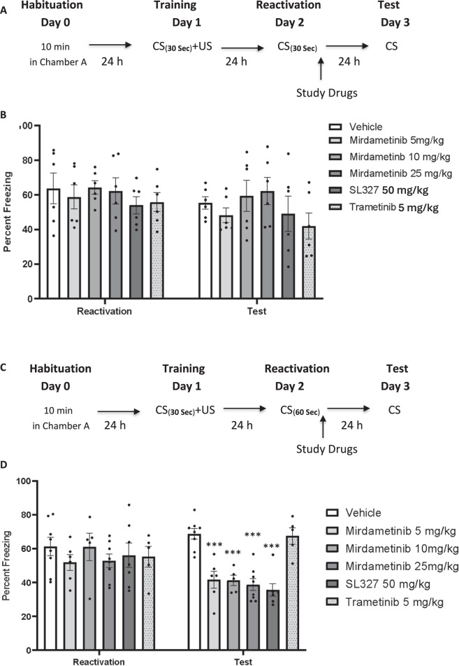
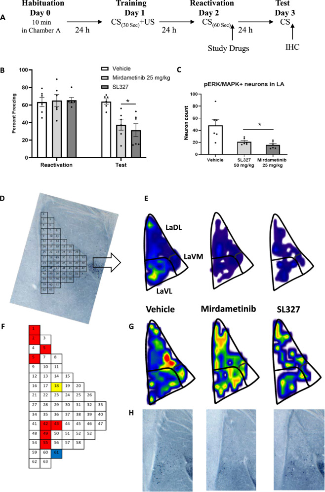
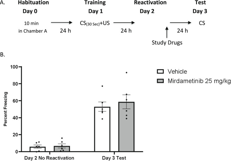
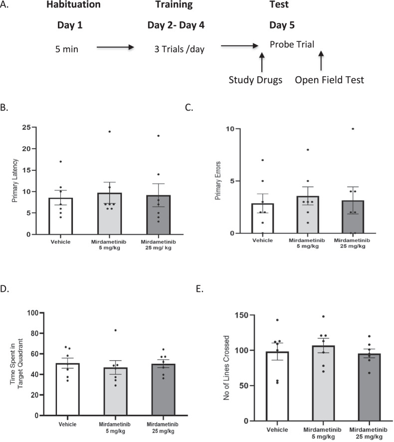
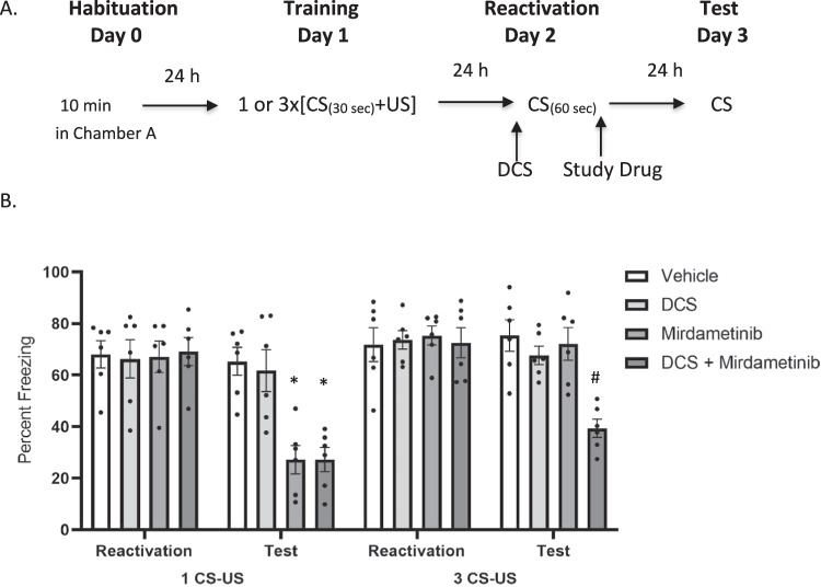
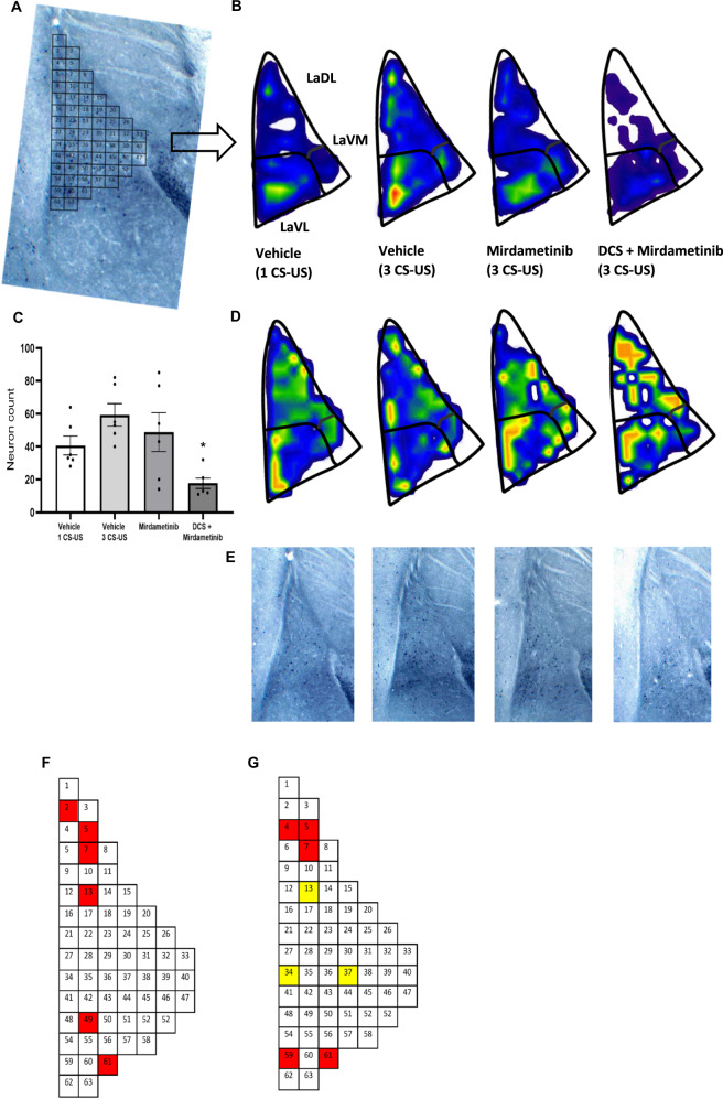
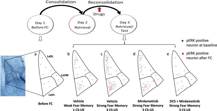
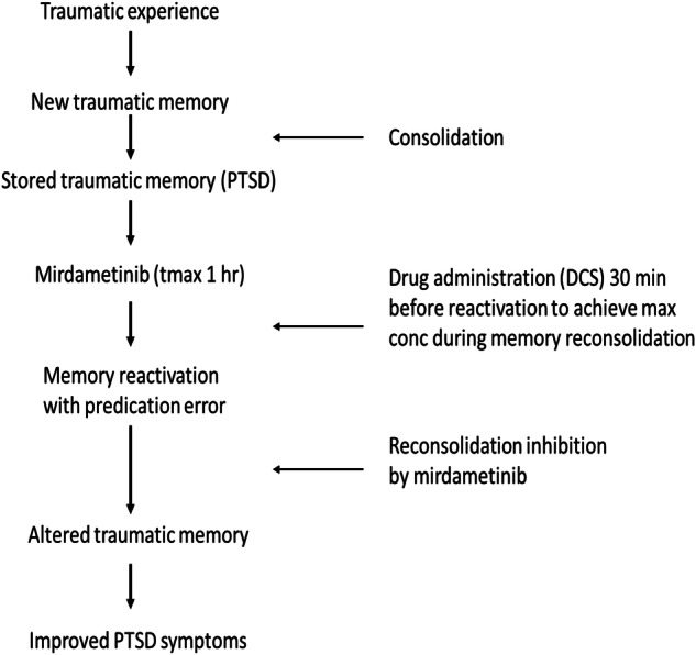
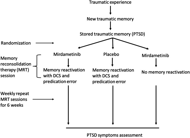
This study establishes mirdametinib as the first MEK inhibitor that can undergo clinical development for psychiatric indications such as post-traumatic stress disorder (PTSD). PTSD is characterized by persistent traumatic memories with limited effective treatment options. A body of evidence suggests that memory storage is dynamic and constantly updated through post-retrieval modification a process termed reconsolidation. Although ERK/MAPK signaling plays a central role in fear memory consolidation, no clinically translatable MEK inhibitor has been tested in experimental models or in clinical trials to disrupt this process. Furthermore, there is need to develop pharmacological and behavioral strategies to labilize the memory to make it susceptible for disruption. Here, we disrupted fear memory reconsolidation with the clinically relevant MEK inhibitor mirdametinib in C57BL/6 mice and tested memory destabilization strategies using an auditory fear conditioning paradigm, with drugs administered following reactivation of memory. We found prediction error effective in labilizing weak fear memory and combined D-cycloserine (DCS) and predication error effective in labilizing strong fear memory. Mirdametinib disrupted the weak fear memory and reduced ERK phosphorylation in lateral amygdala when coupled with prediction error at the time of memory reactivation but required coordinated combination of DCS, prediction error and mirdametinib to disrupt strong fear memory. Barnes maze spatial memory test and open field test revealed that mirdametinib did not affect retrieval of other forms (spatial) of long-term memory and locomotor activity. Furthermore, the effect of mirdametinib was specific to reconsolidation as it had no effect on fear memory when given without reactivation. These translational findings identify a new drug that can be adapted for the treatment of PTSD.


Conflict of interest statement

Competing interests: The authors declare no competing interests.

Figures

Fig. 1
Fig. 1. A single dose of mirdametinib (5, 10, or 25 mg/kg) given after the recall without predication error failed to disrupt fear memory but when given with prediction error at the time of memory reactivation, blocked the reconsolidation of fear memory.
A Schematic representation of experimental protocol used. B Mean percentage of freezing in C57BL/6 mice with vehicle (n = 6), different doses of mirdametinib (5 mg/kg, n = 6; 10 mg/kg, n = 6; 25 mg/kg, n = 6), SL327 (50 mg/kg; n = 6) and trametinib (5 mg/kg; n = 6) in experiment 1. Data: Mean ± SE. C Schematic representation of experimental protocol used. D Mean percentage of freezing in C57BL/6 mice with vehicle (n = 8), different doses of mirdametinib (5 mg/kg, n = 6; 10 mg/kg, n = 6; 25 mg/kg, n = 8), SL327 (50 mg/kg; n = 7) and trametinib (5 mg/kg; n = 6) in experiment 2. Two-way repeated measures ANOVA. * p < 0.05, ** p < 0.01, *** p < 0.001 vs vehicle control Data: Mean ± SE.
Fig. 2
Fig. 2. Mirdametinib high dose (25 mg/kg) and positive control SL327 (50 mg/kg), when given with prediction error at the time of memory reactivation, blocked the reconsolidation of fear memory and reduced pERK activity in LA.
A Schematic representation of experimental protocol used. B Mean percentage of freezing in C57BL/6 mice with vehicle (n = 6), mirdametinib (25 mg/kg, n = 6), SL327 (50 mg/kg, n = 6) in experiment 3. Two-way repeated measures ANOVA. * p < 0.05, ** p < 0.01 vs vehicle. Data Mean ± SE. C Quantitative topographical analysis revealed a reduced number of pERK positive neurons in LA in mirdametinib and SL327 groups compared to vehicle control. D Bin matrix used for dividing the LA superimposed on pERK immunolabeled brain section. E Microanatomical neuron density map depicting the mean spatial distribution of activated neurons in the LA from all study groups at −1.82 Bregma. G Below each map is its coefficient of variance (CV) map, generated by dividing the standard deviation by the mean. F A visual representation of the q value matrix. q < 0.1 were depicted in color for visualization purposes. Multiple comparisons (one-way ANOVA) revealed that 10 out of 63 were statistically different (q < 0.1) between experimental conditions. Subsequently planned comparisons demonstrated that mirdametinib and SL327 reduced a comparable number of neurons (red bins) that were lower than vehicle group in 8/10 bins. In bin 61, mirdametinib was associated with lower neurons (blue bins) than vehicle and SL327 control. Bin 18 was unique to SL327, with a smaller number of pERK positive neurons (yellow bins) in comparison to vehicle and mirdametinib groups. H Representative pictures of each group. IHC Immunohistochemistry.
Fig. 3
Fig. 3. Mirdametinib failed to disrupt fear memory in absence of memory reactivation.
A Schematic representation of experimental protocol used. B Mean percentage of freezing in C57BL/6 mice with vehicle (n = 6) and mirdametinib (25 mg/kg, n = 6) in experiment 4. Two-way repeated measures ANOVA. Data Mean ± SE.
Fig. 4
Fig. 4. Mirdametinib does not cause memory impairment in Barnes maze test or affect locomotor activity in open field test.
A Schematic representation of experimental protocol used. B Primary latency to reach escape box in Barnes maze. C Primary errors before reaching escape box in Barnes maze. D Time spent in target quadrant during probe trial in Barnes maze. E Number of lines crossed in open field test. n = 7/group. One-way ANOVA. Data Mean ± SE.
Fig. 5
Fig. 5. Combined administration of DCS and mirdametinib on the day of memory reactivation, blocked the reconsolidation of strong fear memory.
A Schematic representation of experimental protocol used. B Mean percentage of freezing in C57BL/6 mice in 1 CS-US-saline-vehicle (n = 6), 1 CS-US-DCS-vehicle (DCS 15 mg/kg, n = 6), 1 CS-US-saline-mirdametinib (mirdametinib 25 mg/kg, n = 6), 1 CS-US-DCS-mirdametinib (DCS 15 mg/kg, mirdametinib 25 mg/kg, n = 6), 3 CS-US-saline-vehicle (n = 6), 3 CS-US-DCS-vehicle (DCS 1 mg/kg, n = 6), 3 CS-US-saline-mirdametinib (mirdametinib 25 mg/kg, n = 6), 3 CS-US-DCS-mirdametinib (DCS 15 mg/kg, mirdametinib 25 mg/kg, n = 6) groups. Two-way repeated measures ANOVA. * p < 0.05 vs 1 CS-US-saline-vehicle control. # p < 0.05 vs 3 CS-US-saline-vehicle control. Data Mean ± SE.
Fig. 6
Fig. 6. Combined administration of DCS with mirdametinib reduced pERK positive neurons in LA.
A Bin matrix used for dividing the LA with pERK immunolabeled brain section. B Microanatomical neuron density map showing the mean spatial distribution of pERK expressing neurons in different parts of LA following vehicle administration in weak fear conditioning group and vehicle, mirdametinib and DCS + mirdametinib in strong fear conditioning group at −1.80 Bregma. D Below each map is its coefficient of variance (CV) map, generated by dividing the standard deviation by the mean. C Increase in number of neurons expressing pERK following strong fear conditioning (Group 1: 1 CS-US-saline-vehicle) compared to weak fear conditioning (Group 5: 3 CS-US-saline-vehicle). Combined administration of DCS with mirdametinib (Group 8: 3 CS-US-DCS-mirdametinib) reduced the number of LA neurons with pERK activity compared to vehicle (Group 5: 3 CS-US-saline-vehicle). E Representative pictures of each group. F Post hoc comparison showed increased pERK positive neurons in LaDL and LaVL following strong fear conditioning compared to weak fear conditioning. A visual representation of the q value matrix. q < 0.1 are shown in color for better visualization. Multiple comparisons testing (one way ANOVA) showed that 6 out of 63 were statistically different (q < 0.1) between weak (Group 1: 3 CS-US-sal-vehicle) and strong fear memory groups (Group 5: 3 CS-US- sal-vehicle). Quantitative topographical analysis showed significantly increased pERK immuno-positive neurons in bin 2, 5, 7, 13, 49, 61 in strong fear memory group compared to weak fear memory control. G Post hoc comparison showed combined administration of DCS and mirdametinib reduced pERK positive neurons in LaDL and LaVL. A visual representation of the q value matrix. q < 0.1 are shown in color for better visualization. Multiple comparisons testing (one way ANOVA) showed that 8 out of 63 were statistically different (q < 0.1) between DCS + mirdametinib group and vehicle control (3 CS-US-sal-vehicle). In three out of 63 bins (marked yellow), there was significant difference between both mirdametinib alone (Group 7: 3 CS-US-sal-mirdametinib) and DCS plus mirdametinib group (Group 8: 3 CS-US-DCS-mirdametinib) compared to vehicle control (3 CS-US-sal-vehicle). Quantitative topographical analysis showed significantly reduced pERK immuno-positive neurons in bin 4, 5, 7, 13, 34, 37, 59, 61 in DCS with mirdametinib group compared to vehicle control. LaDL dorsolateral part of LA, LaVL Ventrolateral part of LA, LaVM Ventromedial part of LA.
Fig. 7
Fig. 7. Schematic model showing distribution of pERK-positive neurons in different subregions of LA in different experimental conditions.
a LA neurons with ERK activity at baseline before fear conditioning (FC), b LA neurons with ERK activity after weak fear conditioning (1 CS-US), c LA neurons with ERK activity after strong fear conditioning (3 CS-US), d LA neurons with ERK activity after mirdametinib administration in mice that underwent strong fear conditioning, e LA neurons with ERK activity after combined DCS and mirdametinib administration in mice that underwent strong fear conditioning.
Fig. 8
Fig. 8
Proposed treatment protocol to disrupt trauma memory with mirdametinib to improve symptoms in PTSD patients.
Fig. 9
Fig. 9
Proposed future clinical trial design to evaluate the effect of trauma memory disruption with mirdametinib on symptoms in PTSD patients.

References

    1. Breslau N. The epidemiology of posttraumatic stress disorder: what is the extent of the problem? J Clin Psychiatry. 2001;62:16–22. - PubMed
    1. Yehuda R, Hoge CW, McFarlane AC, Vermetten E, Lanius RA, Nievergelt CM, et al. Post-traumatic stress disorder. Nat Rev Dis Prim. 2015;1:15057. - PubMed
    1. Ressler KJ, Berretta S, Bolshakov VY, Rosso IM, Meloni EG, Rauch SL, et al. Post-traumatic stress disorder: clinical and translational neuroscience from cells to circuits. Nat Rev Neurol. 2022;18:273–88. - PMC - PubMed
    1. Wynn GH, Benedek DM, Johnson L, Choi K, Zhang L, Hu XZ, et al. Chapter 22—Posttraumatic stress disorder. In: Conn PM, editor. Conn’s translational neuroscience. San Diego: Academic Press; 2017. pp. 499–515.
    1. Careaga MBL, Girardi CEN, Suchecki D. Understanding posttraumatic stress disorder through fear conditioning, extinction and reconsolidation. Neurosci Biobehav Rev. 2016;71:48–57. - PubMed