. 2024 Dec 18;15(12):903.
doi: 10.1038/s41419-024-07289-y.

GPx1 deficiency confers increased susceptibility to ferroptosis in macrophages from individuals with active Crohn's disease

Affiliations

GPx1 deficiency confers increased susceptibility to ferroptosis in macrophages from individuals with active Crohn's disease

James A Sousa et al. Cell Death Dis. .

Abstract

Intestinal cell death is a defining feature of Crohn's disease (CD), a major form of inflammatory bowel disease. The focus on this aspect of enteric inflammation has mainly been on epithelial cells, while other cell types such as stromal and myeloid cells have received less attention. Hypothesising that decreased macrophage viability in an oxidative environment could be a contributing factor to the pathophysiology of CD, we found that monocyte-derived macrophages from individuals with active CD (but not those in clinical disease remission) have increased sensitivity to cell death induced by H2O2. Molecular biology and pharmacological studies ruled out apoptosis and necroptosis, while increased lipid peroxidation and surface expression of the transferrin receptor implicated ferroptosis as the mechanism of the H2O2-induced cell death: this was supported by suppression of H2O2-cytotoxicity by liproxstatin-1, a pharmacological inhibitor of ferroptosis. Selenoproteins are important antioxidants, and selenium deficiency can be a feature of CD. Despite normal dietary intake of selenium, monocyte-derived macrophages and intestinal macrophages in individuals with CD had decreased protein and/or mRNA expression of the selenoprotein, glutathione peroxidase (GPx)-1. Knockdown of GPx1 in macrophages from healthy volunteers resulted in increased H2O2-induced cell death reminiscent of that observed with macrophages from CD. In summary, monocyte-derived macrophages from individuals with CD have increased susceptibility to H2O2-induced ferroptosis cell death, that may be facilitated, at least in part, by reduced expression of the antioxidant GPx1. We suggest that reduced GPx1 in monocytes recruited to the gut and intestinal macrophages renders these cells vulnerable to reactive oxygen species-evoked ferroptosis cell death and that unraveling the participation of this pathway in Crohn's disease may reveal novel therapeutic approaches to this chronic condition.


Conflict of interest statement

Competing interests: The authors declare no competing interests. Ethics approval: Ethics approval was issued by the Conjoint Health Research Ethics Board at the University of Calgary (REB24827, REB15-1270, REB19-0402, and REB 20-1534) and the study was performed under accordance with the Declaration of Helsinki.

Figures
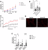
Fig. 1
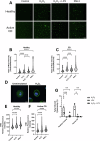
Fig. 1. Macrophages from patients with active CD are more susceptible to H2O2-induced cytotoxicity.
Monocyte-derived macrophages (2.5 × 10⁴ cells) were treated with H2O2, and cell death was assessed using the LDH assay. Macrophages derived from patients with active CD are more susceptible to H2O2-induced cytotoxicity (A) and this difference is statistically significant at 500 µM (B). C A time course of H2O2-induced cell death was performed using SYTOX orange (1 µM) to detect dead cells. H2O2 induced more cell death in macrophages derived from patients with active CD compared with healthy macrophages as indicated by the number of SYTOX-positive cells. N = 3 per group. D Representative images from the 12 h time point. Scale bar = 100 µm. E Macrophages were treated with either IFN-γ (10 ng/mL) or IL-4 (10 ng/mL) for 48 h then cytotoxicity was induced using H2O2 (500 µM, 2 h). Data are means ± SEM (*p < 0.05, **p < 0.005).
Fig. 2
Fig. 2. H2O2 does not induce markers of apoptosis, pyroptosis, or necroptosis.
In healthy macrophages (5 × 10⁵ cells), H2O2 (500 µM, 2 h) does not induce the expression of cleaved caspase-3 (A), IL-1β or IL-18 (B). Necrostatin-1 (25 µM, 30 min pre-treatment) does not affect the cytotoxicity of H2O2 of healthy donor macrophages (C). As determined by qPCR (D), H2O2 (500 µM, 2 h) induces expression of PTGS2, SLC7A11, and TNF (markers of ferroptosis). Data are means ± SEM (*p < 0.05, ***p < 0.0005).
Fig. 3
Fig. 3. H2O2 induces macrophage cytotoxicity through ferroptosis.
Macrophages (62,500 cells) derived from healthy controls and individuals with active CD were treated with H2O2 (500 µM, 2 h) ± liporoxstatin-1 (LPX, 10 µM, 30 min pre-treatment) or RSL3 (1 µM, 2 h) and lipid peroxidation and transferrin receptor 1 (TFR1) staining were assessed. H2O2 induces lipid peroxidation in both healthy macrophages and macrophages derived from patients with active CD (AC; 50 cells per donor were analyzed from 4 healthy controls and 5 patients with active CD. Scale bar = 50 µm). H2O2 and RSL3 induces TFR1 localization to the plasma membrane in healthy (D, E) and active CD patient (F) derived macrophages. (50 cells per donor were analyzed from 4 healthy controls and 3 patients with active CD. Mean fluorescence intensity was normalized to the control. Scale bar = 10 µm. Dashed lines represent the median and dotted lines the quartiles). Liproxstatin-1 blocks the cytotoxic effect of H2O2 in macrophages derived from healthy controls and patients with active CD (G) (*p < 0.05, **p < 0.005, ***p < 0.0005, ****p < 0.0001).
Fig. 4
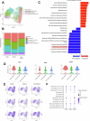
Fig. 4. Macrophages from patients with active CD have reduced selenoprotein mRNA expression in situ.
Published scRNAseq data (SCP1884) was reanalyzed examining colonic macrophage mRNA expression in healthy biopsies and biopsies from patients with CD. (A) UMAP representation of the macrophage clusters. (B) Relative abundance of each cluster between groups. Top differentially expression pathways between scRNAseq data from inflamed biopsies from patients with CD compared with healthy biopsies (C). The same dataset was used to assess selenoprotein mRNA expression of selenoprotein P (SEPP1), glutathione peroxidase (GPx) 1 and GPx4 in macrophages from healthy biopsies and inflamed and non-inflamed biopsies from patients with CD (D) including in the different sub-populations (E, F). (Heal Healthy, Infl Inflamed, NonI Non-inflamed).
Fig. 5
Fig. 5. Lamina propria cells in areas of active inflammation have reduced GPx1 expression.
Colonic biopsies from healthy individuals and those with Crohn’s disease (inflamed areas and non-inflamed areas) were assessed by immunohistochemistry for GPx1 (A, C) and GPx4 (B, D) expression. Representative images show decreased in GPx1-immunoreativity (IR), but not GPx4-IR, in inflamed tissue regions in IBD and as quantified in (C) (n = 6; L, Lumen; LP, lamina propria. Scale bar = 100 μm; *p < 0.05).
Fig. 6
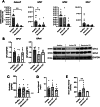
Fig. 6. Monocyte-derived macrophages from patients with active CD have reduced selenoprotein expression despite selenium sufficiency.
Monocyte-derived macrophages from patients with active CD or individuals in disease remission have reduced selective selenoprotein expression as determined by qPCR (A) and immunoblotting (B; densitometry (n = 3) and a representative blot)) as compared to cells from healthy volunteers. Selenium status was assessed by selenium intake using 24 h food recalls (C), serum SelenoP concentration (D), and serum GPx activity (E) by ELISA and enzymatic activity assay, respectively. Data are means ± SEM (*p < 0.05, **p < 0.005; SCLY, Selenocysteine lyase).
Fig. 7
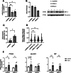
Fig. 7. GPx1 knockout increases susceptibility to H2O2-induced cytotoxicity.
Healthy control monocyte-derived macrophages were transfected with siRNA against GPx1 for 48 h and efficiency of knockdown (KD) was assessed by qPCR (A) and immunoblotting (B) (nsSiRNA, non-specific control SiRNA). GPx1 KD macrophages were treated with H2O2 (500 μM, 2 h) and cell death (C), lipid peroxidation (D; 50 cells per donor were analyzed from 3 healthy) and qPCR of ferroptosis markers (E) was assessed. Data are means ± SEM. Dashed lines represent the media and dotted lines the quartiles (*p < 0.05, **p < 0.005).

References

    1. Moreira Lopes TC, Mosser DM, Goncalves R. Macrophage polarization in intestinal inflammation and gut homeostasis. Inflamm Res. 2020;69:1163–72. - PubMed
    1. Bain CC, Mowat AM. Macrophages in intestinal homeostasis and inflammation. Immunol Rev. 2014;260:102–17. - PMC - PubMed
    1. Du J, Zhang J, Wang L, Wang X, Zhao Y, Lu J, et al. Selective oxidative protection leads to tissue topological changes orchestrated by macrophage during ulcerative colitis. Nat Commun. 2023;14:3675. - PMC - PubMed
    1. Jones GR, Bain CC, Fenton TM, Kelly A, Brown SL, Ivens AC, et al. Dynamics of colon monocyte and macrophage activation during colitis. Front Immunol. 2018;9:2764. - PMC - PubMed
    1. Hunter MM, Wang A, Parhar KS, Johnston MJ, Van Rooijen N, Beck PL, et al. In vitro-derived alternatively activated macrophages reduce colonic inflammation in mice. Gastroenterology. 2010;138:1395–405. - PubMed

MeSH terms