. 2024 Dec 18;15(12):909.
doi: 10.1038/s41419-024-07304-2.

DARPins as a novel tool to detect and degrade p73

Affiliations

DARPins as a novel tool to detect and degrade p73

Philipp Münick et al. Cell Death Dis. .

Abstract

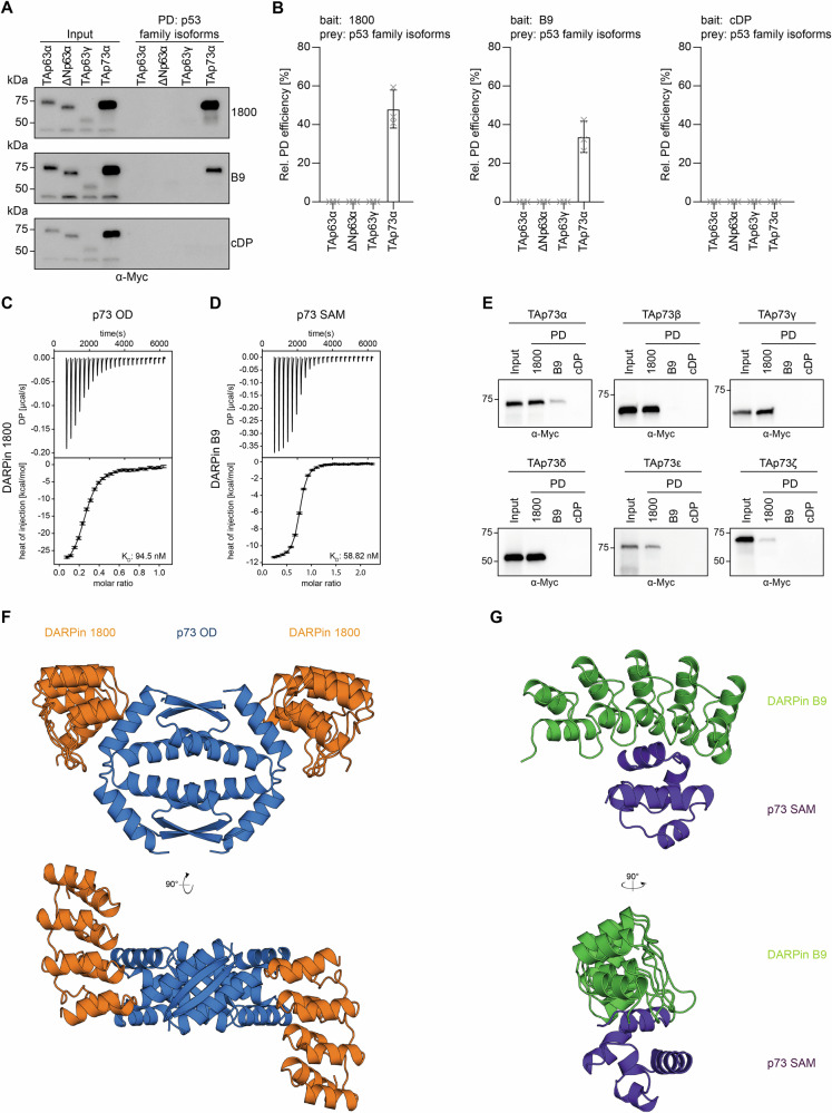
The concept of Targeted Protein Degradation (TPD) has been introduced as an attractive alternative to the development of classical inhibitors. TPD can extend the range of proteins that can be pharmacologically targeted beyond the classical targets for small molecule inhibitors, as a binding pocket is required but its occupancy does not need to lead to inhibition. The method is based on either small molecules that simultaneously bind to a protein of interest and to a cellular E3 ligase and bring them in close proximity (molecular glue) or a bi-functional molecule synthesized from the chemical linkage of a target protein-specific small molecule and one that binds to an E3 ligase (Proteolysis Targeting Chimeras (PROTAC)). The further extension of this approach to bioPROTACs, in which a small protein-based binding module is fused directly to an E3 ligase or an E3 ligase adaptor protein, makes virtually all proteins amenable to targeted degradation, as this method eliminates the requirement for binding pockets for small molecules. Designed Ankyrin Repeat Proteins (DARPins) represent a very attractive class of small protein-based binding modules that can be used for the development of bioPTOTACS. Here we describe the characterization of two DARPins generated against the oligomerization domain and the SAM domain of the transcription factor p73, a member of the p53 protein family. The DARPins can be used for (isoform-)selective pulldown experiments both in cell culture as well as primary tissue lysates. We also demonstrate that they can be used for staining in cell culture experiments. Fusing them to the speckle type POZ protein (SPOP), an adaptor protein for cullin-3 E3 ligase complexes, yields highly selective and effective degraders. We demonstrate that selective degradation of the ΔNp73α isoform reactivates p53.


Conflict of interest statement

Competing interests: Andreas Plückthun is a cofounder and shareholder of Molecular Partners AG, who are commercializing the DARPin technology. All other authors declare that they have no competing interests.

Figures

Fig. 1
Fig. 1. Characterization of DARPins binding to p73 domains.
A Pulldown experiment with stable HeLa cell lines expressing different Myc-tagged p63 or p73 isoforms. Input signals are shown on the left, signals after pulldown on the right. DARPin 1800 and DARPin B9 bind only to p73 but not to p63 isoforms. B Quantification of the pulldown experiments shown in (A). The relative pulldown efficiency normalized to the input samples is shown on the y-axis. The bar diagram shows the mean values and the error bars show the corresponding SD of three biological replicates. An ordinary one-way ANOVA analysis was performed to assess the statistical significance. C Interaction study of the p73 OD and DARPin 1800 using ITC. The top diagram shows the raw measurement and the bottom diagram the integrated heat per titration step. The KD value for the interaction is given in the bottom right corner. The measurement was performed at 25 °C. D Interaction study of the p73 SAM domain and DARPin B9 using ITC. The top diagram shows the raw measurement and the bottom diagram the integrated heat per titration step. The KD value for the interaction is given in the bottom right corner. The measurement was performed at 25 °C. E Pulldown experiments with different Myc-tagged p73 isoforms expressed in Rabbit Reticulocyte Lysate (RRL). Input signals are shown on the left, signals after pulldown on the right. The experiments were performed in biological triplicates with exemplary blots of one replicate shown. F Crystal structure of DARPin 1800 (orange) in complex with the p73 OD (blue) shown in two different orientations rotated by 90°. G Crystal structure of DARPin B9 (green) in complex with the p73 SAM domain (purple) shown in two different orientations rotated by 90°.
Fig. 2
Fig. 2. Dimerization of the selected DARPins increases their functional affinity.
A Interaction study of p73 DBD-OD-SAM and DARPin 1800 (left panel), DARPin 1800 dimerized via a leucine zipper (DARPin 1800 LZ, middle panel) and DARPin 1800 fused to another DARPin 1800 via a (G4S)4 linker (DARPin 1800–1800, right panel) using ITC. The top diagram shows the raw measurement and the bottom diagram the integrated heat per titration step. The KD value for the interaction is given in the bottom right corner. The measurement was performed at 25 °C. B Same experiment as in (A) but with DARPin B9, DARPin B9 LZ and DARPin B9-B9.
Fig. 3
Fig. 3. Detection of p73 isoforms in stable expressing U-2 OS cells.
Cells expressing Myc-tagged TAp73α (A) or Myc-Tagged ∆Np73α (B) were fixed with formaldehyde and incubated with the indicated HA-tagged DARPin followed by incubation with goat anti-HA antibody (a190138a—Bethyl) and the secondary antibody Alexa Fluor 568 anti-goat (A11057—Life Technologies). The same cells were also incubated with mouse anti-myc antibody 4A6 (Millipore) and Alexa Fluor 647 anti-mouse antibody (A31571—Life Technologies) as both p73α isoforms were Myc-tagged. The control DARPin constructs do not show any signal above background.
Fig. 4
Fig. 4. Detection of p73 in primary tissue.
A Pulldown (PD) of p73 from mouse skin extract. The p73 OD-specific DARPin 1800 and its dimerized version 1800 LZ, the p73 SAM-specific DARPin B9 and its dimerized version B9 LZ, the DBD-specific DARPin C14 and its dimerized version C14 LZ as well as the respective control DARPin constructs were biotinylated and immobilized on Streptavidin magnetic beads followed by incubation with the mouse skin extract. Multiple bands corresponding to different isoforms of p73 were detected. However, it is not possible to identify the isoform based on the blot due to the similar molecular weight of several isoforms. The pulldown was analyzed by western blot with an anti-p73 antibody (ab 40658). All DARPins except the control DARPin constructs show pulldown signals. B Same experiment as in (a) but with mouse brain extract. Both experiments were done in biological triplicates with one representative replicate shown.
Fig. 5
Fig. 5. DARPin-based PROTACs degrade p73.
A Schematic representation of the domain structure of the used E3-ligases (left). The natural substrate binding domain is colored in red. On the right side, a schematic representation of the DARPin-E3 chimera constructs is shown in which the substrate binding domain has been replaced by the DARPin (colored in green). B Screening of the efficiency of different DARPin-based PROTACs by a luciferase reporter assay using H1299 cells. The p73 OD-specific DARPin 1800, the p73 SAM-specific DARPin B9 and the DBD-specific DARPin C14 were fused to different E3-ligases or the E3-ligase adaptor protein SPOP. The resulting DARPin fusion constructs were co-transfected with HiBit-tagged TAp73α and the TAp73α protein level was determined 24 h after transfection using the HiBiT dual-luciferase reporter assay (Promega). For normalization, firefly luciferase was expressed from the same plasmid using an IRES. Fusion of the control DARPin to the respective E3-ligase served as negative control. Protein levels were determined by calculating the ratio between NanoLuc luminescence and Firefly luminescence and normalized to a sample not containing any DARPin or degrader. The assay was performed in biological triplicates. C Quantitative analysis of the transcriptional activity of TAp73α in the presence of the respective DARPin or DARPin-SPOP fusion. Triangles indicate increasing amounts of transfected DARPin or DARPin-SPOP plasmid DNA (5–25 ng plasmid DNA). The assay was performed in biological triplicates. The assay was performed in H1299 cells. D Western Blot of the experiment in (C). Myc-tagged p73 was detected using the anti-myc antibody 4A6 (millipore). Vinculin protein level was detected as a loading control using the anti-vinculin antibody 7F9 (Santa Cruz Biotechnology). Triangles indicate increasing amounts of transfected DARPin or DARPin-SPOP plasmid DNA (5–25 ng plasmid DNA). The bar diagrams in (B) and (C) represent mean values of three biological replicates and the error bars the respective standard deviations. Statistical significance was assessed by ordinary one‐way ANOVA (n.s.: P > 0.05, *P ≤ 0.05, **P ≤ 0.01, ***P ≤ 0.001, ****P ≤ 0.0001).
Fig. 6
Fig. 6. DARPin-SPOP fusions restore transcriptional activity of p53 in presence of ΔNp73α and the DARPin-SPOP induced degradation of TAp73α is dependent on the proteosomal pathway.
A Schematic representation of the inhibitory effect of ΔNp73α on the transcriptional activity of p53. High concentrations of ΔNp73α (yellow) displace p53 (orange) from the promoter sequences (left). Degradation of ΔNp73α by DARPin-SPOP fusions enables binding of promotor sequences and thus transcription of the affected genes (right). (adapted from [29]). B Quantitative analysis of the transcriptional activity of Myc-tagged p53 in the presence of Myc-tagged ΔNp73α with or without the respective DARPin-SPOP fusion. Degradation of ΔNp73α by DARPin-SPOP fusions restores p53 transcriptional activity. The assay was performed in H1299 cells. The bar diagrams show the mean values and error bars of the corresponding SD of three biological replicates. Statistical significance was assessed by ordinary one-way ANOVA (n.s.: P > 0.05, *P ≤ 0.05, **P ≤ 0.01, ***P ≤ 0.001, ****P ≤ 0.0001). C Western Blot of the experiment in (B). Myc-tagged p53 and Myc-tagged ΔNp73α were detected using the anti-myc antibody 4A6 (millipore). Vinculin was detected as a loading control using the anti-vinculin antibody 7F9 (Santa Cruz Biotechnology). Triangles indicate increasing amounts of transfected DARPin or DARPin-SPOP plasmid DNA (5–25 ng plasmid DNA). D Proteasome inhibition assay to investigate if the degradation is dependent on the proteosomal pathway. U-2 OS Flp-In/T-REx cells stably expressing Myc-tagged TAp73α were transfected with 5 ng or 25 ng DNA of selected HA-tagged degrader (1800-SPOP, B9-SPOP and C14-SPOP). cDP-SPOP was used as negative control. 12 h after transfection, cells were treated with DMSO or the proteasome inhibitor bortezomib. TAp73α degradation with and without proteasome inhibitor was determined by SDS-PAGE followed by western blot with the anti-myc antibody 4A6 (millipore). The degraders were detected using the anti-HA antibody 16B12 (Biolegend), vinculin protein level was detected as a loading control using the anti-vinculin antibody 7F9 (Santa Cruz Biotechnology). The experiment was performed in biological triplicates with one representative replicate shown.

References

    1. Lemaitre T, Cornu M, Schwalen F, Since M, Kieffer C, Voisin-Chiret AS. Molecular glue degraders: exciting opportunities for novel drug discovery. Expert Opin Drug Discov. 2024;19:433–49. - PubMed
    1. Békés M, Langley DR, Crews CM. PROTAC targeted protein degraders: the past is prologue. Nat Rev Drug Discov. 2022;21:181–200. - PMC - PubMed
    1. Cowan AD, Ciulli A. Driving E3 ligase substrate specificity for targeted protein degradation: lessons from nature and the laboratory. Annu Rev Biochem. 2022;91:295–319. - PubMed
    1. Hanzl A, Winter GE. Targeted protein degradation: current and future challenges. Curr Opin Chem Biol. 2020;56:35–41. - PMC - PubMed
    1. Clift D, McEwan WA, Labzin LI, Konieczny V, Mogessie B, James LC, et al. A method for the acute and rapid degradation of endogenous proteins. Cell. 2017;171:1692. - PMC - PubMed

Substances

LinkOut - more resources